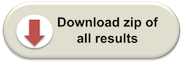
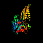
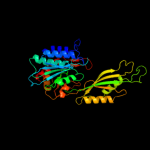
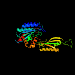
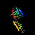
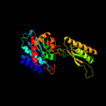
| 1 | c2imoA_
|
|
 |
100.0 |
95 |
PDB header:hydrolase
Chain: A: PDB Molecule:allantoate amidohydrolase;
PDBTitle: crystal structure of allantoate amidohydrolase from escherichia coli2 at ph 4.6
|
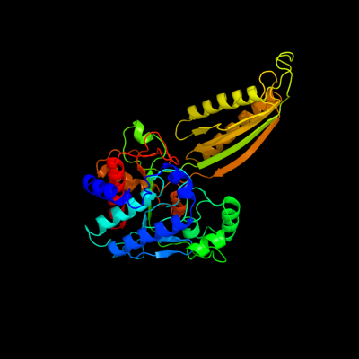
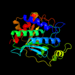
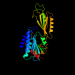
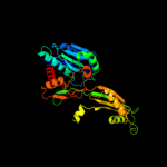
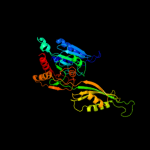
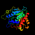
| 2 | c3n5fB_
|
|
 |
100.0 |
31 |
PDB header:hydrolase
Chain: B: PDB Molecule:n-carbamoyl-l-amino acid hydrolase;
PDBTitle: crystal structure of l-n-carbamoylase from geobacillus2 stearothermophilus cect43
|
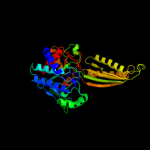
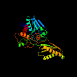
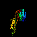
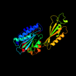
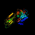
| 3 | c2v8gD_
|
|
 |
100.0 |
27 |
PDB header:hydrolase
Chain: D: PDB Molecule:beta-alanine synthase;
PDBTitle: crystal structure of beta-alanine synthase from2 saccharomyces kluyveri in complex with the product beta-3 alanine
|
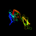
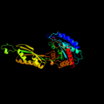
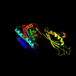
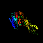
| 4 | d1z2la1
|
|
 |
100.0 |
100 |
Fold:Phosphorylase/hydrolase-like
Superfamily:Zn-dependent exopeptidases
Family:Bacterial dinuclear zinc exopeptidases |
| 5 | c3ramC_
|
|
 |
100.0 |
17 |
PDB header:hydrolase
Chain: C: PDB Molecule:hmra protein;
PDBTitle: crystal structure of hmra
|
| 6 | c3gb0A_
|
|
 |
100.0 |
20 |
PDB header:hydrolase
Chain: A: PDB Molecule:peptidase t;
PDBTitle: crystal structure of aminopeptidase pept (np_980509.1) from bacillus2 cereus atcc 10987 at 2.04 a resolution
|
| 7 | c3pfoB_
|
|
 |
100.0 |
16 |
PDB header:hydrolase
Chain: B: PDB Molecule:putative acetylornithine deacetylase;
PDBTitle: crystal structure of a putative acetylornithine deacetylase (rpa2325)2 from rhodopseudomonas palustris cga009 at 1.90 a resolution
|
| 8 | c2zogA_
|
|
 |
100.0 |
13 |
PDB header:hydrolase
Chain: A: PDB Molecule:cytosolic non-specific dipeptidase;
PDBTitle: crystal structure of mouse carnosinase cn2 complexed with zn and2 bestatin
|
| 9 | c1cg2D_
|
|
 |
100.0 |
18 |
PDB header:metallocarboxypeptidase
Chain: D: PDB Molecule:carboxypeptidase g2;
PDBTitle: carboxypeptidase g2
|
| 10 | c3ifeA_
|
|
 |
100.0 |
17 |
PDB header:hydrolase
Chain: A: PDB Molecule:peptidase t;
PDBTitle: 1.55 angstrom resolution crystal structure of peptidase t (pept-1)2 from bacillus anthracis str. 'ames ancestor'.
|
| 11 | c1vixA_
|
|
 |
100.0 |
17 |
PDB header:hydrolase
Chain: A: PDB Molecule:peptidase t;
PDBTitle: crystal structure of a putative peptidase t
|
| 12 | c2pokB_
|
|
 |
100.0 |
14 |
PDB header:hydrolase
Chain: B: PDB Molecule:peptidase, m20/m25/m40 family;
PDBTitle: crystal structure of a m20 family metallo peptidase from streptococcus2 pneumoniae
|
| 13 | c3dljB_
|
|
 |
100.0 |
15 |
PDB header:hydrolase
Chain: B: PDB Molecule:beta-ala-his dipeptidase;
PDBTitle: crystal structure of human carnosine dipeptidase 1
|
| 14 | c1vgyB_
|
|
 |
100.0 |
15 |
PDB header:structural genomics, unknown function
Chain: B: PDB Molecule:succinyl-diaminopimelate desuccinylase;
PDBTitle: crystal structure of succinyl diaminopimelate desuccinylase
|
| 15 | c3rzaA_
|
|
 |
100.0 |
20 |
PDB header:hydrolase
Chain: A: PDB Molecule:tripeptidase;
PDBTitle: crystal structure of a tripeptidase (sav1512) from staphylococcus2 aureus subsp. aureus mu50 at 2.10 a resolution
|
| 16 | c2rb7A_
|
|
 |
100.0 |
15 |
PDB header:hydrolase
Chain: A: PDB Molecule:peptidase, m20/m25/m40 family;
PDBTitle: crystal structure of co-catalytic metallopeptidase (yp_387682.1) from2 desulfovibrio desulfuricans g20 at 1.60 a resolution
|
| 17 | c2qyvB_
|
|
 |
100.0 |
18 |
PDB header:hydrolase
Chain: B: PDB Molecule:xaa-his dipeptidase;
PDBTitle: crystal structure of putative xaa-his dipeptidase (yp_718209.1) from2 haemophilus somnus 129pt at 2.11 a resolution
|
| 18 | c3mruB_
|
|
 |
100.0 |
16 |
PDB header:hydrolase
Chain: B: PDB Molecule:aminoacyl-histidine dipeptidase;
PDBTitle: crystal structure of aminoacylhistidine dipeptidase from vibrio2 alginolyticus
|
| 19 | c1ysjB_
|
|
 |
100.0 |
14 |
PDB header:hydrolase
Chain: B: PDB Molecule:protein yxep;
PDBTitle: crystal structure of bacillus subtilis yxep protein (apc1829), a2 dinuclear metal binding peptidase from m20 family
|
| 20 | d1r3na1
|
|
 |
100.0 |
31 |
Fold:Phosphorylase/hydrolase-like
Superfamily:Zn-dependent exopeptidases
Family:Bacterial dinuclear zinc exopeptidases |
| 21 | c3ic1A_ |
|
not modelled |
100.0 |
15 |
PDB header:hydrolase
Chain: A: PDB Molecule:succinyl-diaminopimelate desuccinylase;
PDBTitle: crystal structure of zinc-bound succinyl-diaminopimelate desuccinylase2 from haemophilus influenzae
|
| 22 | c3pfeA_ |
|
not modelled |
100.0 |
15 |
PDB header:hydrolase
Chain: A: PDB Molecule:succinyl-diaminopimelate desuccinylase;
PDBTitle: crystal structure of a m20a metallo peptidase (dape, lpg0809) from2 legionella pneumophila subsp. pneumophila str. philadelphia 1 at 1.503 a resolution
|
| 23 | c1lfwA_ |
|
not modelled |
100.0 |
14 |
PDB header:hydrolase
Chain: A: PDB Molecule:pepv;
PDBTitle: crystal structure of pepv
|
| 24 | c3tx8A_ |
|
not modelled |
100.0 |
15 |
PDB header:hydrolase
Chain: A: PDB Molecule:succinyl-diaminopimelate desuccinylase;
PDBTitle: crystal structure of a succinyl-diaminopimelate desuccinylase (arge)2 from corynebacterium glutamicum atcc 13032 at 2.97 a resolution
|
| 25 | c3io1B_ |
|
not modelled |
100.0 |
16 |
PDB header:hydrolase
Chain: B: PDB Molecule:aminobenzoyl-glutamate utilization protein;
PDBTitle: crystal structure of aminobenzoyl-glutamate utilization2 protein from klebsiella pneumoniae
|
| 26 | c2f7vA_ |
|
not modelled |
100.0 |
13 |
PDB header:hydrolase
Chain: A: PDB Molecule:aectylcitrulline deacetylase;
PDBTitle: structure of acetylcitrulline deacetylase complexed with2 one co
|
| 27 | c3ct9B_ |
|
not modelled |
100.0 |
18 |
PDB header:hydrolase
Chain: B: PDB Molecule:acetylornithine deacetylase;
PDBTitle: crystal structure of a putative zinc peptidase (np_812461.1) from2 bacteroides thetaiotaomicron vpi-5482 at 2.31 a resolution
|
| 28 | c2q43A_ |
|
not modelled |
100.0 |
18 |
PDB header:hydrolase
Chain: A: PDB Molecule:iaa-amino acid hydrolase ilr1-like 2;
PDBTitle: ensemble refinement of the protein crystal structure of iaa-aminoacid2 hydrolase from arabidopsis thaliana gene at5g56660
|
| 29 | c3khzA_ |
|
not modelled |
100.0 |
16 |
PDB header:hydrolase
Chain: A: PDB Molecule:putative dipeptidase sacol1801;
PDBTitle: crystal structure of r350a mutant of staphylococcus aureus2 metallopeptidase (sapep/dape) in the apo-form
|
| 30 | c1vheA_ |
|
not modelled |
100.0 |
17 |
PDB header:structural genomics, unknown function
Chain: A: PDB Molecule:aminopeptidase/glucanase homolog;
PDBTitle: crystal structure of a aminopeptidase/glucanase homolog
|
| 31 | c3kl9F_ |
|
not modelled |
100.0 |
14 |
PDB header:hydrolase
Chain: F: PDB Molecule:glutamyl aminopeptidase;
PDBTitle: crystal structure of pepa from streptococcus pneumoniae
|
| 32 | c1yloA_ |
|
not modelled |
100.0 |
15 |
PDB header:structural genomics, unknown function
Chain: A: PDB Molecule:hypothetical protein sf2450;
PDBTitle: crystal structure of protein of unknown function (possible2 aminopeptidase) s2589 from shigella flexneri 2a str. 2457t
|
| 33 | c2cf4A_ |
|
not modelled |
100.0 |
17 |
PDB header:hydrolase
Chain: A: PDB Molecule:protein ph0519;
PDBTitle: pyrococcus horikoshii tet1 peptidase can assemble into a2 tetrahedron or a large octahedral shell
|
| 34 | c1y0yA_ |
|
not modelled |
100.0 |
17 |
PDB header:hydrolase
Chain: A: PDB Molecule:frv operon protein frvx;
PDBTitle: crystal structure of tetrahedral aminopeptidase from p. horikoshii in2 complex with amastatin
|
| 35 | d1vixa1 |
|
not modelled |
100.0 |
22 |
Fold:Phosphorylase/hydrolase-like
Superfamily:Zn-dependent exopeptidases
Family:Bacterial dinuclear zinc exopeptidases |
| 36 | c1vhoA_ |
|
not modelled |
100.0 |
13 |
PDB header:structural genomics, unknown function
Chain: A: PDB Molecule:endoglucanase;
PDBTitle: crystal structure of a putative peptidase/endoglucanase
|
| 37 | c3isxA_ |
|
not modelled |
100.0 |
12 |
PDB header:hydrolase
Chain: A: PDB Molecule:endoglucanase;
PDBTitle: crystal structure of endoglucanase (tm1050) from thermotoga2 maritima at 1.40 a resolution
|
| 38 | c2pe3A_ |
|
not modelled |
100.0 |
19 |
PDB header:hydrolase
Chain: A: PDB Molecule:354aa long hypothetical operon protein frv;
PDBTitle: crystal structure of frv operon protein frvx (ph1821)from pyrococcus2 horikoshii ot3
|
| 39 | d1fnoa4 |
|
not modelled |
100.0 |
22 |
Fold:Phosphorylase/hydrolase-like
Superfamily:Zn-dependent exopeptidases
Family:Bacterial dinuclear zinc exopeptidases |
| 40 | c2fvgA_ |
|
not modelled |
100.0 |
15 |
PDB header:hydrolase
Chain: A: PDB Molecule:endoglucanase;
PDBTitle: crystal structure of endoglucanase (tm1049) from thermotoga maritima2 at 2.01 a resolution
|
| 41 | d1lfwa1 |
|
not modelled |
100.0 |
23 |
Fold:Phosphorylase/hydrolase-like
Superfamily:Zn-dependent exopeptidases
Family:Bacterial dinuclear zinc exopeptidases |
| 42 | c3cpxC_ |
|
not modelled |
99.9 |
13 |
PDB header:hydrolase
Chain: C: PDB Molecule:aminopeptidase, m42 family;
PDBTitle: crystal structure of putative m42 glutamyl aminopeptidase2 (yp_676701.1) from cytophaga hutchinsonii atcc 33406 at 2.39 a3 resolution
|
| 43 | d1xfoa2 |
|
not modelled |
99.9 |
25 |
Fold:Phosphorylase/hydrolase-like
Superfamily:Zn-dependent exopeptidases
Family:Bacterial dinuclear zinc exopeptidases |
| 44 | d1vhoa2 |
|
not modelled |
99.9 |
25 |
Fold:Phosphorylase/hydrolase-like
Superfamily:Zn-dependent exopeptidases
Family:Bacterial dinuclear zinc exopeptidases |
| 45 | d1vhea2 |
|
not modelled |
99.9 |
27 |
Fold:Phosphorylase/hydrolase-like
Superfamily:Zn-dependent exopeptidases
Family:Bacterial dinuclear zinc exopeptidases |
| 46 | d1yloa2 |
|
not modelled |
99.9 |
20 |
Fold:Phosphorylase/hydrolase-like
Superfamily:Zn-dependent exopeptidases
Family:Bacterial dinuclear zinc exopeptidases |
| 47 | c3t6mA_ |
|
not modelled |
99.9 |
20 |
PDB header:hydrolase
Chain: A: PDB Molecule:succinyl-diaminopimelate desuccinylase;
PDBTitle: crystal structure of the catalytic domain of dape protein from2 v.cholerea in the zn bound form
|
| 48 | d1cg2a1 |
|
not modelled |
99.9 |
22 |
Fold:Phosphorylase/hydrolase-like
Superfamily:Zn-dependent exopeptidases
Family:Bacterial dinuclear zinc exopeptidases |
| 49 | c2greC_ |
|
not modelled |
99.9 |
16 |
PDB header:hydrolase
Chain: C: PDB Molecule:deblocking aminopeptidase;
PDBTitle: crystal structure of deblocking aminopeptidase from bacillus cereus
|
| 50 | d2fvga2 |
|
not modelled |
99.9 |
23 |
Fold:Phosphorylase/hydrolase-like
Superfamily:Zn-dependent exopeptidases
Family:Bacterial dinuclear zinc exopeptidases |
| 51 | d1xmba1 |
|
not modelled |
99.9 |
22 |
Fold:Phosphorylase/hydrolase-like
Superfamily:Zn-dependent exopeptidases
Family:Bacterial dinuclear zinc exopeptidases |
| 52 | d1ysja1 |
|
not modelled |
99.8 |
21 |
Fold:Phosphorylase/hydrolase-like
Superfamily:Zn-dependent exopeptidases
Family:Bacterial dinuclear zinc exopeptidases |
| 53 | c1q7lA_ |
|
not modelled |
99.8 |
17 |
PDB header:hydrolase
Chain: A: PDB Molecule:aminoacylase-1;
PDBTitle: zn-binding domain of the t347g mutant of human aminoacylase-2 i
|
| 54 | d1vgya1 |
|
not modelled |
99.8 |
15 |
Fold:Phosphorylase/hydrolase-like
Superfamily:Zn-dependent exopeptidases
Family:Bacterial dinuclear zinc exopeptidases |
| 55 | d2grea2 |
|
not modelled |
99.8 |
13 |
Fold:Phosphorylase/hydrolase-like
Superfamily:Zn-dependent exopeptidases
Family:Bacterial dinuclear zinc exopeptidases |
| 56 | d1z2la2 |
|
not modelled |
99.8 |
100 |
Fold:Ferredoxin-like
Superfamily:Bacterial exopeptidase dimerisation domain
Family:Bacterial exopeptidase dimerisation domain |
| 57 | d1tkja1 |
|
not modelled |
99.7 |
22 |
Fold:Phosphorylase/hydrolase-like
Superfamily:Zn-dependent exopeptidases
Family:Bacterial dinuclear zinc exopeptidases |
| 58 | d1r3na2 |
|
not modelled |
99.6 |
26 |
Fold:Ferredoxin-like
Superfamily:Bacterial exopeptidase dimerisation domain
Family:Bacterial exopeptidase dimerisation domain |
| 59 | d1cg2a2 |
|
not modelled |
99.6 |
15 |
Fold:Ferredoxin-like
Superfamily:Bacterial exopeptidase dimerisation domain
Family:Bacterial exopeptidase dimerisation domain |
| 60 | d1rtqa_ |
|
not modelled |
99.5 |
24 |
Fold:Phosphorylase/hydrolase-like
Superfamily:Zn-dependent exopeptidases
Family:Bacterial dinuclear zinc exopeptidases |
| 61 | d1vgya2 |
|
not modelled |
99.5 |
14 |
Fold:Ferredoxin-like
Superfamily:Bacterial exopeptidase dimerisation domain
Family:Bacterial exopeptidase dimerisation domain |
| 62 | c2glfB_ |
|
not modelled |
99.4 |
13 |
PDB header:hydrolase
Chain: B: PDB Molecule:probable m18-family aminopeptidase 1;
PDBTitle: crystal structure of aminipeptidase (m18 family) from thermotoga2 maritima
|
| 63 | d1ysja2 |
|
not modelled |
99.4 |
13 |
Fold:Ferredoxin-like
Superfamily:Bacterial exopeptidase dimerisation domain
Family:Bacterial exopeptidase dimerisation domain |
| 64 | c1q7lB_ |
|
not modelled |
99.4 |
19 |
PDB header:hydrolase
Chain: B: PDB Molecule:aminoacylase-1;
PDBTitle: zn-binding domain of the t347g mutant of human aminoacylase-2 i
|
| 65 | c3tc8A_ |
|
not modelled |
99.4 |
17 |
PDB header:hydrolase
Chain: A: PDB Molecule:leucine aminopeptidase;
PDBTitle: crystal structure of a hypothetical zn-dependent exopeptidase2 (bdi_3547) from parabacteroides distasonis atcc 8503 at 1.06 a3 resolution
|
| 66 | c2ijzF_ |
|
not modelled |
99.3 |
17 |
PDB header:hydrolase
Chain: F: PDB Molecule:probable m18-family aminopeptidase 2;
PDBTitle: crystal structure of aminopeptidase
|
| 67 | c3l6sA_ |
|
not modelled |
99.3 |
13 |
PDB header:hydrolase
Chain: A: PDB Molecule:aspartyl aminopeptidase;
PDBTitle: crystal structure of human aspartyl aminopeptidase (dnpep),2 in complex with aspartic acid hydroxamate
|
| 68 | c3pb6X_ |
|
not modelled |
99.2 |
22 |
PDB header:transferase
Chain: X: PDB Molecule:glutaminyl-peptide cyclotransferase-like protein;
PDBTitle: crystal structure of the catalytic domain of human golgi-resident2 glutaminyl cyclase at ph 6.5
|
| 69 | d2afwa1 |
|
not modelled |
99.2 |
20 |
Fold:Phosphorylase/hydrolase-like
Superfamily:Zn-dependent exopeptidases
Family:Glutaminyl-peptide cyclotransferase-like |
| 70 | c3guxA_ |
|
not modelled |
99.1 |
14 |
PDB header:hydrolase
Chain: A: PDB Molecule:putative zn-dependent exopeptidase;
PDBTitle: crystal structure of a putative zn-dependent exopeptidase (bvu_1317)2 from bacteroides vulgatus atcc 8482 at 1.80 a resolution
|
| 71 | d1y0ya2 |
|
not modelled |
99.1 |
23 |
Fold:Phosphorylase/hydrolase-like
Superfamily:Zn-dependent exopeptidases
Family:Bacterial dinuclear zinc exopeptidases |
| 72 | d3bi1a3 |
|
not modelled |
99.0 |
23 |
Fold:Phosphorylase/hydrolase-like
Superfamily:Zn-dependent exopeptidases
Family:FolH catalytic domain-like |
| 73 | d1xmba2 |
|
not modelled |
98.9 |
21 |
Fold:Ferredoxin-like
Superfamily:Bacterial exopeptidase dimerisation domain
Family:Bacterial exopeptidase dimerisation domain |
| 74 | d1de4c3 |
|
not modelled |
98.6 |
15 |
Fold:Phosphorylase/hydrolase-like
Superfamily:Zn-dependent exopeptidases
Family:FolH catalytic domain-like |
| 75 | d1lfwa2 |
|
not modelled |
98.6 |
13 |
Fold:Ferredoxin-like
Superfamily:Bacterial exopeptidase dimerisation domain
Family:Bacterial exopeptidase dimerisation domain |
| 76 | c1y7eA_ |
|
not modelled |
98.5 |
23 |
PDB header:hydrolase
Chain: A: PDB Molecule:probable m18-family aminopeptidase 1;
PDBTitle: the crystal structure of aminopeptidase i from borrelia burgdorferi2 b31
|
| 77 | c3iibA_ |
|
not modelled |
98.4 |
29 |
PDB header:hydrolase
Chain: A: PDB Molecule:peptidase m28;
PDBTitle: crystal structure of peptidase m28 precursor (yp_926796.1) from2 shewanella amazonensis sb2b at 1.70 a resolution
|
| 78 | c2ek8A_ |
|
not modelled |
98.3 |
21 |
PDB header:hydrolase
Chain: A: PDB Molecule:aminopeptidase;
PDBTitle: aminopeptidase from aneurinibacillus sp. strain am-1
|
| 79 | d1y7ea2 |
|
not modelled |
98.3 |
23 |
Fold:Phosphorylase/hydrolase-like
Superfamily:Zn-dependent exopeptidases
Family:Bacterial dinuclear zinc exopeptidases |
| 80 | c2gljR_ |
|
not modelled |
98.2 |
22 |
PDB header:hydrolase
Chain: R: PDB Molecule:
PDBTitle: crystal structure of aminopeptidase i from clostridium2 acetobutylicum
|
| 81 | c2ootA_ |
|
not modelled |
98.0 |
26 |
PDB header:hydrolase
Chain: A: PDB Molecule:glutamate carboxypeptidase 2;
PDBTitle: a high resolution structure of ligand-free human glutamate2 carboxypeptidase ii
|
| 82 | c3rbuA_ |
|
not modelled |
98.0 |
25 |
PDB header:hydrolase/hydrolase inhibitor
Chain: A: PDB Molecule:glutamate carboxypeptidase 2;
PDBTitle: n-terminally avitev-tagged human glutamate carboxypeptidase ii in2 complex with 2-pmpa
|
| 83 | c1cx8F_ |
|
not modelled |
97.9 |
14 |
PDB header:metal transport
Chain: F: PDB Molecule:transferrin receptor protein;
PDBTitle: crytal structure of the ectodomain of human transferrin receptor
|
| 84 | d1fnoa3 |
|
not modelled |
97.2 |
11 |
Fold:Ferredoxin-like
Superfamily:Bacterial exopeptidase dimerisation domain
Family:Bacterial exopeptidase dimerisation domain |
| 85 | c3k9tA_ |
|
not modelled |
88.1 |
16 |
PDB header:hydrolase
Chain: A: PDB Molecule:putative peptidase;
PDBTitle: crystal structure of putative peptidase (np_348812.1) from clostridium2 acetobutylicum at 2.37 a resolution
|
| 86 | c2k9lA_ |
|
not modelled |
26.1 |
13 |
PDB header:transcription
Chain: A: PDB Molecule:rna polymerase sigma factor rpon;
PDBTitle: structure of the core binding domain of sigma54
|
| 87 | c3h16A_ |
|
not modelled |
24.3 |
18 |
PDB header:signaling protein
Chain: A: PDB Molecule:tir protein;
PDBTitle: crystal structure of a bacteria tir domain, pdtir from2 paracoccus denitrificans
|
| 88 | d2io8a1 |
|
not modelled |
19.7 |
26 |
Fold:PreATP-grasp domain
Superfamily:PreATP-grasp domain
Family:Glutathionylspermidine synthase substrate-binding domain-like |
| 89 | c2jvfA_ |
|
not modelled |
19.4 |
20 |
PDB header:de novo protein
Chain: A: PDB Molecule:de novo protein m7;
PDBTitle: solution structure of m7, a computationally-designed2 artificial protein
|
| 90 | c1qysA_ |
|
not modelled |
19.4 |
15 |
PDB header:de novo protein
Chain: A: PDB Molecule:top7;
PDBTitle: crystal structure of top7: a computationally designed2 protein with a novel fold
|
| 91 | d1t1ea2 |
|
not modelled |
19.2 |
17 |
Fold:Ferredoxin-like
Superfamily:Protease propeptides/inhibitors
Family:Subtilase propeptides/inhibitors |
| 92 | c3qbuD_ |
|
not modelled |
16.6 |
9 |
PDB header:hydrolase
Chain: D: PDB Molecule:putative uncharacterized protein;
PDBTitle: crystal structure of putative peptidoglyacn deactelyase (hp0310) from2 helicobacter pylori
|
| 93 | d1gxha_ |
|
not modelled |
16.5 |
16 |
Fold:Acyl carrier protein-like
Superfamily:Colicin E immunity proteins
Family:Colicin E immunity proteins |
| 94 | d1ayia_ |
|
not modelled |
15.1 |
14 |
Fold:Acyl carrier protein-like
Superfamily:Colicin E immunity proteins
Family:Colicin E immunity proteins |
| 95 | d1jhfa1 |
|
not modelled |
15.0 |
8 |
Fold:DNA/RNA-binding 3-helical bundle
Superfamily:"Winged helix" DNA-binding domain
Family:LexA repressor, N-terminal DNA-binding domain |
| 96 | c3fdfA_ |
|
not modelled |
14.1 |
23 |
PDB header:structural genomics, unknown function
Chain: A: PDB Molecule:fr253;
PDBTitle: crystal structure of the serine phosphatase of rna2 polymerase ii ctd (ssu72 superfamily) from drosophila3 melanogaster. orthorhombic crystal form. northeast4 structural genomics consortium target fr253.
|
| 97 | c3o2qB_ |
|
not modelled |
13.6 |
19 |
PDB header:hydrolase
Chain: B: PDB Molecule:rna polymerase ii subunit a c-terminal domain phosphatase
PDBTitle: crystal structure of the human symplekin-ssu72-ctd phosphopeptide2 complex
|
| 98 | c3o2sB_ |
|
not modelled |
13.0 |
19 |
PDB header:hydrolase
Chain: B: PDB Molecule:rna polymerase ii subunit a c-terminal domain phosphatase
PDBTitle: crystal structure of the human symplekin-ssu72 complex
|
| 99 | d2cu6a1 |
|
not modelled |
12.5 |
5 |
Fold:Alpha-lytic protease prodomain-like
Superfamily:Fe-S cluster assembly (FSCA) domain-like
Family:PaaD-like |