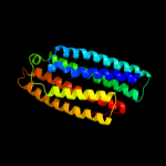
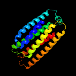
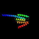
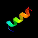
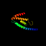
1 d1sumb_
99.9
12
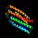
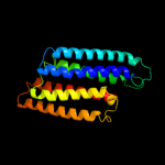
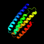
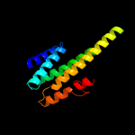
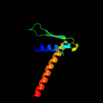
Fold: Spectrin repeat-likeSuperfamily: PhoU-likeFamily: PhoU-like2 d1t72a_
99.9
12
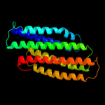
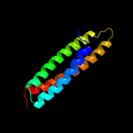
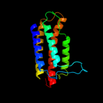
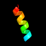
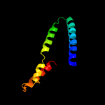
Fold: Spectrin repeat-likeSuperfamily: PhoU-likeFamily: PhoU-like3 d1xwma_
99.9
11
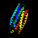
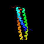
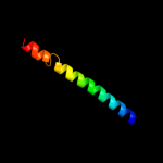
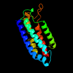
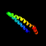
Fold: Spectrin repeat-likeSuperfamily: PhoU-likeFamily: PhoU-like4 c2i0mA_
99.9
15
PDB header: structural genomics, unknown functionChain: A: PDB Molecule: phosphate transport system protein phou;PDBTitle: crystal structure of the phosphate transport system regulatory protein2 phou from streptococcus pneumoniae
5 c3l39A_
99.5
13
PDB header: phosphate-binding proteinChain: A: PDB Molecule: putative phou-like phosphate regulatory protein;PDBTitle: crystal structure of putative phou-like phosphate regulatory2 protein (bt4638) from bacteroides thetaiotaomicron vpi-54823 at 1.93 a resolution
6 c2oltB_
99.3
12
PDB header: unknown functionChain: B: PDB Molecule: hypothetical protein;PDBTitle: crystal structure of a phou-like protein (so_3770) from shewanella2 oneidensis mr-1 at 2.00 a resolution
7 d1vcta1
98.8
18
Fold: Spectrin repeat-likeSuperfamily: PhoU-likeFamily: PhoU-like8 c2bknA_
98.4
17
PDB header: membrane proteinChain: A: PDB Molecule: hypothetical protein ph0236;PDBTitle: structure analysis of unknown function protein
9 c3pjaK_
59.3
12
PDB header: hydrolaseChain: K: PDB Molecule: translin-associated protein x;PDBTitle: crystal structure of human c3po complex
10 c2wb7B_
59.2
13
PDB header: unknown functionChain: B: PDB Molecule: pt26-6p;PDBTitle: pt26-6p
11 c3ke4B_
54.9
11
PDB header: transferaseChain: B: PDB Molecule: hypothetical cytosolic protein;PDBTitle: crystal structure of a pduo-type atp:cob(i)alamin adenosyltransferase2 from bacillus cereus
12 c2z5hB_
48.2
20
PDB header: contractile proteinChain: B: PDB Molecule: general control protein gcn4 and tropomyosinPDBTitle: crystal structure of the head-to-tail junction of2 tropomyosin complexed with a fragment of tnt
13 d1p1ja1
46.2
35
Fold: NAD(P)-binding Rossmann-fold domainsSuperfamily: NAD(P)-binding Rossmann-fold domainsFamily: Glyceraldehyde-3-phosphate dehydrogenase-like, N-terminal domain14 d2o8pa1
44.2
9
Fold: alpha-alpha superhelixSuperfamily: 14-3-3 proteinFamily: 14-3-3 protein15 d1vkoa1
43.5
24
Fold: NAD(P)-binding Rossmann-fold domainsSuperfamily: NAD(P)-binding Rossmann-fold domainsFamily: Glyceraldehyde-3-phosphate dehydrogenase-like, N-terminal domain16 d1rtyb_
43.2
18
Fold: Ferritin-likeSuperfamily: Cobalamin adenosyltransferase-likeFamily: Cobalamin adenosyltransferase17 d1j1ja_
41.0
16
Fold: alpha-alpha superhelixSuperfamily: TranslinFamily: Translin18 d1xppa_
35.3
10
Fold: DCoH-likeSuperfamily: RBP11-like subunits of RNA polymeraseFamily: RBP11/RpoL19 c2hn1A_
33.6
10
PDB header: metal transportChain: A: PDB Molecule: magnesium and cobalt transporter;PDBTitle: crystal structure of a cora soluble domain from a. fulgidus in complex2 with co2+
20 c3qd8M_
33.5
9
PDB header: metal binding proteinChain: M: PDB Molecule: probable bacterioferritin bfrb;PDBTitle: crystal structure of mycobacterium tuberculosis bfrb
21 d1i5na_
not modelled
32.4
9
Fold: Four-helical up-and-down bundleSuperfamily: Histidine-containing phosphotransfer domain, HPT domainFamily: Chemotaxis protein CheA P1 domain22 c3t6gB_
not modelled
32.1
12
PDB header: signaling protein, cell adhesionChain: B: PDB Molecule: breast cancer anti-estrogen resistance protein 1;PDBTitle: structure of the complex between nsp3 (shep1) and p130cas
23 d1s3qa1
not modelled
30.9
8
Fold: Ferritin-likeSuperfamily: Ferritin-likeFamily: Ferritin24 c2yu0A_
not modelled
30.7
25
PDB header: signaling proteinChain: A: PDB Molecule: interferon-activable protein 205;PDBTitle: solution structures of the paad_dapin domain of mus2 musculus interferon-activatable protein 205
25 c3p5nA_
not modelled
30.0
13
PDB header: transport proteinChain: A: PDB Molecule: riboflavin uptake protein;PDBTitle: structure and mechanism of the s component of a bacterial ecf2 transporter
26 c2jd8C_
not modelled
29.8
7
PDB header: metal transportChain: C: PDB Molecule: ferritin homolog;PDBTitle: crystal structure of the zn-soaked ferritin from the2 hyperthermophilic archaeal anaerobe pyrococcus furiosus
27 c1vkoA_
not modelled
29.4
24
PDB header: isomeraseChain: A: PDB Molecule: inositol-3-phosphate synthase;PDBTitle: crystal structure of inositol-3-phosphate synthase (ce21227) from2 caenorhabditis elegans at 2.30 a resolution
28 c2lchA_
not modelled
29.4
11
PDB header: de novo proteinChain: A: PDB Molecule: protein or38;PDBTitle: solution nmr structure of a protein with a redesigned hydrophobic2 core, northeast structural genomics consortium target or38
29 c1p1hD_
not modelled
29.3
35
PDB header: isomeraseChain: D: PDB Molecule: inositol-3-phosphate synthase;PDBTitle: crystal structure of the 1l-myo-inositol/nad+ complex
30 d1k6ka_
not modelled
29.2
14
Fold: Double Clp-N motifSuperfamily: Double Clp-N motifFamily: Double Clp-N motif31 c1kmiZ_
not modelled
28.6
6
PDB header: signaling proteinChain: Z: PDB Molecule: chemotaxis protein chez;PDBTitle: crystal structure of an e.coli chemotaxis protein, chez
32 d2e74f1
not modelled
28.1
29
Fold: Single transmembrane helixSuperfamily: PetM subunit of the cytochrome b6f complexFamily: PetM subunit of the cytochrome b6f complex33 d1zwwa1
not modelled
27.3
11
Fold: BAR/IMD domain-likeSuperfamily: BAR/IMD domain-likeFamily: BAR domain34 c1p58C_
not modelled
27.2
33
PDB header: virusChain: C: PDB Molecule: major envelope protein e;PDBTitle: complex organization of dengue virus membrane proteins as revealed by2 9.5 angstrom cryo-em reconstruction
35 d2o02a1
not modelled
27.1
14
Fold: alpha-alpha superhelixSuperfamily: 14-3-3 proteinFamily: 14-3-3 protein36 c2gzdC_
not modelled
26.8
26
PDB header: protein transportChain: C: PDB Molecule: rab11 family-interacting protein 2;PDBTitle: crystal structure of rab11 in complex with rab11-fip2
37 c2qrxA_
not modelled
25.9
9
PDB header: dna binding proteinChain: A: PDB Molecule: gm27569p;PDBTitle: crystal structure of drosophila melanogaster translin2 protein
38 d1eq1a_
not modelled
25.8
11
Fold: Apolipophorin-IIISuperfamily: Apolipophorin-IIIFamily: Apolipophorin-III39 c2qr4B_
not modelled
25.7
5
PDB header: hydrolaseChain: B: PDB Molecule: peptidase m3b, oligoendopeptidase f;PDBTitle: crystal structure of oligoendopeptidase-f from enterococcus faecium
40 d1vlga_
not modelled
25.6
4
Fold: Ferritin-likeSuperfamily: Ferritin-likeFamily: Ferritin41 c1ha0A_
not modelled
23.9
10
PDB header: viral proteinChain: A: PDB Molecule: protein (hemagglutinin precursor);PDBTitle: hemagglutinin precursor ha0
42 d1u1ia1
not modelled
23.8
18
Fold: NAD(P)-binding Rossmann-fold domainsSuperfamily: NAD(P)-binding Rossmann-fold domainsFamily: Glyceraldehyde-3-phosphate dehydrogenase-like, N-terminal domain43 d1or4a_
not modelled
23.8
13
Fold: Globin-likeSuperfamily: Globin-likeFamily: Globins44 c3fb2B_
not modelled
23.8
8
PDB header: structural proteinChain: B: PDB Molecule: spectrin alpha chain, brain spectrin;PDBTitle: crystal structure of the human brain alpha spectrin repeats2 15 and 16. northeast structural genomics consortium target3 hr5563a.
45 d2elba1
not modelled
23.4
11
Fold: BAR/IMD domain-likeSuperfamily: BAR/IMD domain-likeFamily: BAR domain46 c3kp3B_
not modelled
22.3
17
PDB header: transcription regulator/antibioticChain: B: PDB Molecule: transcriptional regulator tcar;PDBTitle: staphylococcus epidermidis in complex with ampicillin
47 c3hnwB_
not modelled
22.2
15
PDB header: structural genomics, unknown functionChain: B: PDB Molecule: uncharacterized protein;PDBTitle: crystal structure of a basic coiled-coil protein of unknown function2 from eubacterium eligens atcc 27750
48 c2kvcA_
not modelled
22.2
10
PDB header: unknown functionChain: A: PDB Molecule: putative uncharacterized protein;PDBTitle: solution structure of the mycobacterium tuberculosis protein rv0543c,2 a member of the duf3349 superfamily. seattle structural genomics3 center for infectious disease target mytud.17112.a
49 c2wr2B_
not modelled
22.1
10
PDB header: viral proteinChain: B: PDB Molecule: hemagglutinin;PDBTitle: structure of influenza h2 avian hemagglutinin with avian2 receptor
50 d2axti1
not modelled
22.0
14
Fold: Single transmembrane helixSuperfamily: Photosystem II reaction center protein I, PsbIFamily: PsbI-like51 d1dlca3
not modelled
21.9
9
Fold: Toxins' membrane translocation domainsSuperfamily: delta-Endotoxin (insectocide), N-terminal domainFamily: delta-Endotoxin (insectocide), N-terminal domain52 d2d2sa1
not modelled
21.6
10
Fold: alpha-alpha superhelixSuperfamily: Cullin repeat-likeFamily: Exocyst complex component53 c3lk5A_
not modelled
21.6
13
PDB header: transferaseChain: A: PDB Molecule: geranylgeranyl pyrophosphate synthase;PDBTitle: crystal structure of putative geranylgeranyl pyrophosphate synthase2 from corynebacterium glutamicum
54 c2nt8A_
not modelled
19.8
13
PDB header: transferaseChain: A: PDB Molecule: cobalamin adenosyltransferase;PDBTitle: atp bound at the active site of a pduo type atp:co(i)rrinoid2 adenosyltransferase from lactobacillus reuteri
55 c1xnlA_
not modelled
19.8
44
PDB header: viral proteinChain: A: PDB Molecule: membrane protein gp37;PDBTitle: aslv fusion peptide
56 c3kyiA_
not modelled
19.8
13
PDB header: transferaseChain: A: PDB Molecule: putative histidine protein kinase;PDBTitle: crystal structure of the phosphorylated p1 domain of chea3 in complex2 with chey6 from r. sphaeroides
57 d1tqga_
not modelled
19.7
12
Fold: Four-helical up-and-down bundleSuperfamily: Histidine-containing phosphotransfer domain, HPT domainFamily: Chemotaxis protein CheA P1 domain58 c2zhzC_
not modelled
19.6
14
PDB header: transferaseChain: C: PDB Molecule: atp:cob(i)alamin adenosyltransferase, putative;PDBTitle: crystal structure of a pduo-type atp:cobalamin adenosyltransferase2 from burkholderia thailandensis
59 d1twfk_
not modelled
19.5
24
Fold: DCoH-likeSuperfamily: RBP11-like subunits of RNA polymeraseFamily: RBP11/RpoL60 c3nmdA_
not modelled
19.4
17
PDB header: transferaseChain: A: PDB Molecule: cgmp dependent protein kinase;PDBTitle: crystal structure of the leucine zipper domain of cgmp dependent2 protein kinase i beta
61 c1b9uA_
not modelled
19.2
13
PDB header: hydrolaseChain: A: PDB Molecule: protein (atp synthase);PDBTitle: membrane domain of the subunit b of the e.coli atp synthase
62 d1t01a1
not modelled
19.2
14
Fold: Four-helical up-and-down bundleSuperfamily: alpha-catenin/vinculin-likeFamily: alpha-catenin/vinculin63 c2bbjB_
not modelled
18.7
6
PDB header: metal transport/membrane proteinChain: B: PDB Molecule: divalent cation transport-related protein;PDBTitle: crystal structure of the cora mg2+ transporter
64 c3axjB_
not modelled
18.7
14
PDB header: dna binding proteinChain: B: PDB Molecule: translin associated factor x, isoform b;PDBTitle: high resolution crystal structure of c3po
65 d256ba_
not modelled
18.5
17
Fold: Four-helical up-and-down bundleSuperfamily: CytochromesFamily: Cytochrome b56266 c2c5iT_
not modelled
18.5
6
PDB header: protein transportChain: T: PDB Molecule: t-snare affecting a late golgi compartmentPDBTitle: n-terminal domain of tlg1 complexed with n-terminus of2 vps51 in distorted conformation
67 c2ariA_
not modelled
18.4
30
PDB header: viral proteinChain: A: PDB Molecule: envelope polyprotein gp160;PDBTitle: solution structure of micelle-bound fusion domain of hiv-12 gp41
68 d1rkea1
not modelled
18.3
13
Fold: Four-helical up-and-down bundleSuperfamily: alpha-catenin/vinculin-likeFamily: alpha-catenin/vinculin69 c1j1dF_
not modelled
18.3
20
PDB header: contractile proteinChain: F: PDB Molecule: troponin i;PDBTitle: crystal structure of the 46kda domain of human cardiac2 troponin in the ca2+ saturated form
70 c3mp7B_
not modelled
18.3
33
PDB header: protein transportChain: B: PDB Molecule: preprotein translocase subunit sece;PDBTitle: lateral opening of a translocon upon entry of protein suggests the2 mechanism of insertion into membranes
71 d1io1a_
not modelled
18.2
19
Fold: Phase 1 flagellinSuperfamily: Phase 1 flagellinFamily: Phase 1 flagellin72 c2k6sB_
not modelled
18.2
26
PDB header: protein transportChain: B: PDB Molecule: rab11fip2 protein;PDBTitle: structure of rab11-fip2 c-terminal coiled-coil domain
73 c2p90B_
not modelled
18.1
15
PDB header: structural genomics, unknown functionChain: B: PDB Molecule: hypothetical protein cgl1923;PDBTitle: the crystal structure of a protein of unknown function from2 corynebacterium glutamicum atcc 13032
74 c1ykuB_
not modelled
18.1
13
PDB header: unknown functionChain: B: PDB Molecule: hypothetical protein pxo2-61;PDBTitle: crystal structure of a sensor domain homolog
75 c3hjlA_
not modelled
18.0
12
PDB header: proton transportChain: A: PDB Molecule: flagellar motor switch protein flig;PDBTitle: the structure of full-length flig from aquifex aeolicus
76 c1ytzI_
not modelled
18.0
11
PDB header: contractile proteinChain: I: PDB Molecule: troponin i;PDBTitle: crystal structure of skeletal muscle troponin in the ca2+-2 activated state
77 c3sogA_
not modelled
18.0
7
PDB header: structural proteinChain: A: PDB Molecule: amphiphysin;PDBTitle: crystal structure of the bar domain of human amphiphysin, isoform 1
78 d1xata_
not modelled
17.7
20
Fold: Single-stranded left-handed beta-helixSuperfamily: Trimeric LpxA-like enzymesFamily: Galactoside acetyltransferase-like79 c3bt6B_
not modelled
17.5
14
PDB header: viral proteinChain: B: PDB Molecule: influenza b hemagglutinin (ha);PDBTitle: crystal structure of influenza b virus hemagglutinin
80 c3a0bX_
not modelled
17.5
19
PDB header: electron transportChain: X: PDB Molecule: photosystem ii reaction center protein x;PDBTitle: crystal structure of br-substituted photosystem ii complex
81 c3a0bx_
not modelled
17.5
19
PDB header: electron transportChain: X: PDB Molecule: photosystem ii reaction center protein x;PDBTitle: crystal structure of br-substituted photosystem ii complex
82 c3a0hX_
not modelled
17.5
19
PDB header: electron transportChain: X: PDB Molecule: photosystem ii reaction center protein x;PDBTitle: crystal structure of i-substituted photosystem ii complex
83 c3a0hx_
not modelled
17.5
19
PDB header: electron transportChain: X: PDB Molecule: photosystem ii reaction center protein x;PDBTitle: crystal structure of i-substituted photosystem ii complex
84 d1ciia1
not modelled
17.5
15
Fold: Toxins' membrane translocation domainsSuperfamily: ColicinFamily: Colicin85 d1s35a2
not modelled
17.5
7
Fold: Spectrin repeat-likeSuperfamily: Spectrin repeatFamily: Spectrin repeat86 d1t6ua_
not modelled
17.5
13
Fold: Four-helical up-and-down bundleSuperfamily: Nickel-containing superoxide dismutase, NiSODFamily: Nickel-containing superoxide dismutase, NiSOD87 d2iuba1
not modelled
17.3
10
Fold: CorA soluble domain-likeSuperfamily: CorA soluble domain-likeFamily: CorA soluble domain-like88 c3ci9B_
not modelled
17.3
14
PDB header: transcriptionChain: B: PDB Molecule: heat shock factor-binding protein 1;PDBTitle: crystal structure of the human hsbp1
89 c1yv0I_
not modelled
17.3
11
PDB header: contractile proteinChain: I: PDB Molecule: troponin i, fast skeletal muscle;PDBTitle: crystal structure of skeletal muscle troponin in the ca2+-2 free state
90 c1hf9B_
not modelled
17.3
23
PDB header: atpase inhibitorChain: B: PDB Molecule: atpase inhibitor (mitochondrial);PDBTitle: c-terminal coiled-coil domain from bovine if1
91 c1dlcA_
not modelled
17.2
9
PDB header: toxinChain: A: PDB Molecule: delta-endotoxin cryiiia;PDBTitle: crystal structure of insecticidal delta-endotoxin from2 bacillus thuringiensis at 2.5 angstroms resolution
92 c1zxjB_
not modelled
17.2
10
PDB header: structural genomics, unknown functionChain: B: PDB Molecule: hypothetical protein mg377 homolog;PDBTitle: crystal structure of the hypthetical mycoplasma protein,2 mpn555
93 c1t6fA_
not modelled
17.1
19
PDB header: cell cycleChain: A: PDB Molecule: geminin;PDBTitle: crystal structure of the coiled-coil dimerization motif of2 geminin
94 c3o3nA_
not modelled
16.9
12
PDB header: lyaseChain: A: PDB Molecule: alpha-subunit 2-hydroxyisocaproyl-coa dehydratase;PDBTitle: (r)-2-hydroxyisocaproyl-coa dehydratase in complex with its substrate2 (r)-2-hydroxyisocaproyl-coa
95 c3pl4A_
not modelled
16.9
13
PDB header: motor proteinChain: A: PDB Molecule: flagellar motor switch protein;PDBTitle: crystal structure of flig (residue 116-343) from h. pylori
96 c3l8jA_
not modelled
16.8
12
PDB header: protein bindingChain: A: PDB Molecule: programmed cell death protein 10;PDBTitle: crystal structure of ccm3, a cerebral cavernous malformation protein2 critical for vascular integrity
97 d2etsa1
not modelled
16.8
5
Fold: Four-helical up-and-down bundleSuperfamily: YppE-likeFamily: YppE-like98 c1s5lx_
not modelled
16.6
19
PDB header: photosynthesisChain: X: PDB Molecule: photosystem ii psbx protein;PDBTitle: architecture of the photosynthetic oxygen evolving center
99 c2ah6B_
not modelled
16.5
18
PDB header: transferaseChain: B: PDB Molecule: bh1595, unknown conserved protein;PDBTitle: crystal structure of a putative cobalamin adenosyltransferase (bh1595)2 from bacillus halodurans c-125 at 1.60 a resolution


