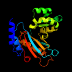
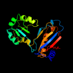
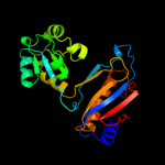
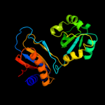
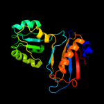
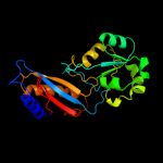
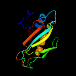
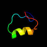
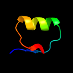
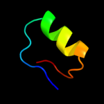
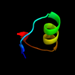
1 c3qoyA_
100.0
45
PDB header: ribosomal proteinChain: A: PDB Molecule: 50s ribosomal protein l1;PDBTitle: crystal structure of ribosomal protein l1 from aquifex aeolicus
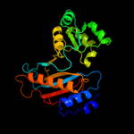
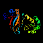
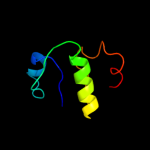
2 d1ad2a_
100.0
50
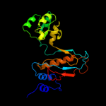
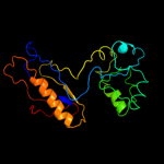
Fold: Ribosomal protein L1Superfamily: Ribosomal protein L1Family: Ribosomal protein L13 c3bboD_
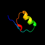
100.0
45
PDB header: ribosomeChain: D: PDB Molecule: ribosomal protein l1;PDBTitle: homology model for the spinach chloroplast 50s subunit2 fitted to 9.4a cryo-em map of the 70s chlororibosome
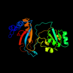
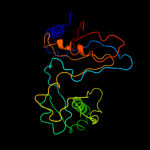
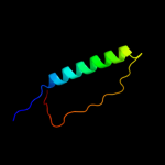
4 c2gyc2_
100.0
100
PDB header: ribosomeChain: 2: PDB Molecule: 50s ribosomal protein l1;PDBTitle: structure of the 50s subunit of a secm-stalled e. coli2 ribosome complex obtained by fitting atomic models for rna3 and protein components into cryo-em map emd-1143
5 d1mzpa_
100.0
26
Fold: Ribosomal protein L1Superfamily: Ribosomal protein L1Family: Ribosomal protein L16 d1dwua_
100.0
29
Fold: Ribosomal protein L1Superfamily: Ribosomal protein L1Family: Ribosomal protein L17 d1i2aa_
100.0
29
Fold: Ribosomal protein L1Superfamily: Ribosomal protein L1Family: Ribosomal protein L18 c2zkr5_
100.0
31
PDB header: ribosomal protein/rnaChain: 5: PDB Molecule: 60s ribosomal protein l10a;PDBTitle: structure of a mammalian ribosomal 60s subunit within an2 80s complex obtained by docking homology models of the rna3 and proteins into an 8.7 a cryo-em map
9 c1s1iA_
100.0
18
PDB header: ribosomeChain: A: PDB Molecule: 60s ribosomal protein l1;PDBTitle: structure of the ribosomal 80s-eef2-sordarin complex from2 yeast obtained by docking atomic models for rna and protein3 components into a 11.7 a cryo-em map. this file, 1s1i,4 contains 60s subunit. the 40s ribosomal subunit is in file5 1s1h.
10 c3iz5A_
100.0
22
PDB header: ribosomeChain: A: PDB Molecule: 60s ribosomal protein l1 (l1p);PDBTitle: localization of the large subunit ribosomal proteins into a 5.5 a2 cryo-em map of triticum aestivum translating 80s ribosome
11 c2ftcA_
100.0
20
PDB header: ribosomeChain: A: PDB Molecule: mitochondrial ribosomal protein l1;PDBTitle: structural model for the large subunit of the mammalian mitochondrial2 ribosome
12 d2j01c1
100.0
49
Fold: Ribosomal protein L1Superfamily: Ribosomal protein L1Family: Ribosomal protein L113 c2ov7C_
100.0
59
PDB header: ribosomal proteinChain: C: PDB Molecule: 50s ribosomal protein l1;PDBTitle: the first domain of the ribosomal protein l1 from thermus2 thermophilus
14 c2kzhA_
57.6
37
PDB header: isomeraseChain: A: PDB Molecule: tryptophan biosynthesis protein trpcf;PDBTitle: three-dimensional structure of a truncated phosphoribosylanthranilate2 isomerase (residues 255-384) from escherichia coli
15 d1piia1
56.9
40
Fold: TIM beta/alpha-barrelSuperfamily: Ribulose-phoshate binding barrelFamily: Tryptophan biosynthesis enzymes16 d1v5xa_
48.6
33
Fold: TIM beta/alpha-barrelSuperfamily: Ribulose-phoshate binding barrelFamily: Tryptophan biosynthesis enzymes17 d1nsja_
43.2
29
Fold: TIM beta/alpha-barrelSuperfamily: Ribulose-phoshate binding barrelFamily: Tryptophan biosynthesis enzymes18 d2i4ra1
42.7
18
Fold: AtpF-likeSuperfamily: AtpF-likeFamily: AtpF-like19 c1piiA_
42.2
38
PDB header: bifunctional(isomerase and synthase)Chain: A: PDB Molecule: n-(5'phosphoribosyl)anthranilate isomerase;PDBTitle: three-dimensional structure of the bifunctional enzyme2 phosphoribosylanthranilate isomerase:3 indoleglycerolphosphate synthase from escherichia coli4 refined at 2.0 angstroms resolution
20 c2w40C_
37.5
22
PDB header: transferaseChain: C: PDB Molecule: glycerol kinase, putative;PDBTitle: crystal structure of plasmodium falciparum glycerol kinase2 with bound glycerol
21 c3nbmA_
not modelled
33.2
15
PDB header: transferaseChain: A: PDB Molecule: pts system, lactose-specific iibc components;PDBTitle: the lactose-specific iib component domain structure of the2 phosphoenolpyruvate:carbohydrate phosphotransferase system (pts) from3 streptococcus pneumoniae.
22 d1qapa1
not modelled
25.6
24
Fold: TIM beta/alpha-barrelSuperfamily: Nicotinate/Quinolinate PRTase C-terminal domain-likeFamily: NadC C-terminal domain-like23 c2ov6A_
not modelled
22.1
11
PDB header: hydrolaseChain: A: PDB Molecule: v-type atp synthase subunit f;PDBTitle: the nmr structure of subunit f of the methanogenic a1ao atp synthase2 and its interaction with the nucleotide-binding subunit b
24 d1o4ua1
not modelled
21.5
25
Fold: TIM beta/alpha-barrelSuperfamily: Nicotinate/Quinolinate PRTase C-terminal domain-likeFamily: NadC C-terminal domain-like25 c2nqqA_
not modelled
21.1
28
PDB header: biosynthetic proteinChain: A: PDB Molecule: molybdopterin biosynthesis protein moea;PDBTitle: moea r137q
26 c2vqpA_
not modelled
19.6
31
PDB header: viral proteinChain: A: PDB Molecule: matrix protein;PDBTitle: structure of the matrix protein from human respiratory2 syncytial virus
27 c3hz6A_
not modelled
19.2
13
PDB header: transferaseChain: A: PDB Molecule: xylulokinase;PDBTitle: crystal structure of xylulokinase from chromobacterium violaceum
28 c2l2qA_
not modelled
18.7
17
PDB header: transferaseChain: A: PDB Molecule: pts system, cellobiose-specific iib component (cela);PDBTitle: solution structure of cellobiose-specific phosphotransferase iib2 component protein from borrelia burgdorferi
29 c2vhmI_
not modelled
16.8
12
PDB header: ribosomeChain: I: PDB Molecule: 50s ribosomal protein l11;PDBTitle: structure of pdf binding helix in complex with the ribosome2 (part 1 of 4)
30 c3g25B_
not modelled
16.3
21
PDB header: transferaseChain: B: PDB Molecule: glycerol kinase;PDBTitle: 1.9 angstrom crystal structure of glycerol kinase (glpk) from2 staphylococcus aureus in complex with glycerol.
31 c3gbtA_
not modelled
16.0
16
PDB header: transferaseChain: A: PDB Molecule: gluconate kinase;PDBTitle: crystal structure of gluconate kinase from lactobacillus acidophilus
32 c1xupO_
not modelled
15.4
15
PDB header: transferaseChain: O: PDB Molecule: glycerol kinase;PDBTitle: enterococcus casseliflavus glycerol kinase complexed with glycerol
33 c2d4wA_
not modelled
13.3
17
PDB header: transferaseChain: A: PDB Molecule: glycerol kinase;PDBTitle: crystal structure of glycerol kinase from cellulomonas sp.2 nt3060
34 c2dpnB_
not modelled
13.1
15
PDB header: transferaseChain: B: PDB Molecule: glycerol kinase;PDBTitle: crystal structure of the glycerol kinase from thermus2 thermophilus hb8
35 c2nlxA_
not modelled
12.8
17
PDB header: transferaseChain: A: PDB Molecule: xylulose kinase;PDBTitle: crystal structure of the apo e. coli xylulose kinase
36 c3flcX_
not modelled
12.7
15
PDB header: transferaseChain: X: PDB Molecule: glycerol kinase;PDBTitle: crystal structure of the his-tagged h232r mutant of glycerol kinase2 from enterococcus casseliflavus with glycerol
37 d2azeb1
not modelled
12.5
20
Fold: E2F-DP heterodimerization regionSuperfamily: E2F-DP heterodimerization regionFamily: E2F dimerization segment38 c2zf5O_
not modelled
12.0
8
PDB header: transferaseChain: O: PDB Molecule: glycerol kinase;PDBTitle: crystal structure of highly thermostable glycerol kinase from a2 hyperthermophilic archaeon
39 d1qpoa1
not modelled
11.6
15
Fold: TIM beta/alpha-barrelSuperfamily: Nicotinate/Quinolinate PRTase C-terminal domain-likeFamily: NadC C-terminal domain-like40 c3ifrB_
not modelled
11.3
21
PDB header: transferaseChain: B: PDB Molecule: carbohydrate kinase, fggy;PDBTitle: the crystal structure of xylulose kinase from rhodospirillum rubrum
41 c2ftcG_
not modelled
10.5
39
PDB header: ribosomeChain: G: PDB Molecule: 39s ribosomal protein l11, mitochondrial;PDBTitle: structural model for the large subunit of the mammalian mitochondrial2 ribosome
42 c3bboK_
not modelled
10.2
15
PDB header: ribosomeChain: K: PDB Molecule: ribosomal protein l11;PDBTitle: homology model for the spinach chloroplast 50s subunit2 fitted to 9.4a cryo-em map of the 70s chlororibosome
43 d1k1ga_
not modelled
10.0
33
Fold: Eukaryotic type KH-domain (KH-domain type I)Superfamily: Eukaryotic type KH-domain (KH-domain type I)Family: Eukaryotic type KH-domain (KH-domain type I)44 d1iiba_
not modelled
9.9
14
Fold: Phosphotyrosine protein phosphatases I-likeSuperfamily: PTS system IIB component-likeFamily: PTS system, Lactose/Cellobiose specific IIB subunit45 d2p3ra1
not modelled
9.9
15
Fold: Ribonuclease H-like motifSuperfamily: Actin-like ATPase domainFamily: Glycerol kinase46 d2nqra3
not modelled
9.5
28
Fold: Molybdenum cofactor biosynthesis proteinsSuperfamily: Molybdenum cofactor biosynthesis proteinsFamily: MoeA central domain-like47 c3jvpA_
not modelled
9.4
16
PDB header: transferaseChain: A: PDB Molecule: ribulokinase;PDBTitle: crystal structure of ribulokinase from bacillus halodurans
48 c3iz5J_
not modelled
9.1
43
PDB header: ribosomeChain: J: PDB Molecule: 60s ribosomal protein l12 (l11p);PDBTitle: localization of the large subunit ribosomal proteins into a 5.5 a2 cryo-em map of triticum aestivum translating 80s ribosome
49 c3enpA_
not modelled
8.7
18
PDB header: hydrolaseChain: A: PDB Molecule: tp53rk-binding protein;PDBTitle: crystal structure of human cgi121
50 d3cjsb1
not modelled
8.2
36
Fold: Ribosomal L11/L12e N-terminal domainSuperfamily: Ribosomal L11/L12e N-terminal domainFamily: Ribosomal L11/L12e N-terminal domain51 c3cjtP_
not modelled
7.9
36
PDB header: transferase/ribosomal proteinChain: P: PDB Molecule: 50s ribosomal protein l11;PDBTitle: ribosomal protein l11 methyltransferase (prma) in complex with2 dimethylated ribosomal protein l11
52 d2d00a1
not modelled
7.6
17
Fold: AtpF-likeSuperfamily: AtpF-likeFamily: AtpF-like53 c1glbG_
not modelled
7.5
15
PDB header: phosphotransferaseChain: G: PDB Molecule: glycerol kinase;PDBTitle: structure of the regulatory complex of escherichia coli iiiglc with2 glycerol kinase
54 c2i4rA_
not modelled
7.5
16
PDB header: hydrolaseChain: A: PDB Molecule: v-type atp synthase subunit f;PDBTitle: crystal structure of the v-type atp synthase subunit f from2 archaeoglobus fulgidus. nesg target gr52a.
55 c3gg4B_
not modelled
6.9
11
PDB header: transferaseChain: B: PDB Molecule: glycerol kinase;PDBTitle: the crystal structure of glycerol kinase from yersinia2 pseudotuberculosis
56 d1r59o1
not modelled
6.9
15
Fold: Ribonuclease H-like motifSuperfamily: Actin-like ATPase domainFamily: Glycerol kinase57 d1gtea2
not modelled
6.9
10
Fold: TIM beta/alpha-barrelSuperfamily: FMN-linked oxidoreductasesFamily: FMN-linked oxidoreductases58 d2bl5a1
not modelled
6.8
25
Fold: Eukaryotic type KH-domain (KH-domain type I)Superfamily: Eukaryotic type KH-domain (KH-domain type I)Family: Eukaryotic type KH-domain (KH-domain type I)59 c3ezwD_
not modelled
6.7
15
PDB header: transferaseChain: D: PDB Molecule: glycerol kinase;PDBTitle: crystal structure of a hyperactive escherichia coli glycerol kinase2 mutant gly230 --> asp obtained using microfluidic crystallization3 devices
60 d1mmsa2
not modelled
6.7
33
Fold: Ribosomal L11/L12e N-terminal domainSuperfamily: Ribosomal L11/L12e N-terminal domainFamily: Ribosomal L11/L12e N-terminal domain61 c1qapA_
not modelled
6.6
24
PDB header: glycosyltransferaseChain: A: PDB Molecule: quinolinic acid phosphoribosyltransferase;PDBTitle: quinolinic acid phosphoribosyltransferase with bound2 quinolinic acid
62 c3gk0H_
not modelled
6.6
39
PDB header: transferaseChain: H: PDB Molecule: pyridoxine 5'-phosphate synthase;PDBTitle: crystal structure of pyridoxal phosphate biosynthetic2 protein from burkholderia pseudomallei
63 c1o4uA_
not modelled
6.5
23
PDB header: transferaseChain: A: PDB Molecule: type ii quinolic acid phosphoribosyltransferase;PDBTitle: crystal structure of a nicotinate nucleotide pyrophosphorylase2 (tm1645) from thermotoga maritima at 2.50 a resolution
64 c2hbpA_
not modelled
6.5
13
PDB header: endocytosis, protein bindingChain: A: PDB Molecule: cytoskeleton assembly control protein sla1;PDBTitle: solution structure of sla1 homology domain 1
65 c2b7pA_
not modelled
6.4
20
PDB header: transferaseChain: A: PDB Molecule: probable nicotinate-nucleotide pyrophosphorylase;PDBTitle: crystal structure of quinolinic acid phosphoribosyltransferase from2 helicobacter pylori
66 c3l0gD_
not modelled
6.4
7
PDB header: transferaseChain: D: PDB Molecule: nicotinate-nucleotide pyrophosphorylase;PDBTitle: crystal structure of nicotinate-nucleotide pyrophosphorylase from2 ehrlichia chaffeensis at 2.05a resolution
67 c1xtzA_
not modelled
6.1
20
PDB header: isomeraseChain: A: PDB Molecule: ribose-5-phosphate isomerase;PDBTitle: crystal structure of the s. cerevisiae d-ribose-5-phosphate isomerase:2 comparison with the archeal and bacterial enzymes
68 d2gycg2
not modelled
6.0
33
Fold: Ribosomal L11/L12e N-terminal domainSuperfamily: Ribosomal L11/L12e N-terminal domainFamily: Ribosomal L11/L12e N-terminal domain69 d1xbpg2
not modelled
5.8
33
Fold: Ribosomal L11/L12e N-terminal domainSuperfamily: Ribosomal L11/L12e N-terminal domainFamily: Ribosomal L11/L12e N-terminal domain70 d1vjpa1
not modelled
5.7
23
Fold: NAD(P)-binding Rossmann-fold domainsSuperfamily: NAD(P)-binding Rossmann-fold domainsFamily: Glyceraldehyde-3-phosphate dehydrogenase-like, N-terminal domain71 c1jqmA_
not modelled
5.6
29
PDB header: ribosomeChain: A: PDB Molecule: 50s ribosomal protein l11;PDBTitle: fitting of l11 protein and elongation factor g (ef-g) in2 the cryo-em map of e. coli 70s ribosome bound with ef-g,3 gdp and fusidic acid
72 d1qk1a2
not modelled
5.2
12
Fold: Glutamine synthetase/guanido kinaseSuperfamily: Glutamine synthetase/guanido kinaseFamily: Guanido kinase catalytic domain73 c2jbmA_
not modelled
5.2
31
PDB header: transferaseChain: A: PDB Molecule: nicotinate-nucleotide pyrophosphorylase;PDBTitle: qprtase structure from human


