1 c3bboI_
100.0
45
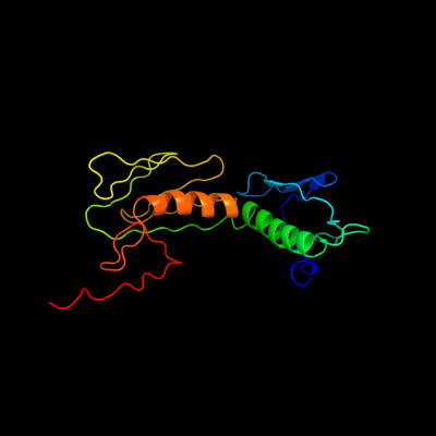
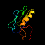
PDB header: ribosomeChain: I: PDB Molecule: ribosomal protein l6;PDBTitle: homology model for the spinach chloroplast 50s subunit2 fitted to 9.4a cryo-em map of the 70s chlororibosome
2 c2i2vG_
100.0
100
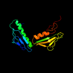
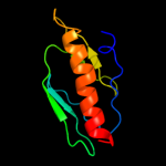
PDB header: ribosomeChain: G: PDB Molecule: 50s ribosomal protein l6;PDBTitle: crystal structure of ribosome with messenger rna and the2 anticodon stem-loop of p-site trna. this file contains the3 50s subunit of one 70s ribosome. the entire crystal4 structure contains two 70s ribosomes and is described in5 remark 400.
3 c2hguH_
100.0
38
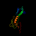
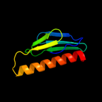
PDB header: ribosomeChain: H: PDB Molecule: 50s ribosomal protein l6;PDBTitle: 70s t.th. ribosome functional complex with mrna and e- and p-site2 trnas at 4.5a. this entry 2hgu contains 50s ribosomal subunit. the3 30s ribosomal subunit can be found in pdb entry 2hgr.
4 c1sm1E_
100.0
39
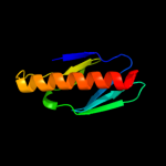
PDB header: ribosome/antibioticChain: E: PDB Molecule: 50s ribosomal protein l6;PDBTitle: complex of the large ribosomal subunit from deinococcus radiodurans2 with quinupristin and dalfopristin
5 c487dJ_
100.0
48
PDB header: ribosomeChain: J: PDB Molecule: protein (50s l6 ribosomal protein);PDBTitle: seven ribosomal proteins fitted to a cryo-electron2 microscopic map of the large 50s subunit at 7.5 angstroms3 resolution
6 c1pnuE_
100.0
39
PDB header: ribosomeChain: E: PDB Molecule: 50s ribosomal protein l6;PDBTitle: crystal structure of a streptomycin dependent ribosome from2 escherichia coli, 50s subunit of 70s ribosome. this file,3 1pnu, contains only molecules of the 50s ribosomal4 subunit. the 30s subunit, mrna, p-site trna, and a-site5 trna are in the pdb file 1pns.
7 c3ccmE_
100.0
25
PDB header: ribosomeChain: E: PDB Molecule: 50s ribosomal protein l6p;PDBTitle: structure of anisomycin resistant 50s ribosomal subunit: 23s rrna2 mutation g2611u
8 c2zkre_
100.0
25
PDB header: ribosomal protein/rnaChain: E: PDB Molecule: rna expansion segment es7 part ii;PDBTitle: structure of a mammalian ribosomal 60s subunit within an2 80s complex obtained by docking homology models of the rna3 and proteins into an 8.7 a cryo-em map
9 c4a1eE_
100.0
21
PDB header: ribosomeChain: E: PDB Molecule: 60s ribosomal protein l9;PDBTitle: t.thermophila 60s ribosomal subunit in complex with2 initiation factor 6. this file contains 5s rrna, 5.8s rrna3 and proteins of molecule 1
10 c3iz5F_
100.0
24
PDB header: ribosomeChain: F: PDB Molecule: 60s ribosomal protein l9 (l6p);PDBTitle: localization of the large subunit ribosomal proteins into a 5.5 a2 cryo-em map of triticum aestivum translating 80s ribosome
11 c1s1iH_
100.0
26
PDB header: ribosomeChain: H: PDB Molecule: 60s ribosomal protein l9-a;PDBTitle: structure of the ribosomal 80s-eef2-sordarin complex from2 yeast obtained by docking atomic models for rna and protein3 components into a 11.7 a cryo-em map. this file, 1s1i,4 contains 60s subunit. the 40s ribosomal subunit is in file5 1s1h.
12 d2qamg2
100.0
100
Fold: Ribosomal protein L6Superfamily: Ribosomal protein L6Family: Ribosomal protein L613 d1rl6a2
100.0
55
Fold: Ribosomal protein L6Superfamily: Ribosomal protein L6Family: Ribosomal protein L614 d2zjre1
100.0
46
Fold: Ribosomal protein L6Superfamily: Ribosomal protein L6Family: Ribosomal protein L615 d2j01h2
100.0
49
Fold: Ribosomal protein L6Superfamily: Ribosomal protein L6Family: Ribosomal protein L616 d2qamg1
99.9
100
Fold: Ribosomal protein L6Superfamily: Ribosomal protein L6Family: Ribosomal protein L617 d1rl6a1
99.9
40
Fold: Ribosomal protein L6Superfamily: Ribosomal protein L6Family: Ribosomal protein L618 d1vqoe1
99.9
29
Fold: Ribosomal protein L6Superfamily: Ribosomal protein L6Family: Ribosomal protein L619 d2zjre2
99.9
32
Fold: Ribosomal protein L6Superfamily: Ribosomal protein L6Family: Ribosomal protein L620 d2cqla1
99.9
26
Fold: Ribosomal protein L6Superfamily: Ribosomal protein L6Family: Ribosomal protein L621 d2j01h1
not modelled
99.8
25
Fold: Ribosomal protein L6Superfamily: Ribosomal protein L6Family: Ribosomal protein L622 d1vqoe2
not modelled
99.7
22
Fold: Ribosomal protein L6Superfamily: Ribosomal protein L6Family: Ribosomal protein L623 d1a0ia1
not modelled
38.5
10
Fold: OB-foldSuperfamily: Nucleic acid-binding proteinsFamily: DNA ligase/mRNA capping enzyme postcatalytic domain24 c3q9qB_
not modelled
35.6
35
PDB header: chaperoneChain: B: PDB Molecule: heat shock protein beta-1;PDBTitle: hspb1 fragment second crystal form
25 c2wj7D_
not modelled
34.5
41
PDB header: chaperoneChain: D: PDB Molecule: alpha-crystallin b chain;PDBTitle: human alphab crystallin
26 c2bolA_
not modelled
31.4
13
PDB header: heat shock proteinChain: A: PDB Molecule: small heat shock protein;PDBTitle: crystal structure and assembly of tsp36, a metazoan small2 heat shock protein
27 c3kd4A_
not modelled
30.7
19
PDB header: hydrolaseChain: A: PDB Molecule: putative protease;PDBTitle: crystal structure of a putative protease (bdi_1141) from2 parabacteroides distasonis atcc 8503 at 2.00 a resolution
28 c2jugB_
not modelled
24.7
24
PDB header: biosynthetic proteinChain: B: PDB Molecule: tubc protein;PDBTitle: multienzyme docking in hybrid megasynthetases
29 c2klrA_
not modelled
22.1
32
PDB header: chaperoneChain: A: PDB Molecule: alpha-crystallin b chain;PDBTitle: solid-state nmr structure of the alpha-crystallin domain in alphab-2 crystallin oligomers
30 c2wj5A_
not modelled
20.5
25
PDB header: chaperoneChain: A: PDB Molecule: heat shock protein beta-6;PDBTitle: rat alpha crystallin domain
31 c3pgeA_
not modelled
17.2
11
PDB header: dna binding proteinChain: A: PDB Molecule: sumo-modified proliferating cell nuclear antigen;PDBTitle: structure of sumoylated pcna
32 d1j5pa3
not modelled
14.4
21
Fold: FwdE/GAPDH domain-likeSuperfamily: Glyceraldehyde-3-phosphate dehydrogenase-like, C-terminal domainFamily: Dihydrodipicolinate reductase-like33 c2gh8B_
not modelled
14.0
11
PDB header: virusChain: B: PDB Molecule: capsid protein;PDBTitle: x-ray structure of a native calicivirus
34 c3nicA_
not modelled
13.2
31
PDB header: hydrolase/dnaChain: A: PDB Molecule: eco29kir;PDBTitle: dna binding and cleavage by the giy-yig endonuclease r.eco29ki2 inactive variant y49f
35 c3l1eA_
not modelled
12.8
29
PDB header: chaperoneChain: A: PDB Molecule: alpha-crystallin a chain;PDBTitle: bovine alphaa crystallin zinc bound
36 c2ktsA_
not modelled
12.3
22
PDB header: chaperoneChain: A: PDB Molecule: heat shock protein hslj;PDBTitle: nmr structure of the protein np_415897.1
37 d1ev13_
not modelled
12.1
11
Fold: Nucleoplasmin-like/VP (viral coat and capsid proteins)Superfamily: Positive stranded ssRNA virusesFamily: Picornaviridae-like VP (VP1, VP2, VP3 and VP4)38 d1oopc_
not modelled
11.8
9
Fold: Nucleoplasmin-like/VP (viral coat and capsid proteins)Superfamily: Positive stranded ssRNA virusesFamily: Picornaviridae-like VP (VP1, VP2, VP3 and VP4)39 d2mev3_
not modelled
11.1
16
Fold: Nucleoplasmin-like/VP (viral coat and capsid proteins)Superfamily: Positive stranded ssRNA virusesFamily: Picornaviridae-like VP (VP1, VP2, VP3 and VP4)40 c2gjhA_
not modelled
11.0
22
PDB header: de novo proteinChain: A: PDB Molecule: designed protein;PDBTitle: nmr structure of cfr (c-terminal fragment of2 computationally designed novel-topology protein top7)
41 c2xseA_
not modelled
10.9
11
PDB header: oxidoreductaseChain: A: PDB Molecule: thymine dioxygenase jbp1;PDBTitle: the structural basis for recognition of j-base containing2 dna by a novel dna-binding domain in jbp1
42 c2d2fA_
not modelled
10.7
15
PDB header: protein bindingChain: A: PDB Molecule: sufc protein;PDBTitle: crystal structure of atypical cytoplasmic abc-atpase sufc from thermus2 thermophilus hb8
43 c3i38C_
not modelled
10.4
37
PDB header: chaperoneChain: C: PDB Molecule: putative chaperone dnaj;PDBTitle: structure of a putative chaperone protein dnaj from klebsiella2 pneumoniae subsp. pneumoniae mgh 78578
44 c3i38E_
not modelled
10.4
37
PDB header: chaperoneChain: E: PDB Molecule: putative chaperone dnaj;PDBTitle: structure of a putative chaperone protein dnaj from klebsiella2 pneumoniae subsp. pneumoniae mgh 78578
45 d1v5ma_
not modelled
9.9
18
Fold: PH domain-like barrelSuperfamily: PH domain-likeFamily: Pleckstrin-homology domain (PH domain)46 d2gv8a2
not modelled
9.8
28
Fold: FAD/NAD(P)-binding domainSuperfamily: FAD/NAD(P)-binding domainFamily: FAD/NAD-linked reductases, N-terminal and central domains47 c2wff3_
not modelled
9.2
18
PDB header: virusChain: 3: PDB Molecule: p1;PDB Fragment: capsid protein vp3, residues 311-536;
PDBTitle: equine rhinitis a virus
48 d1cov3_
not modelled
8.5
11
Fold: Nucleoplasmin-like/VP (viral coat and capsid proteins)Superfamily: Positive stranded ssRNA virusesFamily: Picornaviridae-like VP (VP1, VP2, VP3 and VP4)49 c2wzr3_
not modelled
8.4
11
PDB header: virusChain: 3: PDB Molecule: polyprotein;PDB Fragment: residues 285-503;
PDBTitle: the structure of foot and mouth disease virus serotype sat1
50 d1ncqc_
not modelled
7.0
10
Fold: Nucleoplasmin-like/VP (viral coat and capsid proteins)Superfamily: Positive stranded ssRNA virusesFamily: Picornaviridae-like VP (VP1, VP2, VP3 and VP4)51 d1fpn3_
not modelled
6.3
16
Fold: Nucleoplasmin-like/VP (viral coat and capsid proteins)Superfamily: Positive stranded ssRNA virusesFamily: Picornaviridae-like VP (VP1, VP2, VP3 and VP4)52 c3cjiB_
not modelled
6.2
11
PDB header: virusChain: B: PDB Molecule: polyprotein;PDBTitle: structure of seneca valley virus-001
53 d1eah3_
not modelled
6.1
10
Fold: Nucleoplasmin-like/VP (viral coat and capsid proteins)Superfamily: Positive stranded ssRNA virusesFamily: Picornaviridae-like VP (VP1, VP2, VP3 and VP4)54 d1ejxb_
not modelled
5.9
16
Fold: beta-clipSuperfamily: Urease, beta-subunitFamily: Urease, beta-subunit55 d1h1oa1
not modelled
5.9
28
Fold: Cytochrome cSuperfamily: Cytochrome cFamily: Two-domain cytochrome c56 d1xhmb1
not modelled
5.9
41
Fold: Non-globular all-alpha subunits of globular proteinsSuperfamily: Transducin (heterotrimeric G protein), gamma chainFamily: Transducin (heterotrimeric G protein), gamma chain57 d4ubpb_
not modelled
5.7
13
Fold: beta-clipSuperfamily: Urease, beta-subunitFamily: Urease, beta-subunit58 c1xhmB_
not modelled
5.7
41
PDB header: signaling proteinChain: B: PDB Molecule: guanine nucleotide-binding protein g(i)/g(s)PDBTitle: the crystal structure of a biologically active peptide2 (sigk) bound to a g protein beta:gamma heterodimer
59 d1aym3_
not modelled
5.6
16
Fold: Nucleoplasmin-like/VP (viral coat and capsid proteins)Superfamily: Positive stranded ssRNA virusesFamily: Picornaviridae-like VP (VP1, VP2, VP3 and VP4)60 d1e9ya1
not modelled
5.5
9
Fold: beta-clipSuperfamily: Urease, beta-subunitFamily: Urease, beta-subunit61 d1cuka3
not modelled
5.4
27
Fold: OB-foldSuperfamily: Nucleic acid-binding proteinsFamily: DNA helicase RuvA subunit, N-terminal domain62 d1qfja1
not modelled
5.3
18
Fold: Reductase/isomerase/elongation factor common domainSuperfamily: Riboflavin synthase domain-likeFamily: Ferredoxin reductase FAD-binding domain-like63 c2latA_
not modelled
5.2
40
PDB header: membrane proteinChain: A: PDB Molecule: dolichyl-diphosphooligosaccharide--proteinPDBTitle: solution structure of a human minimembrane protein ost4
64 d1h3fa2
not modelled
5.0
30
Fold: Alpha-L RNA-binding motifSuperfamily: Alpha-L RNA-binding motifFamily: Tyrosyl-tRNA synthetase (TyrRS), C-terminal domain