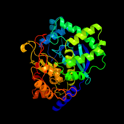
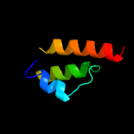
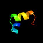
1 c3clqC_
100.0
43
PDB header: structural genomics, unknown functionChain: C: PDB Molecule: uncharacterized protein;PDBTitle: crystal structure of a conserved protein of unknown function from2 enterococcus faecalis v583
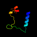
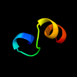
2 d2pw4a1
62.1
18
Fold: Jann2411-likeSuperfamily: Jann2411-likeFamily: Jann2411-like3 c2cqjA_
49.5
33
PDB header: rna binding proteinChain: A: PDB Molecule: u3 small nucleolar ribonucleoprotein proteinPDBTitle: solution structure of the s4 domain of u3 small nucleolar2 ribonucleoprotein protein imp3 homolog
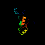
4 c2htmB_
45.8
22
PDB header: biosynthetic proteinChain: B: PDB Molecule: thiazole biosynthesis protein thig;PDBTitle: crystal structure of ttha0676 from thermus thermophilus hb8
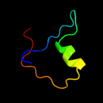
5 d1pdza2
45.3
24
Fold: Enolase N-terminal domain-likeSuperfamily: Enolase N-terminal domain-likeFamily: Enolase N-terminal domain-like6 d2akza2
38.2
18
Fold: Enolase N-terminal domain-likeSuperfamily: Enolase N-terminal domain-likeFamily: Enolase N-terminal domain-like7 d2al1a2
33.9
24
Fold: Enolase N-terminal domain-likeSuperfamily: Enolase N-terminal domain-likeFamily: Enolase N-terminal domain-like8 d1alua_
33.0
15
Fold: 4-helical cytokinesSuperfamily: 4-helical cytokinesFamily: Long-chain cytokines9 c2re2A_
32.2
27
PDB header: oxidoreductaseChain: A: PDB Molecule: uncharacterized protein ta1041;PDBTitle: crystal structure of a putative iron-molybdenum cofactor (femo-co)2 dinitrogenase (ta1041m) from thermoplasma acidophilum dsm 1728 at3 1.30 a resolution
10 d1h3fa2
21.6
11
Fold: Alpha-L RNA-binding motifSuperfamily: Alpha-L RNA-binding motifFamily: Tyrosyl-tRNA synthetase (TyrRS), C-terminal domain11 c2y9xG_
21.2
30
PDB header: oxidoreductaseChain: G: PDB Molecule: lectin-like fold protein;PDBTitle: crystal structure of ppo3, a tyrosinase from agaricus bisporus, in2 deoxy-form that contains additional unknown lectin-like subunit,3 with inhibitor tropolone
12 d1c06a_
21.1
20
Fold: Alpha-L RNA-binding motifSuperfamily: Alpha-L RNA-binding motifFamily: Ribosomal protein S413 c3hp7A_
19.4
21
PDB header: structural genomics, unknown functionChain: A: PDB Molecule: hemolysin, putative;PDBTitle: putative hemolysin from streptococcus thermophilus.
14 c1vjtA_
18.3
7
PDB header: hydrolaseChain: A: PDB Molecule: alpha-glucosidase;PDBTitle: crystal structure of alpha-glucosidase (tm0752) from thermotoga2 maritima at 2.50 a resolution
15 c2d5nB_
17.7
26
PDB header: hydrolase, oxidoreductaseChain: B: PDB Molecule: riboflavin biosynthesis protein ribd;PDBTitle: crystal structure of a bifunctional deaminase and reductase2 involved in riboflavin biosynthesis
16 c2l58A_
16.7
47
PDB header: apoptosisChain: A: PDB Molecule: activator of apoptosis harakiri;PDBTitle: solution structure of the cytosolic fragment 22-53 of bcl-2 member2 harakiri
17 d2gy9d1
16.4
12
Fold: Alpha-L RNA-binding motifSuperfamily: Alpha-L RNA-binding motifFamily: Ribosomal protein S418 d2isba1
15.3
16
Fold: The "swivelling" beta/beta/alpha domainSuperfamily: FumA C-terminal domain-likeFamily: FumA C-terminal domain-like19 c3msuA_
13.8
20
PDB header: transferaseChain: A: PDB Molecule: citrate synthase;PDBTitle: crystal structure of citrate synthase from francisella tularensis
20 d1nuba2
12.4
25
Fold: Knottins (small inhibitors, toxins, lectins)Superfamily: EGF/LamininFamily: Follistatin (FS) module N-terminal domain, FS-N21 d2fyma2
not modelled
12.2
18
Fold: Enolase N-terminal domain-likeSuperfamily: Enolase N-terminal domain-likeFamily: Enolase N-terminal domain-like22 c2ldkA_
not modelled
12.2
25
PDB header: structural genomics, unknown functionChain: A: PDB Molecule: uncharacterized protein;PDBTitle: solution nmr structure of protein aaur_3427 from arthrobacter2 aurescens, northeast structural genomics consortium target aar96
23 c2qnfB_
not modelled
11.7
28
PDB header: hydrolase/dnaChain: B: PDB Molecule: recombination endonuclease vii;PDBTitle: crystal structure of t4 endonuclease vii h43n mutant in2 complex with heteroduplex dna containing base mismatches
24 c2xzmD_
not modelled
11.5
9
PDB header: ribosomeChain: D: PDB Molecule: ribosomal protein s4 containing protein;PDBTitle: crystal structure of the eukaryotic 40s ribosomal2 subunit in complex with initiation factor 1. this file3 contains the 40s subunit and initiation factor for4 molecule 1
25 c3d0jA_
not modelled
11.4
18
PDB header: structural genomics, unknown functionChain: A: PDB Molecule: uncharacterized protein ca_c3497;PDBTitle: crystal structure of conserved protein of unknown function ca_c34972 from clostridium acetobutylicum atcc 824
26 c2bibA_
not modelled
11.1
12
PDB header: hydrolaseChain: A: PDB Molecule: teichoic acid phosphorylcholine esterase/ choline bindingPDBTitle: crystal structure of the complete modular teichioic acid2 phosphorylcholine esterase pce (cbpe) from streptococcus3 pneumoniae
27 d1jh3a_
not modelled
10.4
18
Fold: Alpha-L RNA-binding motifSuperfamily: Alpha-L RNA-binding motifFamily: Tyrosyl-tRNA synthetase (TyrRS), C-terminal domain28 d1kfia4
not modelled
9.9
29
Fold: TBP-likeSuperfamily: Phosphoglucomutase, C-terminal domainFamily: Phosphoglucomutase, C-terminal domain29 d1duvg1
not modelled
9.7
19
Fold: ATC-likeSuperfamily: Aspartate/ornithine carbamoyltransferaseFamily: Aspartate/ornithine carbamoyltransferase30 d1t6la2
not modelled
9.5
67
Fold: DNA clampSuperfamily: DNA clampFamily: DNA polymerase processivity factor31 d2o34a1
not modelled
9.4
43
Fold: T-foldSuperfamily: ApbE-likeFamily: DVU1097-like32 d2jeka1
not modelled
9.4
17
Fold: Rv1873-likeSuperfamily: Rv1873-likeFamily: Rv1873-like33 c2bs5A_
not modelled
9.2
20
PDB header: sugar binding proteinChain: A: PDB Molecule: fucose-binding lectin protein;PDBTitle: lectin from ralstonia solanacearum complexed with 2-2 fucosyllactose
34 c2hxvA_
not modelled
8.9
29
PDB header: biosynthetic proteinChain: A: PDB Molecule: diaminohydroxyphosphoribosylaminopyrimidine deaminase/ 5-PDBTitle: crystal structure of a diaminohydroxyphosphoribosylaminopyrimidine2 deaminase/ 5-amino-6-(5-phosphoribosylamino)uracil reductase (tm1828)3 from thermotoga maritima at 1.80 a resolution
35 d1s4da_
not modelled
8.7
19
Fold: Tetrapyrrole methylaseSuperfamily: Tetrapyrrole methylaseFamily: Tetrapyrrole methylase36 d1p9ka_
not modelled
8.3
38
Fold: Alpha-L RNA-binding motifSuperfamily: Alpha-L RNA-binding motifFamily: YbcJ-like37 d3pmga4
not modelled
8.0
19
Fold: TBP-likeSuperfamily: Phosphoglucomutase, C-terminal domainFamily: Phosphoglucomutase, C-terminal domain38 c2i7uA_
not modelled
7.6
48
PDB header: de novo protein/ligand binding proteinChain: A: PDB Molecule: four-alpha-helix bundle;PDBTitle: structural and dynamical analysis of a four-alpha-helix2 bundle with designed anesthetic binding pockets
39 c1zc1A_
not modelled
7.4
20
PDB header: protein turnoverChain: A: PDB Molecule: ubiquitin fusion degradation protein 1;PDBTitle: ufd1 exhibits the aaa-atpase fold with two distinct2 ubiquitin interaction sites
40 d2uubd1
not modelled
7.3
25
Fold: Alpha-L RNA-binding motifSuperfamily: Alpha-L RNA-binding motifFamily: Ribosomal protein S441 d2hsja1
not modelled
7.1
19
Fold: Flavodoxin-likeSuperfamily: SGNH hydrolaseFamily: Acetylhydrolase42 c1s1hD_
not modelled
7.0
23
PDB header: ribosomeChain: D: PDB Molecule: 40s ribosomal protein s9-a;PDBTitle: structure of the ribosomal 80s-eef2-sordarin complex from2 yeast obtained by docking atomic models for rna and protein3 components into a 11.7 a cryo-em map. this file, 1s1h,4 contains 40s subunit. the 60s ribosomal subunit is in file5 1s1i.
43 c2yujA_
not modelled
6.9
20
PDB header: protein bindingChain: A: PDB Molecule: ubiquitin fusion degradation 1-like;PDBTitle: solution structure of human ubiquitin fusion degradation2 protein 1 homolog ufd1
44 d1twda_
not modelled
6.6
17
Fold: TIM beta/alpha-barrelSuperfamily: CutC-likeFamily: CutC-like45 c3bbnD_
not modelled
6.3
15
PDB header: ribosomeChain: D: PDB Molecule: ribosomal protein s4;PDBTitle: homology model for the spinach chloroplast 30s subunit2 fitted to 9.4a cryo-em map of the 70s chlororibosome.
46 c2iunD_
not modelled
6.3
25
PDB header: viral proteinChain: D: PDB Molecule: avian adenovirus celo long fibre;PDBTitle: structure of the c-terminal head domain of the avian2 adenovirus celo long fibre (p21 crystal form)
47 d1dxha1
not modelled
6.1
19
Fold: ATC-likeSuperfamily: Aspartate/ornithine carbamoyltransferaseFamily: Aspartate/ornithine carbamoyltransferase48 c2zt9F_
not modelled
6.0
37
PDB header: photosynthesisChain: F: PDB Molecule: cytochrome b6-f complex subunit 7;PDBTitle: crystal structure of the cytochrome b6f complex from nostoc sp. pcc2 7120
49 c2e0wA_
not modelled
6.0
14
PDB header: transferaseChain: A: PDB Molecule: gamma-glutamyltranspeptidase;PDBTitle: t391a precursor mutant protein of gamma-glutamyltranspeptidase from2 escherichia coli
50 c3l2eB_
not modelled
5.9
16
PDB header: transferaseChain: B: PDB Molecule: glycocyamine kinase beta chain;PDBTitle: glycocyamine kinase, alpha-beta heterodimer from marine worm2 namalycastis sp.
51 c2lf2A_
not modelled
5.7
18
PDB header: structural genomics, unknown functionChain: A: PDB Molecule: uncharacterized protein;PDBTitle: solution nmr structure of the ahsa1-like protein chu_1110 from2 cytophaga hutchinsonii, northeast structural genomics consortium3 target chr152
52 c2w40C_
not modelled
5.7
18
PDB header: transferaseChain: C: PDB Molecule: glycerol kinase, putative;PDBTitle: crystal structure of plasmodium falciparum glycerol kinase2 with bound glycerol
53 c2f6sA_
not modelled
5.6
14
PDB header: structural genomics, unknown functionChain: A: PDB Molecule: cell filamentation protein, putative;PDBTitle: structure of cell filamentation protein (fic) from helicobacter pylori
54 c3btpA_
not modelled
5.5
36
PDB header: dna binding protein, chaperoneChain: A: PDB Molecule: single-strand dna-binding protein;PDBTitle: crystal structure of agrobacterium tumefaciens vire2 in complex with2 its chaperone vire1: a novel fold and implications for dna binding
55 d2c42a2
not modelled
5.3
44
Fold: Thiamin diphosphate-binding fold (THDP-binding)Superfamily: Thiamin diphosphate-binding fold (THDP-binding)Family: PFOR PP module56 c1yzxB_
not modelled
5.2
45
PDB header: transferaseChain: B: PDB Molecule: glutathione s-transferase kappa 1;PDBTitle: crystal structure of human kappa class glutathione2 transferase