1 c2gqsA_
100.0
100
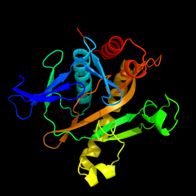
PDB header: ligaseChain: A: PDB Molecule: phosphoribosylaminoimidazole-succinocarboxamidePDBTitle: saicar synthetase complexed with cair-mg2+ and adp
2 c2z02A_
100.0
43
PDB header: ligaseChain: A: PDB Molecule: phosphoribosylaminoimidazole-succinocarboxamidePDBTitle: crystal structure of2 phosphoribosylaminoimidazolesuccinocarboxamide synthase3 wit atp from methanocaldococcus jannaschii
3 c3kreA_
100.0
41
PDB header: ligaseChain: A: PDB Molecule: phosphoribosylaminoimidazole-succinocarboxamide synthase;PDBTitle: crystal structure of phosphoribosylaminoimidazole-succinocarboxamide2 synthase from ehrlichia chaffeensis at 1.8a resolution
4 c3nuaB_
100.0
42
PDB header: ligaseChain: B: PDB Molecule: phosphoribosylaminoimidazole-succinocarboxamide synthase;PDBTitle: crystal structure of phosphoribosylaminoimidazole-succinocarboxamide2 synthase from clostridium perfringens
5 c2ywvB_
100.0
44
PDB header: ligaseChain: B: PDB Molecule: phosphoribosylaminoimidazole succinocarboxamide synthetase;PDBTitle: crystal structure of saicar synthetase from geobacillus kaustophilus
6 d2cnqa1
100.0
30
Fold: SAICAR synthase-likeSuperfamily: SAICAR synthase-likeFamily: SAICAR synthase7 c3r9rA_
100.0
30
PDB header: ligaseChain: A: PDB Molecule: phosphoribosylaminoimidazole-succinocarboxamide synthase;PDBTitle: structure of a phosphoribosylaminoimidazole-succinocarboxamide2 synthase from mycobacterium abscessus atcc 19977 / dsm 44196
8 d1kuta_
100.0
39
Fold: SAICAR synthase-likeSuperfamily: SAICAR synthase-likeFamily: SAICAR synthase9 c2h31A_
100.0
32
PDB header: ligase, lyaseChain: A: PDB Molecule: multifunctional protein ade2;PDBTitle: crystal structure of human paics, a bifunctional carboxylase and2 synthetase in purine biosynthesis
10 c1zp9A_
57.1
14
PDB header: transferaseChain: A: PDB Molecule: rio1 kinase;PDBTitle: crystal structure of full-legnth a.fulgidus rio1 serine kinase bound2 to atp and mn2+ ions.
11 d2h3ka1
36.5
14
Fold: Immunoglobulin-like beta-sandwichSuperfamily: NEAT domain-likeFamily: NEAT domain12 c3nj2B_
33.8
16
PDB header: unknown functionChain: B: PDB Molecule: duf269-containing protein;PDBTitle: crystal structure of cce_0566 from the cyanobacterium cyanothece2 51142, a protein associated with nitrogen fixation from the duf2693 family
13 d1kjqa3
19.8
34
Fold: ATP-graspSuperfamily: Glutathione synthetase ATP-binding domain-likeFamily: BC ATP-binding domain-like14 d2a5za1
18.7
12
Fold: Concanavalin A-like lectins/glucanasesSuperfamily: Concanavalin A-like lectins/glucanasesFamily: SO2946-like15 d1i4ya_
18.4
9
Fold: Four-helical up-and-down bundleSuperfamily: Hemerythrin-likeFamily: Hemerythrin-like16 c3g7pA_
17.5
18
PDB header: unknown functionChain: A: PDB Molecule: nitrogen fixation protein;PDBTitle: crystal structure of a nifx-associated protein of unknown function2 (afe_1514) from acidithiobacillus ferrooxidans atcc at 2.00 a3 resolution
17 c2pvpB_
16.8
34
PDB header: ligaseChain: B: PDB Molecule: d-alanine-d-alanine ligase;PDBTitle: crystal structure of d-alanine-d-alanine ligase from helicobacter2 pylori
18 d1pmia_
15.3
15
Fold: Double-stranded beta-helixSuperfamily: RmlC-like cupinsFamily: Type I phosphomannose isomerase19 c3lzcA_
14.1
15
PDB header: biosynthetic proteinChain: A: PDB Molecule: dph2;PDBTitle: crystal structure of dph2 from pyrococcus horikoshii
20 c2auwB_
12.7
23
PDB header: unknown functionChain: B: PDB Molecule: hypothetical protein ne0471;PDBTitle: crystal structure of putative dna binding protein ne0471 from2 nitrosomonas europaea atcc 19718
21 c3h1yA_
not modelled
11.7
35
PDB header: isomeraseChain: A: PDB Molecule: mannose-6-phosphate isomerase;PDBTitle: crystal structure of mannose 6-phosphate isomerase from2 salmonella typhimurium bound to substrate (f6p)and metal3 atom (zn)
22 d1hrba_
not modelled
11.4
9
Fold: Four-helical up-and-down bundleSuperfamily: Hemerythrin-likeFamily: Hemerythrin-like23 c3cegA_
not modelled
11.0
13
PDB header: ligaseChain: A: PDB Molecule: baculoviral iap repeat-containing protein 6;PDBTitle: crystal structure of the ubc domain of baculoviral iap2 repeat-containing protein 6
24 c3eplA_
not modelled
10.6
24
PDB header: transferase/rnaChain: A: PDB Molecule: trna isopentenyltransferase;PDBTitle: crystallographic snapshots of eukaryotic2 dimethylallyltransferase acting on trna: insight into trna3 recognition and reaction mechanism
25 c3s6bA_
not modelled
10.5
7
PDB header: hydrolaseChain: A: PDB Molecule: methionine aminopeptidase;PDBTitle: crystal structure of methionine aminopeptidase 1b from plasmodium2 falciparum, pf10_0150
26 c1tqmA_
not modelled
10.3
13
PDB header: ribosomeChain: A: PDB Molecule: conserved hypothetical protein;PDBTitle: crystal structure of a. fulgidus rio2 serine protein kinase bound to2 amppnp
27 c3exaD_
not modelled
9.6
18
PDB header: transferaseChain: D: PDB Molecule: trna delta(2)-isopentenylpyrophosphatePDBTitle: crystal structure of the full-length trna2 isopentenylpyrophosphate transferase (bh2366) from3 bacillus halodurans, northeast structural genomics4 consortium target bhr41.
28 c3r23B_
not modelled
9.5
20
PDB header: ligaseChain: B: PDB Molecule: d-alanine--d-alanine ligase;PDBTitle: crystal structure of d-alanine--d-alanine ligase from bacillus2 anthracis
29 d1zara2
not modelled
9.5
15
Fold: Protein kinase-like (PK-like)Superfamily: Protein kinase-like (PK-like)Family: RIO1-like kinases30 c3idwA_
not modelled
9.4
6
PDB header: endocytosisChain: A: PDB Molecule: actin cytoskeleton-regulatory complex protein sla1;PDBTitle: crystal structure of sla1 homology domain 2
31 c2vd2A_
not modelled
8.8
16
PDB header: transferaseChain: A: PDB Molecule: atp phosphoribosyltransferase;PDBTitle: the crystal structure of hisg from b. subtilis
32 c2qeaB_
not modelled
8.3
24
PDB header: oxidoreductaseChain: B: PDB Molecule: putative general stress protein 26;PDBTitle: crystal structure of a putative general stress protein 26 (jann_0955)2 from jannaschia sp. ccs1 at 2.46 a resolution
33 c1ylaB_
not modelled
8.3
18
PDB header: ligaseChain: B: PDB Molecule: ubiquitin-conjugating enzyme e2-25 kda;PDBTitle: ubiquitin-conjugating enzyme e2-25 kda (huntington interacting protein2 2)
34 d2hq7a1
not modelled
8.3
7
Fold: Split barrel-likeSuperfamily: FMN-binding split barrelFamily: PNP-oxidase like35 d1a62a2
not modelled
8.2
18
Fold: OB-foldSuperfamily: Nucleic acid-binding proteinsFamily: Cold shock DNA-binding domain-like36 c3a8tA_
not modelled
8.1
13
PDB header: transferaseChain: A: PDB Molecule: adenylate isopentenyltransferase;PDBTitle: plant adenylate isopentenyltransferase in complex with atp
37 c3d3qB_
not modelled
7.8
24
PDB header: transferaseChain: B: PDB Molecule: trna delta(2)-isopentenylpyrophosphatePDBTitle: crystal structure of trna delta(2)-isopentenylpyrophosphate2 transferase (se0981) from staphylococcus epidermidis.3 northeast structural genomics consortium target ser100
38 d2cdqa3
not modelled
7.6
11
Fold: Ferredoxin-likeSuperfamily: ACT-likeFamily: Aspartokinase allosteric domain-like39 c2dvzA_
not modelled
7.6
14
PDB header: transport proteinChain: A: PDB Molecule: putative exported protein;PDBTitle: structure of a periplasmic transporter
40 c3dmbA_
not modelled
7.6
16
PDB header: oxidoreductaseChain: A: PDB Molecule: putative general stress protein 26 with a pnp-oxidase likePDBTitle: crystal structure of a putative general stress family protein2 (xcc2264) from xanthomonas campestris pv. campestris at 2.30 a3 resolution
41 d2hmfa2
not modelled
7.5
17
Fold: Ferredoxin-likeSuperfamily: ACT-likeFamily: Aspartokinase allosteric domain-like42 d1rz4a1
not modelled
7.5
25
Fold: DNA/RNA-binding 3-helical bundleSuperfamily: "Winged helix" DNA-binding domainFamily: Eukaryotic translation initiation factor 3 subunit 12, eIF3k, C-terminal domain43 d2d6fa1
not modelled
7.5
30
Fold: Sm-like foldSuperfamily: GatD N-terminal domain-likeFamily: GatD N-terminal domain-like44 d1tf5a1
not modelled
6.9
42
Fold: Pre-protein crosslinking domain of SecASuperfamily: Pre-protein crosslinking domain of SecAFamily: Pre-protein crosslinking domain of SecA45 d1nkta1
not modelled
6.9
50
Fold: Pre-protein crosslinking domain of SecASuperfamily: Pre-protein crosslinking domain of SecAFamily: Pre-protein crosslinking domain of SecA46 d2r7ka2
not modelled
6.7
20
Fold: ATP-graspSuperfamily: Glutathione synthetase ATP-binding domain-likeFamily: PurP ATP-binding domain-like47 c3dzoA_
not modelled
6.7
21
PDB header: transferaseChain: A: PDB Molecule: rhoptry kinase domain;PDBTitle: crystal structure of a rhoptry kinase from toxoplasma gondii
48 c3fozB_
not modelled
6.5
6
PDB header: transferase/rnaChain: B: PDB Molecule: trna delta(2)-isopentenylpyrophosphate transferase;PDBTitle: structure of e. coli isopentenyl-trna transferase in complex with e.2 coli trna(phe)
49 c2pr1B_
not modelled
6.5
16
PDB header: transferaseChain: B: PDB Molecule: uncharacterized n-acetyltransferase ylbp;PDBTitle: crystal structure of the bacillus subtilis n-acetyltransferase ylbp2 protein in complex with coenzyme-a
50 c3e46A_
not modelled
6.1
18
PDB header: ligaseChain: A: PDB Molecule: ubiquitin-conjugating enzyme e2-25 kda;PDBTitle: crystal structure of ubiquitin-conjugating enzyme e2-25kda2 (huntington interacting protein 2) m172a mutant
51 d1g57a_
not modelled
6.0
18
Fold: YrdC/RibBSuperfamily: YrdC/RibBFamily: 3,4-dihydroxy-2-butanone 4-phosphate synthase, DHBP synthase, RibB52 d1uj4a2
not modelled
5.9
71
Fold: Ferredoxin-likeSuperfamily: D-ribose-5-phosphate isomerase (RpiA), lid domainFamily: D-ribose-5-phosphate isomerase (RpiA), lid domain53 d2es9a1
not modelled
5.9
15
Fold: YoaC-likeSuperfamily: YoaC-likeFamily: YoaC-like54 d2ozla1
not modelled
5.9
15
Fold: Thiamin diphosphate-binding fold (THDP-binding)Superfamily: Thiamin diphosphate-binding fold (THDP-binding)Family: Branched-chain alpha-keto acid dehydrogenase PP module55 c3lwbA_
not modelled
5.7
30
PDB header: ligaseChain: A: PDB Molecule: d-alanine--d-alanine ligase;PDBTitle: crystal structure of apo d-alanine:d-alanine ligase (ddl) from2 mycobacterium tuberculosis
56 c1zdbA_
not modelled
5.7
50
PDB header: igg binding domainChain: A: PDB Molecule: mini protein a domain, z38;PDBTitle: phage-selected mini protein a domain, z38, nmr, minimized2 mean structure
57 c3byvA_
not modelled
5.5
23
PDB header: transferaseChain: A: PDB Molecule: rhoptry kinase;PDBTitle: crystal structure of toxoplasma gondii specific rhoptry2 antigen kinase domain
58 d2cyya1
not modelled
5.4
11
Fold: DNA/RNA-binding 3-helical bundleSuperfamily: "Winged helix" DNA-binding domainFamily: Lrp/AsnC-like transcriptional regulator N-terminal domain59 d1q46a1
not modelled
5.4
18
Fold: SAM domain-likeSuperfamily: eIF2alpha middle domain-likeFamily: eIF2alpha middle domain-like