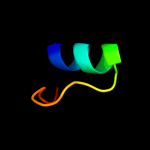
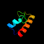
1 d1xg7a_
48.1
30
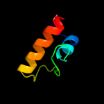
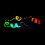
Fold: DNA-glycosylaseSuperfamily: DNA-glycosylaseFamily: AgoG-like2 d1ee8a1
44.6
21
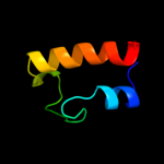
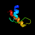
Fold: S13-like H2TH domainSuperfamily: S13-like H2TH domainFamily: Middle domain of MutM-like DNA repair proteins3 d1k3xa1
42.5
13
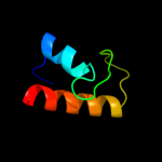
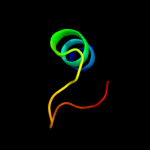
Fold: S13-like H2TH domainSuperfamily: S13-like H2TH domainFamily: Middle domain of MutM-like DNA repair proteins4 d1tdza1
37.5
17
Fold: S13-like H2TH domainSuperfamily: S13-like H2TH domainFamily: Middle domain of MutM-like DNA repair proteins5 d1r2za1
37.5
19
Fold: S13-like H2TH domainSuperfamily: S13-like H2TH domainFamily: Middle domain of MutM-like DNA repair proteins6 c3edyA_
36.5
22
PDB header: hydrolaseChain: A: PDB Molecule: tripeptidyl-peptidase 1;PDBTitle: crystal structure of the precursor form of human tripeptidyl-peptidase2 1
7 d1k82a1
35.3
10
Fold: S13-like H2TH domainSuperfamily: S13-like H2TH domainFamily: Middle domain of MutM-like DNA repair proteins8 d1xqoa_
30.6
30
Fold: DNA-glycosylaseSuperfamily: DNA-glycosylaseFamily: AgoG-like9 c2kncA_
29.9
28
PDB header: cell adhesionChain: A: PDB Molecule: integrin alpha-iib;PDBTitle: platelet integrin alfaiib-beta3 transmembrane-cytoplasmic2 heterocomplex
10 c3ee6A_
29.7
22
PDB header: hydrolaseChain: A: PDB Molecule: tripeptidyl-peptidase 1;PDBTitle: crystal structure analysis of tripeptidyl peptidase -i
11 d1ug3a1
29.0
15
Fold: alpha-alpha superhelixSuperfamily: ARM repeatFamily: MIF4G domain-like12 c2lbgA_
27.8
46
PDB header: membrane proteinChain: A: PDB Molecule: major prion protein;PDBTitle: structure of the chr of the prion protein in dpc micelles
13 d1t1ea2
25.9
12
Fold: Ferredoxin-likeSuperfamily: Protease propeptides/inhibitorsFamily: Subtilase propeptides/inhibitors14 c2k1aA_
25.9
28
PDB header: cell adhesionChain: A: PDB Molecule: integrin alpha-iib;PDBTitle: bicelle-embedded integrin alpha(iib) transmembrane segment
15 c3h2zA_
25.4
18
PDB header: oxidoreductaseChain: A: PDB Molecule: mannitol-1-phosphate 5-dehydrogenase;PDBTitle: the crystal structure of mannitol-1-phosphate dehydrogenase from2 shigella flexneri
16 d1t5ja_
24.6
18
Fold: ADP-ribosylglycohydrolaseSuperfamily: ADP-ribosylglycohydrolaseFamily: ADP-ribosylglycohydrolase17 c3g9dB_
24.3
23
PDB header: hydrolaseChain: B: PDB Molecule: dinitrogenase reductase activactingPDBTitle: crystal structure glycohydrolase
18 d2aq0a1
23.6
8
Fold: SAM domain-likeSuperfamily: RuvA domain 2-likeFamily: Hef domain-like19 c2qtyB_
22.1
27
PDB header: hydrolaseChain: B: PDB Molecule: poly(adp-ribose) glycohydrolase arh3;PDBTitle: crystal structure of mouse adp-ribosylhydrolase 3 (marh3)
20 c2r6cG_
21.6
15
PDB header: replicationChain: G: PDB Molecule: dnag primase, helicase binding domain;PDBTitle: crystal form bh2
21 c2k7rA_
not modelled
20.3
16
PDB header: replicationChain: A: PDB Molecule: primosomal protein dnai;PDBTitle: n-terminal domain of the bacillus subtilis helicase-loading2 protein dnai
22 c2wocA_
not modelled
19.4
27
PDB header: hydrolaseChain: A: PDB Molecule: adp-ribosyl-[dinitrogen reductase] glycohydrolase;PDBTitle: crystal structure of the dinitrogenase reductase-activating2 glycohydrolase (drag) from rhodospirillum rubrum
23 c2yzwA_
not modelled
19.2
27
PDB header: hydrolaseChain: A: PDB Molecule: adp-ribosylglycohydrolase;PDBTitle: adp-ribosylglycohydrolase-related protein complex
24 c1b9uA_
not modelled
17.8
28
PDB header: hydrolaseChain: A: PDB Molecule: protein (atp synthase);PDBTitle: membrane domain of the subunit b of the e.coli atp synthase
25 d2gp4a2
not modelled
16.9
13
Fold: IlvD/EDD N-terminal domain-likeSuperfamily: IlvD/EDD N-terminal domain-likeFamily: lvD/EDD N-terminal domain-like26 d1rh5b_
not modelled
16.8
22
Fold: Single transmembrane helixSuperfamily: Preprotein translocase SecE subunitFamily: Preprotein translocase SecE subunit27 c1ug3A_
not modelled
15.7
15
PDB header: translationChain: A: PDB Molecule: eukaryotic protein synthesis initiation factorPDBTitle: c-terminal portion of human eif4gi
28 d2piha1
not modelled
15.0
5
Fold: YheA-likeSuperfamily: YheA/YmcA-likeFamily: YmcA-like29 c2pihA_
not modelled
15.0
5
PDB header: structural genomics, unknown functionChain: A: PDB Molecule: protein ymca;PDBTitle: crystal structure of protein ymca from bacillus subtilis,2 northeast structural genomics target sr375
30 c2kncB_
not modelled
14.8
14
PDB header: cell adhesionChain: B: PDB Molecule: integrin beta-3;PDBTitle: platelet integrin alfaiib-beta3 transmembrane-cytoplasmic2 heterocomplex
31 c2f5qA_
not modelled
14.6
19
PDB header: hydrolase/dnaChain: A: PDB Molecule: formamidopyrimidine-dna glycosidase;PDBTitle: catalytically inactive (e3q) mutm crosslinked to oxog:c2 containing dna cc2
32 c3hd7A_
not modelled
13.9
6
PDB header: exocytosisChain: A: PDB Molecule: vesicle-associated membrane protein 2;PDBTitle: helical extension of the neuronal snare complex into the membrane,2 spacegroup c 1 2 1
33 d1v54d_
not modelled
13.9
13
Fold: Single transmembrane helixSuperfamily: Mitochondrial cytochrome c oxidase subunit IVFamily: Mitochondrial cytochrome c oxidase subunit IV34 c3qnqD_
not modelled
13.8
16
PDB header: membrane protein, transport proteinChain: D: PDB Molecule: pts system, cellobiose-specific iic component;PDBTitle: crystal structure of the transporter chbc, the iic component from the2 n,n'-diacetylchitobiose-specific phosphotransferase system
35 c2y69Q_
not modelled
13.7
13
PDB header: electron transportChain: Q: PDB Molecule: cytochrome c oxidase subunit 4 isoform 1;PDBTitle: bovine heart cytochrome c oxidase re-refined with molecular2 oxygen
36 d2a1ja1
not modelled
13.6
8
Fold: SAM domain-likeSuperfamily: RuvA domain 2-likeFamily: Hef domain-like37 d2crga1
not modelled
13.2
29
Fold: DNA/RNA-binding 3-helical bundleSuperfamily: Homeodomain-likeFamily: Myb/SANT domain38 c2kluA_
not modelled
12.8
26
PDB header: immune system, membrane proteinChain: A: PDB Molecule: t-cell surface glycoprotein cd4;PDBTitle: nmr structure of the transmembrane and cytoplasmic domains2 of human cd4
39 c2w2eA_
not modelled
12.7
10
PDB header: membrane proteinChain: A: PDB Molecule: aquaporin;PDBTitle: 1.15 angstrom crystal structure of p.pastoris aquaporin,2 aqy1, in a closed conformation at ph 3.5
40 d1xi8a3
not modelled
12.5
20
Fold: Molybdenum cofactor biosynthesis proteinsSuperfamily: Molybdenum cofactor biosynthesis proteinsFamily: MoeA central domain-like41 c3hfwA_
not modelled
12.4
20
PDB header: hydrolaseChain: A: PDB Molecule: protein adp-ribosylarginine hydrolase;PDBTitle: crystal structure of human adp-ribosylhydrolase 1 (harh1)
42 c2opfA_
not modelled
12.2
15
PDB header: hydrolase/dnaChain: A: PDB Molecule: endonuclease viii;PDBTitle: crystal structure of the dna repair enzyme endonuclease-viii (nei)2 from e. coli (r252a) in complex with ap-site containing dna substrate
43 c2gp4A_
not modelled
12.0
15
PDB header: lyaseChain: A: PDB Molecule: 6-phosphogluconate dehydratase;PDBTitle: structure of [fes]cluster-free apo form of 6-phosphogluconate2 dehydratase from shewanella oneidensis
44 c3a46B_
not modelled
11.3
13
PDB header: hydrolaseChain: B: PDB Molecule: formamidopyrimidine-dna glycosylase;PDBTitle: crystal structure of mvnei1/thf complex
45 c2l23A_
not modelled
11.0
13
PDB header: transcriptionChain: A: PDB Molecule: mediator of rna polymerase ii transcription subunit 25;PDBTitle: nmr structure of the acid (activator interacting domain) of the human2 mediator med25 protein
46 c1wazA_
not modelled
11.0
18
PDB header: transport proteinChain: A: PDB Molecule: merf;PDBTitle: nmr structure determination of the bacterial mercury2 transporter, merf, in micelles
47 c3h3pT_
not modelled
10.8
43
PDB header: immune systemChain: T: PDB Molecule: 4e10_s0_1tjlc_004_n;PDBTitle: crystal structure of hiv epitope-scaffold 4e10 fv complex
48 c1q90L_
not modelled
10.7
30
PDB header: photosynthesisChain: L: PDB Molecule: cytochrome b6f complex subunit petl;PDBTitle: structure of the cytochrome b6f (plastohydroquinone : plastocyanin2 oxidoreductase) from chlamydomonas reinhardtii
49 d1q90l_
not modelled
10.7
30
Fold: Single transmembrane helixSuperfamily: PetL subunit of the cytochrome b6f complexFamily: PetL subunit of the cytochrome b6f complex50 c3kvhA_
not modelled
10.7
17
PDB header: rna binding proteinChain: A: PDB Molecule: protein syndesmos;PDBTitle: crystal structure of human protein syndesmos (nudt16-like protein)
51 c1ee8A_
not modelled
10.7
21
PDB header: dna binding proteinChain: A: PDB Molecule: mutm (fpg) protein;PDBTitle: crystal structure of mutm (fpg) protein from thermus thermophilus hb8
52 c1nnjA_
not modelled
10.2
15
PDB header: hydrolaseChain: A: PDB Molecule: formamidopyrimidine-dna glycosylase;PDBTitle: crystal structure complex between the lactococcus lactis fpg and an2 abasic site containing dna
53 d1ymga1
not modelled
10.1
28
Fold: Aquaporin-likeSuperfamily: Aquaporin-likeFamily: Aquaporin-like54 c1ymgA_
not modelled
10.1
28
PDB header: membrane proteinChain: A: PDB Molecule: lens fiber major intrinsic protein;PDBTitle: the channel architecture of aquaporin o at 2.2 angstrom resolution
55 c2hjmB_
not modelled
9.8
21
PDB header: structural genomics, unknown functionChain: B: PDB Molecule: hypothetical protein pf1176;PDBTitle: crystal structure of a singleton protein pf1176 from p. furiosus
56 d1k6za_
not modelled
9.7
26
Fold: Secretion chaperone-likeSuperfamily: Type III secretory system chaperone-likeFamily: Type III secretory system chaperone57 c2vt2A_
not modelled
9.5
15
PDB header: transcriptionChain: A: PDB Molecule: redox-sensing transcriptional repressor rex;PDBTitle: structure and functional properties of the bacillus2 subtilis transcriptional repressor rex
58 c3d9sB_
not modelled
9.4
33
PDB header: membrane proteinChain: B: PDB Molecule: aquaporin-5;PDBTitle: human aquaporin 5 (aqp5) - high resolution x-ray structure
59 d1rfza_
not modelled
9.4
11
Fold: YutG-likeSuperfamily: YutG-likeFamily: YutG-like60 c2zu6E_
not modelled
9.4
13
PDB header: hydrolaseChain: E: PDB Molecule: programmed cell death protein 4;PDBTitle: crystal structure of the eif4a-pdcd4 complex
61 c1k82D_
not modelled
9.3
10
PDB header: hydrolase/dnaChain: D: PDB Molecule: formamidopyrimidine-dna glycosylase;PDBTitle: crystal structure of e.coli formamidopyrimidine-dna2 glycosylase (fpg) covalently trapped with dna
62 c2kqzA_
not modelled
9.3
4
PDB header: protein bindingChain: A: PDB Molecule: proteasomal ubiquitin receptor adrm1;PDBTitle: fragment of proteasome protein
63 d1xova2
not modelled
9.1
13
Fold: Phosphorylase/hydrolase-likeSuperfamily: Zn-dependent exopeptidasesFamily: N-acetylmuramoyl-L-alanine amidase-like64 d1jlja_
not modelled
9.1
18
Fold: Molybdenum cofactor biosynthesis proteinsSuperfamily: Molybdenum cofactor biosynthesis proteinsFamily: MogA-like65 c2qtqB_
not modelled
9.0
9
PDB header: transcriptionChain: B: PDB Molecule: transcriptional regulator, tetr family;PDBTitle: crystal structure of a predicted dna-binding transcriptional regulator2 (saro_1072) from novosphingobium aromaticivorans dsm at 1.85 a3 resolution
66 c3c02A_
not modelled
8.9
17
PDB header: membrane proteinChain: A: PDB Molecule: aquaglyceroporin;PDBTitle: x-ray structure of the aquaglyceroporin from plasmodium falciparum
67 d1ug8a_
not modelled
8.8
22
Fold: IF3-likeSuperfamily: R3H domainFamily: R3H domain68 c2bcxB_
not modelled
8.8
86
PDB header: calcium binding proteinChain: B: PDB Molecule: ryanodine receptor 1;PDBTitle: crystal structure of calmodulin in complex with a ryanodine2 receptor peptide
69 d1jyaa_
not modelled
8.6
26
Fold: Secretion chaperone-likeSuperfamily: Type III secretory system chaperone-likeFamily: Type III secretory system chaperone70 d2a4ha1
not modelled
8.6
20
Fold: Thioredoxin foldSuperfamily: Thioredoxin-likeFamily: Selenoprotein W-related71 c2zmeA_
not modelled
8.5
5
PDB header: protein transportChain: A: PDB Molecule: vacuolar-sorting protein snf8;PDBTitle: integrated structural and functional model of the human escrt-ii2 complex
72 c3cuqA_
not modelled
8.3
5
PDB header: protein transportChain: A: PDB Molecule: vacuolar-sorting protein snf8;PDBTitle: integrated structural and functional model of the human escrt-ii2 complex
73 d2cdxa_
not modelled
8.3
20
Fold: Snake toxin-likeSuperfamily: Snake toxin-likeFamily: Snake venom toxins74 d1rc2a_
not modelled
8.3
33
Fold: Aquaporin-likeSuperfamily: Aquaporin-likeFamily: Aquaporin-like75 d1y0na_
not modelled
8.2
11
Fold: YehU-likeSuperfamily: YehU-likeFamily: YehU-like76 c1u0bB_
not modelled
8.2
13
PDB header: ligase/rnaChain: B: PDB Molecule: cysteinyl trna;PDBTitle: crystal structure of cysteinyl-trna synthetase binary2 complex with trnacys
77 d1dd4c_
not modelled
8.2
18
Fold: Ribosomal protein L7/12, oligomerisation (N-terminal) domainSuperfamily: Ribosomal protein L7/12, oligomerisation (N-terminal) domainFamily: Ribosomal protein L7/12, oligomerisation (N-terminal) domain78 c1amlA_
not modelled
8.1
27
PDB header: serine protease inhibitorChain: A: PDB Molecule: amyloid a4;PDBTitle: the alzheimer`s disease amyloid a4 peptide (residues 1-40)
79 d1j4na_
not modelled
8.1
22
Fold: Aquaporin-likeSuperfamily: Aquaporin-likeFamily: Aquaporin-like80 d1cksa_
not modelled
8.1
67
Fold: Cell cycle regulatory proteinsSuperfamily: Cell cycle regulatory proteinsFamily: Cell cycle regulatory proteins81 c2dzrA_
not modelled
8.1
29
PDB header: transcriptionChain: A: PDB Molecule: general transcription factor ii-i repeat domain-PDBTitle: solution structure of rsgi ruh-067, a gtf2i domain in human2 cdna
82 c1dvpA_
not modelled
8.0
8
PDB header: transferaseChain: A: PDB Molecule: hepatocyte growth factor-regulated tyrosinePDBTitle: crystal structure of the vhs and fyve tandem domains of hrs,2 a protein involved in membrane trafficking and signal3 transduction
83 c2jesG_
not modelled
8.0
20
PDB header: viral proteinChain: G: PDB Molecule: portal protein;PDBTitle: portal protein from bacteriophage spp1
84 d1u5ta1
not modelled
8.0
16
Fold: DNA/RNA-binding 3-helical bundleSuperfamily: "Winged helix" DNA-binding domainFamily: Vacuolar sorting protein domain85 d2hxja1
not modelled
7.9
22
Fold: FinO-likeSuperfamily: FinO-likeFamily: FinO-like86 c2f2bA_
not modelled
7.9
33
PDB header: membrane proteinChain: A: PDB Molecule: aquaporin aqpm;PDBTitle: crystal structure of integral membrane protein aquaporin aqpm at 1.68a2 resolution
87 c3ahhA_
not modelled
7.8
12
PDB header: lyaseChain: A: PDB Molecule: xylulose 5-phosphate/fructose 6-phosphate phosphoketolase;PDBTitle: h142a mutant of phosphoketolase from bifidobacterium breve complexed2 with acetyl thiamine diphosphate
88 c1m5iA_
not modelled
7.8
13
PDB header: antitumor proteinChain: A: PDB Molecule: apc protein;PDBTitle: crystal structure of the coiled coil region 129-250 of the2 tumor suppressor gene product apc
89 d1hq1a_
not modelled
7.7
35
Fold: Signal peptide-binding domainSuperfamily: Signal peptide-binding domainFamily: Signal peptide-binding domain90 c2yqkA_
not modelled
7.7
26
PDB header: transcription/apoptosisChain: A: PDB Molecule: arginine-glutamic acid dipeptide repeats protein;PDBTitle: solution structure of the sant domain in arginine-glutamic2 acid dipeptide (re) repeats
91 c1zaxZ_
not modelled
7.7
18
PDB header: structural proteinChain: Z: PDB Molecule: 50s ribosomal protein l7/l12;PDBTitle: ribosomal protein l10-l12(ntd) complex, space group p212121,2 form b
92 c1zawV_
not modelled
7.7
18
PDB header: structural proteinChain: V: PDB Molecule: 50s ribosomal protein l7/l12;PDBTitle: ribosomal protein l10-l12(ntd) complex, space group p212121,2 form a
93 d1rkta2
not modelled
7.6
12
Fold: Tetracyclin repressor-like, C-terminal domainSuperfamily: Tetracyclin repressor-like, C-terminal domainFamily: Tetracyclin repressor-like, C-terminal domain94 c1dfwA_
not modelled
7.6
54
PDB header: immune systemChain: A: PDB Molecule: lung surfactant protein b;PDBTitle: conformational mapping of the n-terminal segment of2 surfactant protein b in lipid using 13c-enhanced fourier3 transform infrared spectroscopy (ftir)
95 d1qb2a_
not modelled
7.6
18
Fold: Signal peptide-binding domainSuperfamily: Signal peptide-binding domainFamily: Signal peptide-binding domain96 d1sb0a_
not modelled
7.6
14
Fold: Kix domain of CBP (creb binding protein)Superfamily: Kix domain of CBP (creb binding protein)Family: Kix domain of CBP (creb binding protein)97 c3llqB_
not modelled
7.5
25
PDB header: membrane proteinChain: B: PDB Molecule: aquaporin z 2;PDBTitle: aquaporin structure from plant pathogen agrobacterium tumerfaciens
98 c2krxA_
not modelled
7.5
33
PDB header: structural genomics, unknown functionChain: A: PDB Molecule: asl3597 protein;PDBTitle: solution nmr structure of asl3597 from nostoc sp. pcc7120. northeast2 structural genomics consortium target id nsr244.
99 c1jauA_
not modelled
7.5
33
PDB header: viral proteinChain: A: PDB Molecule: transmembrane glycoprotein (gp41);PDBTitle: nmr solution structure of the trp-rich peptide of hiv gp412 bound to dpc micelles


