| 1 | c1ldaA_
|
|
 |
100.0 |
100 |
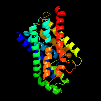
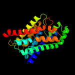
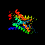
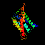
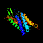
PDB header:transport protein
Chain: A: PDB Molecule:glycerol uptake facilitator protein;
PDBTitle: crystal structure of the e. coli glycerol facilitator (glpf) without2 substrate glycerol
|
| 2 | d1fx8a_
|
|
 |
100.0 |
100 |
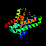
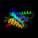
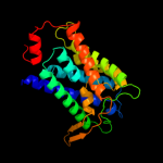
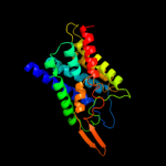
Fold:Aquaporin-like
Superfamily:Aquaporin-like
Family:Aquaporin-like |
| 3 | c3c02A_
|
|
 |
100.0 |
36 |
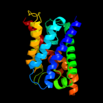
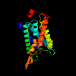
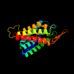
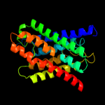
PDB header:membrane protein
Chain: A: PDB Molecule:aquaglyceroporin;
PDBTitle: x-ray structure of the aquaglyceroporin from plasmodium falciparum
|
| 4 | c2f2bA_
|
|
 |
100.0 |
36 |
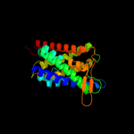
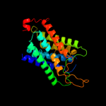
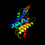
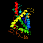
PDB header:membrane protein
Chain: A: PDB Molecule:aquaporin aqpm;
PDBTitle: crystal structure of integral membrane protein aquaporin aqpm at 1.68a2 resolution
|
| 5 | c2w2eA_
|
|
 |
100.0 |
27 |
PDB header:membrane protein
Chain: A: PDB Molecule:aquaporin;
PDBTitle: 1.15 angstrom crystal structure of p.pastoris aquaporin,2 aqy1, in a closed conformation at ph 3.5
|
| 6 | d1j4na_
|
|
 |
100.0 |
29 |
Fold:Aquaporin-like
Superfamily:Aquaporin-like
Family:Aquaporin-like |
| 7 | c3llqB_
|
|
 |
100.0 |
30 |
PDB header:membrane protein
Chain: B: PDB Molecule:aquaporin z 2;
PDBTitle: aquaporin structure from plant pathogen agrobacterium tumerfaciens
|
| 8 | c1ymgA_
|
|
 |
100.0 |
27 |
PDB header:membrane protein
Chain: A: PDB Molecule:lens fiber major intrinsic protein;
PDBTitle: the channel architecture of aquaporin o at 2.2 angstrom resolution
|
| 9 | d1ymga1
|
|
 |
100.0 |
27 |
Fold:Aquaporin-like
Superfamily:Aquaporin-like
Family:Aquaporin-like |
| 10 | c2d57A_
|
|
 |
100.0 |
28 |
PDB header:transport protein
Chain: A: PDB Molecule:aquaporin-4;
PDBTitle: double layered 2d crystal structure of aquaporin-4 (aqp4m23) at 3.2 a2 resolution by electron crystallography
|
| 11 | c3d9sB_
|
|
 |
100.0 |
31 |
PDB header:membrane protein
Chain: B: PDB Molecule:aquaporin-5;
PDBTitle: human aquaporin 5 (aqp5) - high resolution x-ray structure
|
| 12 | c3gd8A_
|
|
 |
100.0 |
27 |
PDB header:membrane protein
Chain: A: PDB Molecule:aquaporin-4;
PDBTitle: crystal structure of human aquaporin 4 at 1.8 and its mechanism of2 conductance
|
| 13 | d1h6ia_
|
|
 |
100.0 |
30 |
Fold:Aquaporin-like
Superfamily:Aquaporin-like
Family:Aquaporin-like |
| 14 | d1rc2a_
|
|
 |
100.0 |
29 |
Fold:Aquaporin-like
Superfamily:Aquaporin-like
Family:Aquaporin-like |
| 15 | c3iyzA_
|
|
 |
100.0 |
28 |
PDB header:transport protein
Chain: A: PDB Molecule:aquaporin-4;
PDBTitle: structure of aquaporin-4 s180d mutant at 10.0 a resolution from2 electron micrograph
|
| 16 | c2b5fD_
|
|
 |
100.0 |
29 |
PDB header:transport protein,membrane protein
Chain: D: PDB Molecule:aquaporin;
PDBTitle: crystal structure of the spinach aquaporin sopip2;1 in an2 open conformation to 3.9 resolution
|
| 17 | c3kcvG_
|
|
 |
72.7 |
12 |
PDB header:transport protein
Chain: G: PDB Molecule:probable formate transporter 1;
PDBTitle: structure of formate channel
|
| 18 | c2kncA_
|
|
 |
58.1 |
24 |
PDB header:cell adhesion
Chain: A: PDB Molecule:integrin alpha-iib;
PDBTitle: platelet integrin alfaiib-beta3 transmembrane-cytoplasmic2 heterocomplex
|
| 19 | d1f0ka_
|
|
 |
33.6 |
33 |
Fold:UDP-Glycosyltransferase/glycogen phosphorylase
Superfamily:UDP-Glycosyltransferase/glycogen phosphorylase
Family:Peptidoglycan biosynthesis glycosyltransferase MurG |
| 20 | c3qnqD_
|
|
 |
27.6 |
12 |
PDB header:membrane protein, transport protein
Chain: D: PDB Molecule:pts system, cellobiose-specific iic component;
PDBTitle: crystal structure of the transporter chbc, the iic component from the2 n,n'-diacetylchitobiose-specific phosphotransferase system
|
| 21 | d1pn3a_ |
|
not modelled |
21.5 |
28 |
Fold:UDP-Glycosyltransferase/glycogen phosphorylase
Superfamily:UDP-Glycosyltransferase/glycogen phosphorylase
Family:Gtf glycosyltransferase |
| 22 | d1rrva_ |
|
not modelled |
19.2 |
22 |
Fold:UDP-Glycosyltransferase/glycogen phosphorylase
Superfamily:UDP-Glycosyltransferase/glycogen phosphorylase
Family:Gtf glycosyltransferase |
| 23 | d1iira_ |
|
not modelled |
18.5 |
22 |
Fold:UDP-Glycosyltransferase/glycogen phosphorylase
Superfamily:UDP-Glycosyltransferase/glycogen phosphorylase
Family:Gtf glycosyltransferase |
| 24 | c3orgB_ |
|
not modelled |
11.6 |
11 |
PDB header:transport protein
Chain: B: PDB Molecule:cmclc;
PDBTitle: crystal structure of a eukaryotic clc transporter
|
| 25 | c1s4iC_ |
|
not modelled |
9.4 |
43 |
PDB header:oxidoreductase
Chain: C: PDB Molecule:superoxide dismutase-like protein yojm;
PDBTitle: crystal structure of a sod-like protein from bacillus subtilis
|
| 26 | c3d0qB_ |
|
not modelled |
9.2 |
22 |
PDB header:transferase
Chain: B: PDB Molecule:protein calg3;
PDBTitle: crystal structure of calg3 from micromonospora echinospora determined2 in space group i222
|
| 27 | d1esoa_ |
|
not modelled |
8.3 |
67 |
Fold:Immunoglobulin-like beta-sandwich
Superfamily:Cu,Zn superoxide dismutase-like
Family:Cu,Zn superoxide dismutase-like |
| 28 | d1eqwa_ |
|
not modelled |
8.1 |
57 |
Fold:Immunoglobulin-like beta-sandwich
Superfamily:Cu,Zn superoxide dismutase-like
Family:Cu,Zn superoxide dismutase-like |
| 29 | d1oala_ |
|
not modelled |
7.4 |
50 |
Fold:Immunoglobulin-like beta-sandwich
Superfamily:Cu,Zn superoxide dismutase-like
Family:Cu,Zn superoxide dismutase-like |
| 30 | c2aqmA_ |
|
not modelled |
7.2 |
50 |
PDB header:oxidoreductase
Chain: A: PDB Molecule:superoxide dismutase [cu-zn];
PDBTitle: cu/zn superoxide dismutase from brucella abortus
|
| 31 | d2apsa_ |
|
not modelled |
6.9 |
50 |
Fold:Immunoglobulin-like beta-sandwich
Superfamily:Cu,Zn superoxide dismutase-like
Family:Cu,Zn superoxide dismutase-like |
| 32 | d1yrra2 |
|
not modelled |
6.5 |
40 |
Fold:TIM beta/alpha-barrel
Superfamily:Metallo-dependent hydrolases
Family:N-acetylglucosamine-6-phosphate deacetylase, NagA, catalytic domain |
| 33 | c1qupA_ |
|
not modelled |
6.2 |
17 |
PDB header:chaperone
Chain: A: PDB Molecule:superoxide dismutase 1 copper chaperone;
PDBTitle: crystal structure of the copper chaperone for superoxide2 dismutase
|
| 34 | c1jk9D_ |
|
not modelled |
6.2 |
17 |
PDB header:oxidoreductase
Chain: D: PDB Molecule:copper chaperone for superoxide dismutase;
PDBTitle: heterodimer between h48f-ysod1 and yccs
|
| 35 | d1f1ga_ |
|
not modelled |
6.0 |
57 |
Fold:Immunoglobulin-like beta-sandwich
Superfamily:Cu,Zn superoxide dismutase-like
Family:Cu,Zn superoxide dismutase-like |
| 36 | d1to4a_ |
|
not modelled |
6.0 |
71 |
Fold:Immunoglobulin-like beta-sandwich
Superfamily:Cu,Zn superoxide dismutase-like
Family:Cu,Zn superoxide dismutase-like |
| 37 | d1un7a2 |
|
not modelled |
5.9 |
30 |
Fold:TIM beta/alpha-barrel
Superfamily:Metallo-dependent hydrolases
Family:N-acetylglucosamine-6-phosphate deacetylase, NagA, catalytic domain |
| 38 | d2c9va1 |
|
not modelled |
5.8 |
57 |
Fold:Immunoglobulin-like beta-sandwich
Superfamily:Cu,Zn superoxide dismutase-like
Family:Cu,Zn superoxide dismutase-like |
| 39 | d1ej8a_ |
|
not modelled |
5.8 |
17 |
Fold:Immunoglobulin-like beta-sandwich
Superfamily:Cu,Zn superoxide dismutase-like
Family:Cu,Zn superoxide dismutase-like |
| 40 | c2qvtA_ |
|
not modelled |
5.8 |
38 |
PDB header:unknown function
Chain: A: PDB Molecule:avrl567-d;
PDBTitle: structure of melampsora lini avirulence protein, avrl567-d
|
| 41 | d1jk9b1 |
|
not modelled |
5.8 |
17 |
Fold:Immunoglobulin-like beta-sandwich
Superfamily:Cu,Zn superoxide dismutase-like
Family:Cu,Zn superoxide dismutase-like |
| 42 | c2q2lB_ |
|
not modelled |
5.7 |
57 |
PDB header:oxidoreductase
Chain: B: PDB Molecule:superoxide dismutase;
PDBTitle: crystal structure of superoxide dismutase from p.2 atrosanguina
|
| 43 | c2jlpA_ |
|
not modelled |
5.6 |
67 |
PDB header:oxidoreductase
Chain: A: PDB Molecule:extracellular superoxide dismutase (cu-zn);
PDBTitle: crystal structure of human extracellular copper-zinc2 superoxide dismutase.
|
| 44 | d1do5a_ |
|
not modelled |
5.6 |
57 |
Fold:Immunoglobulin-like beta-sandwich
Superfamily:Cu,Zn superoxide dismutase-like
Family:Cu,Zn superoxide dismutase-like |
| 45 | c3ia7A_ |
|
not modelled |
5.4 |
17 |
PDB header:transferase
Chain: A: PDB Molecule:calg4;
PDBTitle: crystal structure of calg4, the calicheamicin glycosyltransferase
|
| 46 | c3klzE_ |
|
not modelled |
5.3 |
14 |
PDB header:membrane protein
Chain: E: PDB Molecule:putative formate transporter 1;
PDBTitle: pentameric formate channel with formate bound
|
| 47 | c3iaaB_ |
|
not modelled |
5.3 |
22 |
PDB header:transferase
Chain: B: PDB Molecule:calg2;
PDBTitle: crystal structure of calg2, calicheamicin glycosyltransferase, tdp2 bound form
|