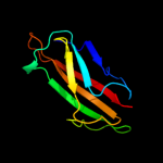
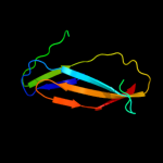
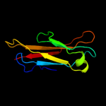
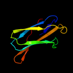
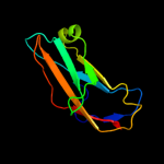
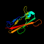
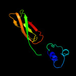
1 d2c9qa1
99.9
39
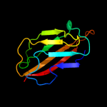
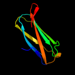
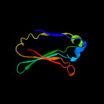
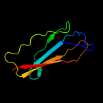
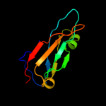
Fold: Immunoglobulin-like beta-sandwichSuperfamily: E set domainsFamily: Copper resistance protein C (CopC, PcoC)2 d1ix2a_
99.9
41
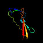
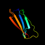
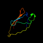
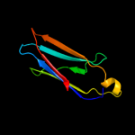
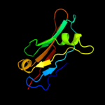
Fold: Immunoglobulin-like beta-sandwichSuperfamily: E set domainsFamily: Copper resistance protein C (CopC, PcoC)3 c3isyA_
96.9
19
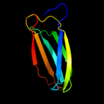
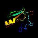
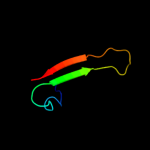
PDB header: protein bindingChain: A: PDB Molecule: intracellular proteinase inhibitor;PDBTitle: crystal structure of an intracellular proteinase inhibitor (ipi,2 bsu11130) from bacillus subtilis at 2.61 a resolution
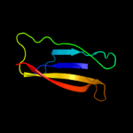
4 c2xwxB_
96.3
12
PDB header: chitin-binding proteinChain: B: PDB Molecule: glcnac-binding protein a;PDBTitle: vibrio cholerae colonization factor gbpa crystal structure
5 c2r5oA_
95.7
12
PDB header: transport proteinChain: A: PDB Molecule: putative atp binding component of abc-PDBTitle: crystal structure of the c-terminal domain of wzt
6 c3c12A_
95.4
19
PDB header: biosynthetic proteinChain: A: PDB Molecule: flagellar protein;PDBTitle: crystal structure of flgd from xanthomonas campestris:2 insights into the hook capping essential for flagellar3 assembly
7 c2p9rA_
95.1
21
PDB header: signaling proteinChain: A: PDB Molecule: alpha-2-macroglobulin;PDBTitle: human alpha2-macroglogulin is composed of multiple domains,2 as predicted by homology with complement component c3
8 c2x5pA_
94.6
25
PDB header: protein bindingChain: A: PDB Molecule: fibronectin binding protein;PDBTitle: crystal structure of the streptococcus pyogenes fibronectin binding2 protein fbab-b
9 c3pe9B_
94.2
12
PDB header: unknown functionChain: B: PDB Molecule: fibronectin(iii)-like module;PDBTitle: structures of clostridium thermocellum cbha fibronectin(iii)-like2 modules
10 c3d33B_
93.3
15
PDB header: unknown functionChain: B: PDB Molecule: domain of unknown function with an immunoglobulin-likePDBTitle: crystal structure of a duf3244 family protein with an immunoglobulin-2 like beta-sandwich fold (bvu_0276) from bacteroides vulgatus atcc3 8482 at 1.70 a resolution
11 c2e59A_
93.0
13
PDB header: lipid binding proteinChain: A: PDB Molecule: lymphocyte antigen 96;PDBTitle: crystal structure of human md-2 in complex with lipid iva
12 c3osvC_
92.3
9
PDB header: structural proteinChain: C: PDB Molecule: flagellar basal-body rod modification protein flgd;PDBTitle: the crytsal structure of flgd from p. aeruginosa
13 c3pdgA_
91.3
17
PDB header: unknown functionChain: A: PDB Molecule: fibronectin(iii)-like module;PDBTitle: structures of clostridium thermocellum cbha fibronectin(iii)-like2 modules
14 c3cu7A_
91.2
13
PDB header: immune systemChain: A: PDB Molecule: complement c5;PDBTitle: human complement component 5
15 c3jqxA_
90.1
13
PDB header: cell adhesionChain: A: PDB Molecule: colh protein;PDBTitle: crystal structure of clostridium histolyticum colh collagenase2 collagen binding domain 3 at 2.2 angstrom resolution in the presence3 of calcium and cademium
16 c3mu3A_
89.6
22
PDB header: immune systemChain: A: PDB Molecule: protein md-1;PDBTitle: crystal structure of chicken md-1 complexed with lipid iva
17 c3pe9D_
88.3
13
PDB header: unknown functionChain: D: PDB Molecule: fibronectin(iii)-like module;PDBTitle: structures of clostridium thermocellum cbha fibronectin(iii)-like2 modules
18 c3pe9C_
88.1
12
PDB header: unknown functionChain: C: PDB Molecule: fibronectin(iii)-like module;PDBTitle: structures of clostridium thermocellum cbha fibronectin(iii)-like2 modules
19 c3pe9A_
88.1
12
PDB header: unknown functionChain: A: PDB Molecule: fibronectin(iii)-like module;PDBTitle: structures of clostridium thermocellum cbha fibronectin(iii)-like2 modules
20 c2pn5A_
87.9
20
PDB header: immune systemChain: A: PDB Molecule: thioester-containing protein i;PDBTitle: crystal structure of tep1r
21 d1nepa_
not modelled
87.9
9
Fold: Immunoglobulin-like beta-sandwichSuperfamily: E set domainsFamily: ML domain22 d1w8oa1
not modelled
87.8
12
Fold: Immunoglobulin-like beta-sandwichSuperfamily: E set domainsFamily: E-set domains of sugar-utilizing enzymes23 d1nqjb_
not modelled
87.1
20
Fold: CUB-likeSuperfamily: Collagen-binding domainFamily: Collagen-binding domain24 c2b39B_
not modelled
86.8
15
PDB header: immune systemChain: B: PDB Molecule: c3;PDBTitle: structure of mammalian c3 with an intact thioester at 3a resolution
25 c2kpnA_
not modelled
85.5
36
PDB header: hydrolaseChain: A: PDB Molecule: bacillolysin;PDBTitle: solution nmr structure of a bacterial ig-like (big_3) domain from2 bacillus cereus. northeast structural genomics consortium target3 bcr147a
26 c2ra1A_
not modelled
84.5
11
PDB header: sugar binding proteinChain: A: PDB Molecule: surface layer protein;PDBTitle: crystal structure of the n-terminal part of the bacterial s-layer2 protein sbsc
27 c2qkiA_
not modelled
83.8
13
PDB header: immune system/hydrolase inhibitorChain: A: PDB Molecule: complement c3;PDBTitle: human c3c in complex with the inhibitor compstatin
28 c3ottB_
not modelled
83.4
16
PDB header: transcriptionChain: B: PDB Molecule: two-component system sensor histidine kinase;PDBTitle: crystal structure of the extracellular domain of the putative one2 component system bt4673 from b. thetaiotaomicron
29 c3m7oB_
not modelled
81.9
10
PDB header: immune systemChain: B: PDB Molecule: lymphocyte antigen 86;PDBTitle: crystal structure of mouse md-1 in complex with phosphatidylcholine
30 c3kptA_
not modelled
81.2
16
PDB header: cell adhesionChain: A: PDB Molecule: collagen adhesion protein;PDBTitle: crystal structure of bcpa, the major pilin subunit of2 bacillus cereus
31 d2a9da1
not modelled
80.1
8
Fold: Immunoglobulin-like beta-sandwichSuperfamily: E set domainsFamily: Molybdenum-containing oxidoreductases-like dimerisation domain32 c2k7pA_
not modelled
79.7
17
PDB header: structural proteinChain: A: PDB Molecule: filamin-a;PDBTitle: filamin a ig-like domains 16-17
33 c3pddA_
not modelled
75.6
16
PDB header: unknown functionChain: A: PDB Molecule: glycoside hydrolase, family 9;PDBTitle: structures of clostridium thermocellum cbha fibronectin(iii)-like2 modules
34 d1owwa_
not modelled
75.6
13
Fold: Immunoglobulin-like beta-sandwichSuperfamily: Fibronectin type IIIFamily: Fibronectin type III35 c2z64C_
not modelled
74.7
11
PDB header: immune systemChain: C: PDB Molecule: lymphocyte antigen 96;PDBTitle: crystal structure of mouse tlr4 and mouse md-2 complex
36 c2bpbA_
not modelled
73.8
11
PDB header: oxidoreductaseChain: A: PDB Molecule: sulfite\:cytochrome c oxidoreductase subunit a;PDBTitle: sulfite dehydrogenase from starkeya novella
37 d1nqja_
not modelled
73.8
20
Fold: CUB-likeSuperfamily: Collagen-binding domainFamily: Collagen-binding domain38 c3sd2A_
not modelled
70.9
15
PDB header: unknown functionChain: A: PDB Molecule: putative member of duf3244 protein family;PDBTitle: crystal structure of a putative member of duf3244 protein family2 (bt_3571) from bacteroides thetaiotaomicron vpi-5482 at 1.40 a3 resolution
39 d2bvya1
not modelled
70.7
18
Fold: Immunoglobulin-like beta-sandwichSuperfamily: E set domainsFamily: E-set domains of sugar-utilizing enzymes40 d1pfsa_
not modelled
68.7
19
Fold: OB-foldSuperfamily: Nucleic acid-binding proteinsFamily: Phage ssDNA-binding proteins41 d2diba1
not modelled
64.7
14
Fold: Immunoglobulin-like beta-sandwichSuperfamily: E set domainsFamily: Filamin repeat (rod domain)42 c2di7A_
not modelled
64.6
10
PDB header: structural proteinChain: A: PDB Molecule: bk158_1;PDBTitle: solution structure of the filamin domain from human bk158_12 protein
43 c3hrzA_
not modelled
64.0
14
PDB header: immune systemChain: A: PDB Molecule: cobra venom factor;PDBTitle: cobra venom factor (cvf) in complex with human factor b
44 c3qhtC_
not modelled
63.3
13
PDB header: de novo proteinChain: C: PDB Molecule: monobody ysmb-1;PDBTitle: crystal structure of the monobody ysmb-1 bound to yeast sumo
45 c2yrlA_
not modelled
62.9
23
PDB header: structural genomics, unknown functionChain: A: PDB Molecule: kiaa1837 protein;PDBTitle: solution structure of the pkd domain from kiaa 1837 protein
46 c3payB_
not modelled
60.8
16
PDB header: cell adhesionChain: B: PDB Molecule: putative adhesin;PDBTitle: crystal structure of a putative adhesin (bacova_04077) from2 bacteroides ovatus at 2.50 a resolution
47 c3nmeA_
59.1
20
PDB header: hydrolaseChain: A: PDB Molecule: sex4 glucan phosphatase;PDBTitle: structure of a plant phosphatase
48 c2ds4A_
not modelled
57.0
18
PDB header: protein bindingChain: A: PDB Molecule: tripartite motif protein 45;PDBTitle: solution structure of the filamin domain from human2 tripartite motif protein 45
49 c3a0oB_
not modelled
56.9
14
PDB header: lyaseChain: B: PDB Molecule: oligo alginate lyase;PDBTitle: crystal structure of alginate lyase from agrobacterium tumefaciens c58
50 c3gf8A_
not modelled
56.2
10
PDB header: carbohydrate binding proteinChain: A: PDB Molecule: putative polysaccharide binding proteins (duf1812);PDBTitle: crystal structure of putative polysaccharide binding proteins2 (duf1812) (np_809975.1) from bacteroides thetaiotaomicron vpi-5482 at3 2.20 a resolution
51 c3irpX_
not modelled
54.9
16
PDB header: cell adhesionChain: X: PDB Molecule: uro-adherence factor a;PDBTitle: crystal structure of functional region of uafa from staphylococcus2 saprophyticus at 1.50 angstrom resolution
52 d3csba1
not modelled
54.7
13
Fold: Immunoglobulin-like beta-sandwichSuperfamily: Fibronectin type IIIFamily: Fibronectin type III53 c1ttfA_
not modelled
54.5
17
PDB header: glycoproteinChain: A: PDB Molecule: fibronectin;PDBTitle: the three-dimensional structure of the tenth type iii2 module of fibronectin: an insight into rgd-mediated3 interactions
54 c1ttgA_
not modelled
54.5
17
PDB header: glycoproteinChain: A: PDB Molecule: fibronectin;PDBTitle: the three-dimensional structure of the tenth type iii2 module of fibronectin: an insight into rgd-mediated3 interactions
55 d2d7na1
not modelled
54.3
17
Fold: Immunoglobulin-like beta-sandwichSuperfamily: E set domainsFamily: Filamin repeat (rod domain)56 c3pvmB_
not modelled
53.3
10
PDB header: immune systemChain: B: PDB Molecule: cobra venom factor;PDBTitle: structure of complement c5 in complex with cvf
57 c2w1wB_
not modelled
52.2
13
PDB header: hydrolaseChain: B: PDB Molecule: lipolytic enzyme, g-d-s-l;PDBTitle: native structure of a family 35 carbohydrate binding module2 from clostridium thermocellum
58 d1uwya1
not modelled
51.7
11
Fold: Prealbumin-likeSuperfamily: Carboxypeptidase regulatory domain-likeFamily: Carboxypeptidase regulatory domain59 c3jquA_
not modelled
50.0
32
PDB header: cell adhesionChain: A: PDB Molecule: collagenase;PDBTitle: crystal structure of clostridium histolyticum colg collagenase2 polycystic kidney disease domain at 1.4 angstrom resolution
60 c2yetB_
not modelled
50.0
11
PDB header: hydrolaseChain: B: PDB Molecule: gh61 isozyme a;PDBTitle: thermoascus gh61 isozyme a
61 d2diaa1
not modelled
49.5
13
Fold: Immunoglobulin-like beta-sandwichSuperfamily: E set domainsFamily: Filamin repeat (rod domain)62 c3l48B_
not modelled
49.1
14
PDB header: transport proteinChain: B: PDB Molecule: outer membrane usher protein papc;PDBTitle: crystal structure of the c-terminal domain of the papc usher
63 c1qfhB_
not modelled
48.8
25
PDB header: actin binding proteinChain: B: PDB Molecule: protein (gelation factor);PDBTitle: dimerization of gelation factor from dictyostelium2 discoideum: crystal structure of rod domains 5 and 6
64 d1xwva_
not modelled
48.6
11
Fold: Immunoglobulin-like beta-sandwichSuperfamily: E set domainsFamily: ML domain65 c3rzwA_
not modelled
48.6
15
PDB header: protein bindingChain: A: PDB Molecule: monobody ysmb-9;PDBTitle: crystal structure of the monobody ysmb-9 bound to human sumo1
66 d2ag4a1
not modelled
48.0
24
Fold: Ganglioside M2 (gm2) activatorSuperfamily: Ganglioside M2 (gm2) activatorFamily: Ganglioside M2 (gm2) activator67 c2xetB_
not modelled
47.6
16
PDB header: transport proteinChain: B: PDB Molecule: f1 capsule-anchoring protein;PDBTitle: conserved hydrophobic clusters on the surface of the caf1a usher2 c-terminal domain are important for f1 antigen assembly
68 c3ejaB_
not modelled
47.4
19
PDB header: unknown functionChain: B: PDB Molecule: protein gh61e;PDBTitle: magnesium-bound glycoside hydrolase 61 isoform e from thielavia2 terrestris
69 c3mn8A_
not modelled
46.7
17
PDB header: hydrolaseChain: A: PDB Molecule: lp15968p;PDBTitle: structure of drosophila melanogaster carboxypeptidase d isoform 1b2 short
70 d1fnfa2
not modelled
46.5
13
Fold: Immunoglobulin-like beta-sandwichSuperfamily: Fibronectin type IIIFamily: Fibronectin type III71 d1lmia_
not modelled
46.5
36
Fold: Immunoglobulin-like beta-sandwichSuperfamily: Antigen MPT63/MPB63 (immunoprotective extracellular protein)Family: Antigen MPT63/MPB63 (immunoprotective extracellular protein)72 c2o6dB_
not modelled
46.3
20
PDB header: membrane protein, protein bindingChain: B: PDB Molecule: 34 kda membrane antigen;PDBTitle: structure of native rtp34 from treponema pallidum
73 c2o0i1_
not modelled
46.0
15
PDB header: surface active proteinChain: 1: PDB Molecule: c protein alpha-antigen;PDBTitle: crystal structure of the r185a mutant of the n-terminal domain of the2 group b streptococcus alpha c protein
74 d1tdqa2
not modelled
44.9
14
Fold: Immunoglobulin-like beta-sandwichSuperfamily: Fibronectin type IIIFamily: Fibronectin type III75 d1v5ja_
not modelled
43.1
12
Fold: Immunoglobulin-like beta-sandwichSuperfamily: Fibronectin type IIIFamily: Fibronectin type III76 c2v4vA_
not modelled
42.7
14
PDB header: hydrolaseChain: A: PDB Molecule: gh59 galactosidase;PDBTitle: crystal structure of a family 6 carbohydrate-binding module2 from clostridium cellulolyticum in complex with xylose
77 d2dmca1
not modelled
41.4
12
Fold: Immunoglobulin-like beta-sandwichSuperfamily: E set domainsFamily: Filamin repeat (rod domain)78 d1ex0a1
not modelled
41.2
11
Fold: Immunoglobulin-like beta-sandwichSuperfamily: E set domainsFamily: Transglutaminase N-terminal domain79 c2ocfD_
not modelled
41.1
17
PDB header: hormone/growth factorChain: D: PDB Molecule: fibronectin;PDBTitle: human estrogen receptor alpha ligand-binding domain in complex with2 estradiol and the e2#23 fn3 monobody
80 c2a9dB_
not modelled
40.3
10
PDB header: oxidoreductaseChain: B: PDB Molecule: sulfite oxidase;PDBTitle: crystal structure of recombinant chicken sulfite oxidase with arg at2 residue 161
81 c2k7qA_
not modelled
39.6
15
PDB header: structural proteinChain: A: PDB Molecule: filamin-a;PDBTitle: filamin a ig-like domains 18-19
82 c2e9jA_
not modelled
39.6
12
PDB header: structural proteinChain: A: PDB Molecule: filamin-b;PDBTitle: solution structure of the 14th filamin domain from human2 filamin-b
83 c2vtcB_
not modelled
39.4
18
PDB header: hydrolaseChain: B: PDB Molecule: cel61b;PDBTitle: the structure of a glycoside hydrolase family 61 member,2 cel61b from the hypocrea jecorina.
84 d1ktja_
not modelled
39.2
11
Fold: Immunoglobulin-like beta-sandwichSuperfamily: E set domainsFamily: ML domain85 d1qfha1
not modelled
38.9
25
Fold: Immunoglobulin-like beta-sandwichSuperfamily: E set domainsFamily: Filamin repeat (rod domain)86 c2dtgE_
not modelled
38.8
17
PDB header: hormone receptor/immune systemChain: E: PDB Molecule: insulin receptor;PDBTitle: insulin receptor (ir) ectodomain in complex with fab's
87 d2fnba_
not modelled
37.6
15
Fold: Immunoglobulin-like beta-sandwichSuperfamily: Fibronectin type IIIFamily: Fibronectin type III88 d1tfpa_
not modelled
36.9
8
Fold: Prealbumin-likeSuperfamily: Transthyretin (synonym: prealbumin)Family: Transthyretin (synonym: prealbumin)89 d1cwva3
not modelled
36.5
13
Fold: Immunoglobulin-like beta-sandwichSuperfamily: Invasin/intimin cell-adhesion fragmentsFamily: Invasin/intimin cell-adhesion fragments90 c2xtsC_
not modelled
35.9
9
PDB header: oxidoreductase/electron transportChain: C: PDB Molecule: sulfite dehydrogenase;PDBTitle: crystal structure of the sulfane dehydrogenase soxcd from paracoccus2 pantotrophus
91 d1fnha3
not modelled
35.2
14
Fold: Immunoglobulin-like beta-sandwichSuperfamily: Fibronectin type IIIFamily: Fibronectin type III92 d2d7oa1
not modelled
34.2
22
Fold: Immunoglobulin-like beta-sandwichSuperfamily: E set domainsFamily: Filamin repeat (rod domain)93 c2jxpA_
not modelled
34.2
8
PDB header: lipoproteinChain: A: PDB Molecule: putative lipoprotein b;PDBTitle: solution nmr structure of uncharacterized lipoprotein b2 from nitrosomonas europaea. northeast structural genomics3 target ner45a
94 c3qhtD_
not modelled
33.6
13
PDB header: de novo proteinChain: D: PDB Molecule: monobody ysmb-1;PDBTitle: crystal structure of the monobody ysmb-1 bound to yeast sumo
95 c3nrqB_
not modelled
33.3
11
PDB header: transport proteinChain: B: PDB Molecule: periplasmic protein-probably involved in high-affinity fe2+PDBTitle: crystal structure of copper-reconstituted fetp from uropathogenic2 escherichia coli strain f11
96 c2kzwA_
not modelled
32.9
16
PDB header: structural genomics, unknown functionChain: A: PDB Molecule: uncharacterized protein;PDBTitle: solution nmr structure of q8psa4 from methanosarcina mazei, northeast2 structural genomics consortium target mar143a
97 c3iswA_
not modelled
32.9
17
PDB header: structural proteinChain: A: PDB Molecule: filamin-a;PDBTitle: crystal structure of filamin-a immunoglobulin-like repeat 21 bound to2 an n-terminal peptide of cftr
98 c1uwyA_
not modelled
32.5
11
PDB header: hydrolaseChain: A: PDB Molecule: carboxypeptidase m;PDBTitle: crystal structure of human carboxypeptidase m
99 d1ut9a2
not modelled
31.2
12
Fold: Immunoglobulin-like beta-sandwichSuperfamily: E set domainsFamily: E-set domains of sugar-utilizing enzymes100 c3rghA_
not modelled
31.2
27
PDB header: cell adhesionChain: A: PDB Molecule: filamin-a;PDBTitle: structure of filamin a immunoglobulin-like repeat 10 from homo sapiens
101 c2xicB_
not modelled
30.7
25
PDB header: cell adhesionChain: B: PDB Molecule: ancillary protein 1;PDBTitle: pilus-presented adhesin, spy0125 (cpa), p212121 form (esrf data)
102 c3lxuX_
not modelled
30.6
18
PDB header: hydrolaseChain: X: PDB Molecule: tripeptidyl-peptidase 2;PDBTitle: crystal structure of tripeptidyl peptidase 2 (tpp ii)
103 c2lllA_
not modelled
28.4
10
PDB header: structural proteinChain: A: PDB Molecule: lamin-b2;PDBTitle: solution nmr structure of c-terminal globular domain of human lamin-2 b2, northeast structural genomics consortium target hr8546a
104 d1qfha2
not modelled
28.2
20
Fold: Immunoglobulin-like beta-sandwichSuperfamily: E set domainsFamily: Filamin repeat (rod domain)105 d1ogpa1
not modelled
27.8
14
Fold: Immunoglobulin-like beta-sandwichSuperfamily: E set domainsFamily: Molybdenum-containing oxidoreductases-like dimerisation domain106 d1w9sa_
not modelled
27.3
14
Fold: Galactose-binding domain-likeSuperfamily: Galactose-binding domain-likeFamily: Family 6 carbohydrate binding module, CBM6107 d2qfra1
not modelled
26.7
14
Fold: Immunoglobulin-like beta-sandwichSuperfamily: Purple acid phosphatase, N-terminal domainFamily: Purple acid phosphatase, N-terminal domain108 c2gpzC_
not modelled
26.7
17
PDB header: hydrolaseChain: C: PDB Molecule: transthyretin-like protein;PDBTitle: transthyretin-like protein from salmonella dublin
109 c3gzkA_
not modelled
26.5
18
PDB header: hydrolaseChain: A: PDB Molecule: cellulase;PDBTitle: structure of a. acidocaldarius cellulase cela
110 c3uyoD_
not modelled
26.5
15
PDB header: signaling protein/protein bindingChain: D: PDB Molecule: monobody sh13;PDBTitle: crystal structure of monobody sh13/abl1 sh2 domain complex
111 d2e9ia1
not modelled
26.3
17
Fold: Immunoglobulin-like beta-sandwichSuperfamily: E set domainsFamily: Filamin repeat (rod domain)112 d2dj4a1
not modelled
25.9
14
Fold: Immunoglobulin-like beta-sandwichSuperfamily: E set domainsFamily: Filamin repeat (rod domain)113 c2nsmA_
not modelled
25.8
17
PDB header: hydrolaseChain: A: PDB Molecule: carboxypeptidase n catalytic chain;PDBTitle: crystal structure of the human carboxypeptidase n (kininase i)2 catalytic domain
114 d1ufga_
not modelled
25.8
16
Fold: Immunoglobulin-like beta-sandwichSuperfamily: Lamin A/C globular tail domainFamily: Lamin A/C globular tail domain115 c2e7mA_
not modelled
25.0
22
PDB header: structural proteinChain: A: PDB Molecule: protein kiaa0319;PDBTitle: solution structure of the pkd domain (329-428) from human2 kiaa0319
116 d1tdqa1
not modelled
24.5
13
Fold: Immunoglobulin-like beta-sandwichSuperfamily: Fibronectin type IIIFamily: Fibronectin type III117 c2w3jA_
not modelled
24.4
15
PDB header: sugar-binding proteinChain: A: PDB Molecule: carbohydrate binding module;PDBTitle: structure of a family 35 carbohydrate binding module from2 an environmental isolate
118 c3d30A_
not modelled
23.8
30
PDB header: peptidoglycan-binding proteinChain: A: PDB Molecule: expansin like protein;PDBTitle: structure of an expansin like protein from bacillus subtilis at 1.9a2 resolution
119 d1kmta_
not modelled
23.7
17
Fold: Immunoglobulin-like beta-sandwichSuperfamily: E set domainsFamily: RhoGDI-like120 d1doab_
not modelled
22.9
17
Fold: Immunoglobulin-like beta-sandwichSuperfamily: E set domainsFamily: RhoGDI-like


