1 c1y7mB_
100.0
33
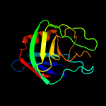
PDB header: structural genomics, unknown functionChain: B: PDB Molecule: hypothetical protein bsu14040;PDBTitle: crystal structure of the b. subtilis ykud protein at 2 a2 resolution
2 d1y7ma1
100.0
35
Fold: L,D-transpeptidase catalytic domain-likeSuperfamily: L,D-transpeptidase catalytic domain-likeFamily: L,D-transpeptidase catalytic domain-like3 c2hklB_
100.0
25
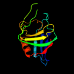
PDB header: transferaseChain: B: PDB Molecule: l,d-transpeptidase;PDBTitle: crystal structure of enterococcus faecium l,d-2 transpeptidase c442s mutant
4 d1zata1
100.0
26
Fold: L,D-transpeptidase catalytic domain-likeSuperfamily: L,D-transpeptidase catalytic domain-likeFamily: L,D-transpeptidase catalytic domain-like5 c2l9yA_
98.2
15
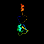
PDB header: sugar binding proteinChain: A: PDB Molecule: cvnh-lysm lectin;PDBTitle: solution structure of the mocvnh-lysm module from the rice blast2 fungus magnaporthe oryzae protein (mgg_03307)
6 c2djpA_
98.0
25
PDB header: structural genomics, unknown functionChain: A: PDB Molecule: hypothetical protein sb145;PDBTitle: the solution structure of the lysm domain of human2 hypothetical protein sb145
7 d1y7ma2
97.7
34
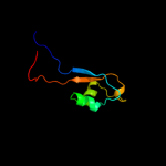
Fold: LysM domainSuperfamily: LysM domainFamily: LysM domain8 d1e0ga_
97.5
24
Fold: LysM domainSuperfamily: LysM domainFamily: LysM domain9 c2gu1A_
93.0
17
PDB header: hydrolaseChain: A: PDB Molecule: zinc peptidase;PDBTitle: crystal structure of a zinc containing peptidase from2 vibrio cholerae
10 c1h5nC_
61.2
20
PDB header: oxidoreductaseChain: C: PDB Molecule: dmso reductase;PDBTitle: dmso reductase modified by the presence of dms and air
11 c3mcaB_
59.7
21
PDB header: translation regulation/hydrolaseChain: B: PDB Molecule: protein dom34;PDBTitle: structure of the dom34-hbs1 complex and implications for its role in2 no-go decay
12 c1y5iA_
47.2
13
PDB header: oxidoreductaseChain: A: PDB Molecule: respiratory nitrate reductase 1 alpha chain;PDBTitle: the crystal structure of the narghi mutant nari-k86a
13 c2vdaB_
26.9
36
PDB header: protein transportChain: B: PDB Molecule: maltoporin;PDBTitle: solution structure of the seca-signal peptide complex
14 d1y5ia1
16.4
13
Fold: Double psi beta-barrelSuperfamily: ADC-likeFamily: Formate dehydrogenase/DMSO reductase, C-terminal domain15 d2hthb1
15.8
19
Fold: PH domain-like barrelSuperfamily: PH domain-likeFamily: VPS36 N-terminal domain-like16 c2k50A_
14.5
14
PDB header: structural genomics, unknown functionChain: A: PDB Molecule: replication factor a related protein;PDBTitle: solution nmr structure of the replication factor a related2 protein from methanobacterium thermoautotrophicum.3 northeast structural genomics target tr91a.
17 d1wjja_
14.2
23
Fold: OB-foldSuperfamily: Nucleic acid-binding proteinsFamily: Single strand DNA-binding domain, SSB18 c2kkeA_
13.5
42
PDB header: structural genomics, unknown functionChain: A: PDB Molecule: uncharacterized protein;PDBTitle: solution nmr structure of a dimeric protein of unknown2 function from methanobacterium thermoautotrophicum,3 northeast structural genomics consortium target tr5
19 c1eu1A_
13.4
15
PDB header: oxidoreductaseChain: A: PDB Molecule: dimethyl sulfoxide reductase;PDBTitle: the crystal structure of rhodobacter sphaeroides dimethylsulfoxide2 reductase reveals two distinct molybdenum coordination environments.
20 c1tmoA_
13.2
16
PDB header: oxidoreductaseChain: A: PDB Molecule: trimethylamine n-oxide reductase;PDBTitle: trimethylamine n-oxide reductase from shewanella massilia
21 d2vgna1
not modelled
7.4
17
Fold: Sm-like foldSuperfamily: Dom34/Pelota N-terminal domain-likeFamily: Dom34/Pelota N-terminal domain-like22 d1k78a1
not modelled
7.2
71
Fold: DNA/RNA-binding 3-helical bundleSuperfamily: Homeodomain-likeFamily: Paired domain23 d2qi2a1
not modelled
7.1
26
Fold: Sm-like foldSuperfamily: Dom34/Pelota N-terminal domain-likeFamily: Dom34/Pelota N-terminal domain-like24 c3d12A_
not modelled
6.9
38
PDB header: hydrolase/membrane proteinChain: A: PDB Molecule: hemagglutinin-neuraminidase;PDBTitle: crystal structures of nipah virus g attachment glycoprotein in complex2 with its receptor ephrin-b3
25 d1ogya1
not modelled
6.9
15
Fold: Double psi beta-barrelSuperfamily: ADC-likeFamily: Formate dehydrogenase/DMSO reductase, C-terminal domain26 c3obyB_
not modelled
6.8
21
PDB header: hydrolaseChain: B: PDB Molecule: protein pelota homolog;PDBTitle: crystal structure of archaeoglobus fulgidus pelota reveals inter-2 domain structural plasticity
27 d1t3la1
not modelled
6.7
30
Fold: SH3-like barrelSuperfamily: SH3-domainFamily: SH3-domain28 d1eu1a1
not modelled
6.3
12
Fold: Double psi beta-barrelSuperfamily: ADC-likeFamily: Formate dehydrogenase/DMSO reductase, C-terminal domain29 c3rf1B_
not modelled
6.1
21
PDB header: ligaseChain: B: PDB Molecule: glycyl-trna synthetase alpha subunit;PDBTitle: the crystal structure of glycyl-trna synthetase subunit alpha from2 campylobacter jejuni subsp. jejuni nctc 11168
30 d6paxa1
not modelled
6.1
57
Fold: DNA/RNA-binding 3-helical bundleSuperfamily: Homeodomain-likeFamily: Paired domain31 c3nadB_
not modelled
5.8
14
PDB header: lyaseChain: B: PDB Molecule: ferulate decarboxylase;PDBTitle: crystal structure of phenolic acid decarboxylase from bacillus pumilus2 ui-670
32 c3agjD_
not modelled
5.6
28
PDB header: translation/hydrolaseChain: D: PDB Molecule: protein pelota homolog;PDBTitle: crystal structure of archaeal pelota and gtp-bound ef1 alpha complex
33 c3agjB_
not modelled
5.6
28
PDB header: translation/hydrolaseChain: B: PDB Molecule: protein pelota homolog;PDBTitle: crystal structure of archaeal pelota and gtp-bound ef1 alpha complex
34 d1r1ga_
not modelled
5.4
55
Fold: Knottins (small inhibitors, toxins, lectins)Superfamily: Scorpion toxin-likeFamily: Short-chain scorpion toxins35 c1r1gA_
not modelled
5.4
55
PDB header: toxinChain: A: PDB Molecule: neurotoxin bmk37;PDBTitle: crystal structure of the scorpion toxin bmbkttx1
36 c1r1gB_
not modelled
5.4
55
PDB header: toxinChain: B: PDB Molecule: neurotoxin bmk37;PDBTitle: crystal structure of the scorpion toxin bmbkttx1
37 c2bfuL_
not modelled
5.4
33
PDB header: virusChain: L: PDB Molecule: cowpea mosaic virus, large (l) subunit;PDBTitle: x-ray structure of cpmv top component