1 c2l26A_
100.0
24
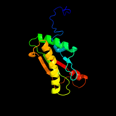
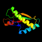
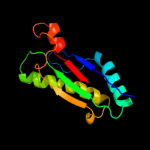
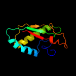
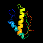
PDB header: membrane proteinChain: A: PDB Molecule: uncharacterized protein rv0899/mt0922;PDBTitle: rv0899 from mycobacterium tuberculosis contains two separated domains
2 d2aizp1
100.0
55
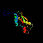
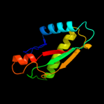
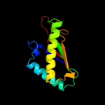
Fold: Bacillus chorismate mutase-likeSuperfamily: OmpA-likeFamily: OmpA-like3 d2hqsc1
100.0
100
Fold: Bacillus chorismate mutase-likeSuperfamily: OmpA-likeFamily: OmpA-like4 c2kgwA_
100.0
25
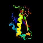
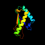
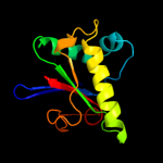
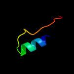
PDB header: membrane proteinChain: A: PDB Molecule: outer membrane protein a;PDBTitle: solution structure of the carboxy-terminal domain of ompatb, a pore2 forming protein from mycobacterium tuberculosis
5 c3khnB_
100.0
17
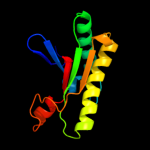
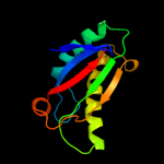
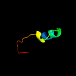
PDB header: structural genomics, unknown functionChain: B: PDB Molecule: motb protein, putative;PDBTitle: crystal structure of putative motb like protein dvu_22282 from desulfovibrio vulgaris.
6 c2k1sA_
100.0
24
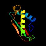
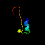
PDB header: lipoproteinChain: A: PDB Molecule: inner membrane lipoprotein yiad;PDBTitle: solution nmr structure of the folded c-terminal fragment of yiad from2 escherichia coli. northeast structural genomics consortium target3 er553.
7 c3td4D_
100.0
25
PDB header: membrane protein,peptide binding proteinChain: D: PDB Molecule: outer membrane protein omp38;PDBTitle: crystal structure of ompa-like domain from acinetobacter baumannii in2 complex with diaminopimelate
8 c3cyqM_
100.0
24
PDB header: membrane proteinChain: M: PDB Molecule: chemotaxis protein motb;PDBTitle: the crystal structure of the complex of the c-terminal domain of2 helicobacter pylori motb (residues 125-256) with n-acetylmuramic acid
9 c1r1mA_
100.0
34
PDB header: membrane proteinChain: A: PDB Molecule: outer membrane protein class 4;PDBTitle: structure of the ompa-like domain of rmpm from neisseria2 meningitidis
10 d1r1ma_
100.0
34
Fold: Bacillus chorismate mutase-likeSuperfamily: OmpA-likeFamily: OmpA-like11 c2zvyB_
100.0
23
PDB header: membrane proteinChain: B: PDB Molecule: chemotaxis protein motb;PDBTitle: structure of the periplasmic domain of motb from salmonella2 (crystal form ii)
12 c3ldtA_
99.9
16
PDB header: membrane proteinChain: A: PDB Molecule: outer membrane protein, ompa family protein;PDBTitle: crystal structure of an outer membrane protein(ompa)from2 legionella pneumophila
13 c3oonA_
99.9
28
PDB header: membrane proteinChain: A: PDB Molecule: outer membrane protein (tpn50);PDBTitle: the structure of an outer membrance protein from borrelia burgdorferi2 b31
14 c2zovA_
99.9
26
PDB header: membrane proteinChain: A: PDB Molecule: chemotaxis protein motb;PDBTitle: structure of the periplasmic domain of motb from salmonella2 (crystal form i)
15 c2zf8A_
99.8
27
PDB header: structural proteinChain: A: PDB Molecule: component of sodium-driven polar flagellar motor;PDBTitle: crystal structure of moty
16 c2r6hC_
48.4
38
PDB header: oxidoreductaseChain: C: PDB Molecule: nadh:ubiquinone oxidoreductase, na translocating, fPDBTitle: crystal structure of the domain comprising the nad binding and the fad2 binding regions of the nadh:ubiquinone oxidoreductase, na3 translocating, f subunit from porphyromonas gingivalis
17 d1gvha3
45.4
22
Fold: Ferredoxin reductase-like, C-terminal NADP-linked domainSuperfamily: Ferredoxin reductase-like, C-terminal NADP-linked domainFamily: Flavohemoglobin, C-terminal domain18 c1gvhA_
37.0
23
PDB header: oxidoreductaseChain: A: PDB Molecule: flavohemoprotein;PDBTitle: the x-ray structure of ferric escherichia coli2 flavohemoglobin reveals an unespected geometry of the3 distal heme pocket
19 d1tiaa_
36.8
27
Fold: alpha/beta-HydrolasesSuperfamily: alpha/beta-HydrolasesFamily: Fungal lipases20 c3ngmB_
34.8
36
PDB header: hydrolaseChain: B: PDB Molecule: extracellular lipase;PDBTitle: crystal structure of lipase from gibberella zeae
21 d1tiba_
not modelled
33.6
35
Fold: alpha/beta-HydrolasesSuperfamily: alpha/beta-HydrolasesFamily: Fungal lipases22 c3g7nA_
not modelled
32.0
16
PDB header: hydrolaseChain: A: PDB Molecule: lipase;PDBTitle: crystal structure of a triacylglycerol lipase from2 penicillium expansum at 1.3
23 c1tvcA_
not modelled
24.9
14
PDB header: oxidoreductaseChain: A: PDB Molecule: methane monooxygenase component c;PDBTitle: fad and nadh binding domain of methane monooxygenase2 reductase from methylococcus capsulatus (bath)
24 d1bwvs_
not modelled
23.8
19
Fold: RuBisCO, small subunitSuperfamily: RuBisCO, small subunitFamily: RuBisCO, small subunit25 c3o0dF_
not modelled
23.3
26
PDB header: hydrolaseChain: F: PDB Molecule: triacylglycerol lipase;PDBTitle: crystal structure of lip2 lipase from yarrowia lipolytica at 1.7 a2 resolution
26 d1uzhc1
not modelled
22.5
16
Fold: RuBisCO, small subunitSuperfamily: RuBisCO, small subunitFamily: RuBisCO, small subunit27 d1bxni_
not modelled
20.4
16
Fold: RuBisCO, small subunitSuperfamily: RuBisCO, small subunitFamily: RuBisCO, small subunit28 d1uwca_
not modelled
20.1
25
Fold: alpha/beta-HydrolasesSuperfamily: alpha/beta-HydrolasesFamily: Fungal lipases29 d1cqxa3
not modelled
19.8
13
Fold: Ferredoxin reductase-like, C-terminal NADP-linked domainSuperfamily: Ferredoxin reductase-like, C-terminal NADP-linked domainFamily: Flavohemoglobin, C-terminal domain30 d1tvca2
not modelled
19.7
11
Fold: Ferredoxin reductase-like, C-terminal NADP-linked domainSuperfamily: Ferredoxin reductase-like, C-terminal NADP-linked domainFamily: Aromatic dioxygenase reductase-like31 c1cqxB_
not modelled
19.6
15
PDB header: lipid binding proteinChain: B: PDB Molecule: flavohemoprotein;PDBTitle: crystal structure of the flavohemoglobin from alcaligenes eutrophus at2 1.75 a resolution
32 d1krha2
not modelled
17.5
15
Fold: Ferredoxin reductase-like, C-terminal NADP-linked domainSuperfamily: Ferredoxin reductase-like, C-terminal NADP-linked domainFamily: Aromatic dioxygenase reductase-like33 c3l7pA_
not modelled
17.1
17
PDB header: transcriptionChain: A: PDB Molecule: putative nitrogen regulatory protein pii;PDBTitle: crystal structure of smu.1657c, putative nitrogen regulatory protein2 pii from streptococcus mutans
34 d1rbli_
not modelled
16.5
17
Fold: RuBisCO, small subunitSuperfamily: RuBisCO, small subunitFamily: RuBisCO, small subunit35 d1xdpa4
not modelled
16.1
9
Fold: Phospholipase D/nucleaseSuperfamily: Phospholipase D/nucleaseFamily: Polyphosphate kinase C-terminal domain36 d3tgla_
not modelled
16.0
23
Fold: alpha/beta-HydrolasesSuperfamily: alpha/beta-HydrolasesFamily: Fungal lipases37 d1gk8i_
not modelled
15.6
16
Fold: RuBisCO, small subunitSuperfamily: RuBisCO, small subunitFamily: RuBisCO, small subunit38 d1svdm1
not modelled
14.2
12
Fold: RuBisCO, small subunitSuperfamily: RuBisCO, small subunitFamily: RuBisCO, small subunit39 d1ir1s_
not modelled
14.2
19
Fold: RuBisCO, small subunitSuperfamily: RuBisCO, small subunitFamily: RuBisCO, small subunit40 d1ej7s_
not modelled
13.8
16
Fold: RuBisCO, small subunitSuperfamily: RuBisCO, small subunitFamily: RuBisCO, small subunit41 d8ruci_
not modelled
13.8
19
Fold: RuBisCO, small subunitSuperfamily: RuBisCO, small subunitFamily: RuBisCO, small subunit42 d2o8ra4
not modelled
13.3
9
Fold: Phospholipase D/nucleaseSuperfamily: Phospholipase D/nucleaseFamily: Polyphosphate kinase C-terminal domain43 d4pgaa_
not modelled
13.2
18
Fold: Glutaminase/AsparaginaseSuperfamily: Glutaminase/AsparaginaseFamily: Glutaminase/Asparaginase44 d2v6ai1
not modelled
13.1
16
Fold: RuBisCO, small subunitSuperfamily: RuBisCO, small subunitFamily: RuBisCO, small subunit45 c2gpjA_
not modelled
12.7
12
PDB header: fad-binding proteinChain: A: PDB Molecule: siderophore-interacting protein;PDBTitle: crystal structure of a siderophore-interacting protein (sputcn32_0076)2 from shewanella putrefaciens cn-32 at 2.20 a resolution
46 d1uzdc1
not modelled
12.4
13
Fold: RuBisCO, small subunitSuperfamily: RuBisCO, small subunitFamily: RuBisCO, small subunit47 c2ckcA_
not modelled
12.2
17
PDB header: hydrolaseChain: A: PDB Molecule: chromodomain-helicase-dna-binding protein 7;PDBTitle: solution structures of the brk domains of the human chromo2 helicase domain 7 and 8, reveals structural similarity3 with gyf domain suggesting a role in protein interaction
48 d2ckca1
not modelled
12.2
17
Fold: GYF/BRK domain-likeSuperfamily: BRK domain-likeFamily: BRK domain-like49 d1wdds_
not modelled
11.7
19
Fold: RuBisCO, small subunitSuperfamily: RuBisCO, small subunitFamily: RuBisCO, small subunit50 d2v0ea1
not modelled
11.1
20
Fold: GYF/BRK domain-likeSuperfamily: BRK domain-likeFamily: BRK domain-like51 c2rodB_
not modelled
10.2
17
PDB header: apoptosisChain: B: PDB Molecule: noxa;PDBTitle: solution structure of mcl-1 complexed with noxaa
52 c2bgjB_
not modelled
9.2
17
PDB header: oxidoreductaseChain: B: PDB Molecule: ferredoxin-nadp(h) reductase;PDBTitle: x-ray structure of the ferredoxin-nadp(h) reductase from2 rhodobacter capsulatus at 2.1 angstroms
53 d1o7ja_
not modelled
8.8
18
Fold: Glutaminase/AsparaginaseSuperfamily: Glutaminase/AsparaginaseFamily: Glutaminase/Asparaginase54 c3qbwA_
not modelled
8.1
16
PDB header: transferaseChain: A: PDB Molecule: anhydro-n-acetylmuramic acid kinase;PDBTitle: crystal structure of pseudomonas aeruginosa 1,6-anhydro-n-2 actetylmuramic acid kinase (anmk) bound to adenosine diphosphate
55 c3cqyA_
not modelled
8.1
24
PDB header: transferaseChain: A: PDB Molecule: anhydro-n-acetylmuramic acid kinase;PDBTitle: crystal structure of a functionally unknown protein (so_1313) from2 shewanella oneidensis mr-1
56 c1jrjA_
not modelled
8.0
17
PDB header: hormone/growth factorChain: A: PDB Molecule: exendin-4;PDBTitle: solution structure of exendin-4 in 30-vol% trifluoroethanol
57 c2ckaA_
not modelled
7.9
17
PDB header: hydrolaseChain: A: PDB Molecule: chromodomain-helicase-dna-binding protein 8;PDBTitle: solution structures of the brk domains of the human chromo2 helicase domain 7 and 8, reveals structural similarity3 with gyf domain suggesting a role in protein interaction
58 d2ckaa1
not modelled
7.9
17
Fold: GYF/BRK domain-likeSuperfamily: BRK domain-likeFamily: BRK domain-like59 d1agxa_
not modelled
7.7
23
Fold: Glutaminase/AsparaginaseSuperfamily: Glutaminase/AsparaginaseFamily: Glutaminase/Asparaginase60 c2o8rA_
not modelled
7.1
9
PDB header: transferaseChain: A: PDB Molecule: polyphosphate kinase;PDBTitle: crystal structure of polyphosphate kinase from2 porphyromonas gingivalis
61 d1lgya_
not modelled
7.1
22
Fold: alpha/beta-HydrolasesSuperfamily: alpha/beta-HydrolasesFamily: Fungal lipases62 d1wsaa_
not modelled
6.8
13
Fold: Glutaminase/AsparaginaseSuperfamily: Glutaminase/AsparaginaseFamily: Glutaminase/Asparaginase63 d2ibge1
not modelled
6.7
13
Fold: Hedgehog/DD-peptidaseSuperfamily: Hedgehog/DD-peptidaseFamily: Hedgehog (development protein), N-terminal signaling domain64 c2ogsA_
not modelled
6.7
24
PDB header: hydrolaseChain: A: PDB Molecule: thermostable carboxylesterase est50;PDBTitle: crystal structure of the geobacillus stearothermophilus2 carboxylesterase est55 at ph 6.2
65 d1uc8a1
not modelled
6.6
26
Fold: PreATP-grasp domainSuperfamily: PreATP-grasp domainFamily: Lysine biosynthesis enzyme LysX, N-terminal domain66 d1tkja1
not modelled
6.5
15
Fold: Phosphorylase/hydrolase-likeSuperfamily: Zn-dependent exopeptidasesFamily: Bacterial dinuclear zinc exopeptidases67 c3pe3D_
not modelled
5.8
22
PDB header: transferaseChain: D: PDB Molecule: udp-n-acetylglucosamine--peptide n-PDBTitle: structure of human o-glcnac transferase and its complex with a peptide2 substrate
68 c3pa8A_
not modelled
5.5
14
PDB header: toxin/peptide inhibitorChain: A: PDB Molecule: toxin b;PDBTitle: structure of the c. difficile tcdb cysteine protease domain in complex2 with a peptide inhibitor
69 c3a9lB_
not modelled
5.5
22
PDB header: hydrolaseChain: B: PDB Molecule: poly-gamma-glutamate hydrolase;PDBTitle: structure of bacteriophage poly-gamma-glutamate hydrolase
70 c2oghA_
not modelled
5.4
18
PDB header: translationChain: A: PDB Molecule: eukaryotic translation initiation factor eif-1;PDBTitle: solution structure of yeast eif1
71 c1nauA_
not modelled
5.3
36
PDB header: hormone/growth factorChain: A: PDB Molecule: glucagon;PDBTitle: nmr solution structure of the glucagon antagonist [deshis1,2 desphe6, glu9]glucagon amide in the presence of3 perdeuterated dodecylphosphocholine micelles