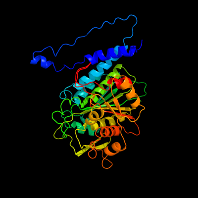
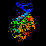
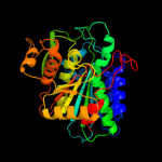
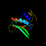
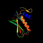
| 1 | c3kzwD_
|
|
 |
100.0 |
33 |
PDB header:hydrolase
Chain: D: PDB Molecule:cytosol aminopeptidase;
PDBTitle: crystal structure of cytosol aminopeptidase from staphylococcus aureus2 col
|
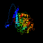
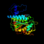
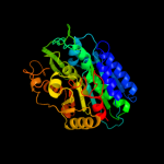
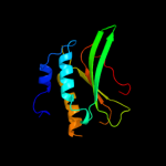
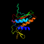
| 2 | c3h8gC_
|
|
 |
100.0 |
36 |
PDB header:hydrolase
Chain: C: PDB Molecule:cytosol aminopeptidase;
PDBTitle: bestatin complex structure of leucine aminopeptidase from pseudomonas2 putida
|
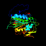
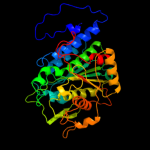
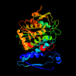
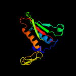
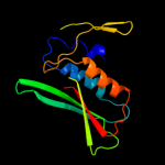
| 3 | c3jruB_
|
|
 |
100.0 |
33 |
PDB header:hydrolase
Chain: B: PDB Molecule:probable cytosol aminopeptidase;
PDBTitle: crystal structure of leucyl aminopeptidase (pepa) from xoo0834,2 xanthomonas oryzae pv. oryzae kacc10331
|
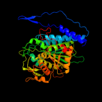
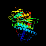
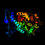
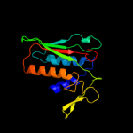
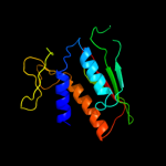
| 4 | c1lanA_
|
|
 |
100.0 |
33 |
PDB header:hydrolase (alpha-aminoacylpeptide)
Chain: A: PDB Molecule:leucine aminopeptidase;
PDBTitle: leucine aminopeptidase complex with l-leucinal
|
| 5 | c1gytG_
|
|
 |
100.0 |
34 |
PDB header:hydrolase
Chain: G: PDB Molecule:cytosol aminopeptidase;
PDBTitle: e. coli aminopeptidase a (pepa)
|
| 6 | c3ij3A_
|
|
 |
100.0 |
37 |
PDB header:hydrolase
Chain: A: PDB Molecule:cytosol aminopeptidase;
PDBTitle: 1.8 angstrom resolution crystal structure of cytosol aminopeptidase2 from coxiella burnetii
|
| 7 | c3kr5E_
|
|
 |
100.0 |
31 |
PDB header:hydrolase
Chain: E: PDB Molecule:m17 leucyl aminopeptidase;
PDBTitle: structure of a protease 4
|
| 8 | d1gyta2
|
|
 |
100.0 |
38 |
Fold:Phosphorylase/hydrolase-like
Superfamily:Zn-dependent exopeptidases
Family:Leucine aminopeptidase, C-terminal domain |
| 9 | d1lama1
|
|
 |
100.0 |
36 |
Fold:Phosphorylase/hydrolase-like
Superfamily:Zn-dependent exopeptidases
Family:Leucine aminopeptidase, C-terminal domain |
| 10 | c2hc9A_
|
|
 |
100.0 |
27 |
PDB header:hydrolase
Chain: A: PDB Molecule:leucine aminopeptidase 1;
PDBTitle: structure of caenorhabditis elegans leucine aminopeptidase-zinc2 complex (lap1)
|
| 11 | c3peiA_
|
|
 |
100.0 |
30 |
PDB header:hydrolase
Chain: A: PDB Molecule:cytosol aminopeptidase;
PDBTitle: crystal structure of cytosol aminopeptidase from francisella2 tularensis
|
| 12 | d1vhea2
|
|
 |
96.0 |
12 |
Fold:Phosphorylase/hydrolase-like
Superfamily:Zn-dependent exopeptidases
Family:Bacterial dinuclear zinc exopeptidases |
| 13 | d2grea2
|
|
 |
92.0 |
13 |
Fold:Phosphorylase/hydrolase-like
Superfamily:Zn-dependent exopeptidases
Family:Bacterial dinuclear zinc exopeptidases |
| 14 | d1yloa2
|
|
 |
91.5 |
13 |
Fold:Phosphorylase/hydrolase-like
Superfamily:Zn-dependent exopeptidases
Family:Bacterial dinuclear zinc exopeptidases |
| 15 | c3rzaA_
|
|
 |
90.2 |
10 |
PDB header:hydrolase
Chain: A: PDB Molecule:tripeptidase;
PDBTitle: crystal structure of a tripeptidase (sav1512) from staphylococcus2 aureus subsp. aureus mu50 at 2.10 a resolution
|
| 16 | d1vhoa2
|
|
 |
83.9 |
9 |
Fold:Phosphorylase/hydrolase-like
Superfamily:Zn-dependent exopeptidases
Family:Bacterial dinuclear zinc exopeptidases |
| 17 | c3ct9B_
|
|
 |
83.6 |
11 |
PDB header:hydrolase
Chain: B: PDB Molecule:acetylornithine deacetylase;
PDBTitle: crystal structure of a putative zinc peptidase (np_812461.1) from2 bacteroides thetaiotaomicron vpi-5482 at 2.31 a resolution
|
| 18 | d2fvga2
|
|
 |
81.9 |
15 |
Fold:Phosphorylase/hydrolase-like
Superfamily:Zn-dependent exopeptidases
Family:Bacterial dinuclear zinc exopeptidases |
| 19 | c3gb0A_
|
|
 |
78.6 |
9 |
PDB header:hydrolase
Chain: A: PDB Molecule:peptidase t;
PDBTitle: crystal structure of aminopeptidase pept (np_980509.1) from bacillus2 cereus atcc 10987 at 2.04 a resolution
|
| 20 | c3t6mA_
|
|
 |
78.4 |
13 |
PDB header:hydrolase
Chain: A: PDB Molecule:succinyl-diaminopimelate desuccinylase;
PDBTitle: crystal structure of the catalytic domain of dape protein from2 v.cholerea in the zn bound form
|
| 21 | d1vgya1 |
|
not modelled |
76.4 |
9 |
Fold:Phosphorylase/hydrolase-like
Superfamily:Zn-dependent exopeptidases
Family:Bacterial dinuclear zinc exopeptidases |
| 22 | d2hgaa1 |
|
not modelled |
73.8 |
13 |
Fold:PDZ domain-like
Superfamily:PDZ domain-like
Family:MTH1368 C-terminal domain-like |
| 23 | d1xmba1 |
|
not modelled |
71.0 |
19 |
Fold:Phosphorylase/hydrolase-like
Superfamily:Zn-dependent exopeptidases
Family:Bacterial dinuclear zinc exopeptidases |
| 24 | d1cg2a1 |
|
not modelled |
64.3 |
14 |
Fold:Phosphorylase/hydrolase-like
Superfamily:Zn-dependent exopeptidases
Family:Bacterial dinuclear zinc exopeptidases |
| 25 | c3pfoB_ |
|
not modelled |
63.1 |
9 |
PDB header:hydrolase
Chain: B: PDB Molecule:putative acetylornithine deacetylase;
PDBTitle: crystal structure of a putative acetylornithine deacetylase (rpa2325)2 from rhodopseudomonas palustris cga009 at 1.90 a resolution
|
| 26 | c1ysjB_ |
|
not modelled |
61.9 |
13 |
PDB header:hydrolase
Chain: B: PDB Molecule:protein yxep;
PDBTitle: crystal structure of bacillus subtilis yxep protein (apc1829), a2 dinuclear metal binding peptidase from m20 family
|
| 27 | c2q43A_ |
|
not modelled |
60.5 |
14 |
PDB header:hydrolase
Chain: A: PDB Molecule:iaa-amino acid hydrolase ilr1-like 2;
PDBTitle: ensemble refinement of the protein crystal structure of iaa-aminoacid2 hydrolase from arabidopsis thaliana gene at5g56660
|
| 28 | d1sota1 |
|
not modelled |
58.4 |
19 |
Fold:PDZ domain-like
Superfamily:PDZ domain-like
Family:HtrA-like serine proteases |
| 29 | c3i18A_ |
|
not modelled |
56.1 |
20 |
PDB header:structural genomics, unknown function
Chain: A: PDB Molecule:lmo2051 protein;
PDBTitle: crystal structure of the pdz domain of the sdrc-like protein2 (lmo2051) from listeria monocytogenes, northeast structural3 genomics consortium target lmr166b
|
| 30 | c1vhoA_ |
|
not modelled |
54.6 |
8 |
PDB header:structural genomics, unknown function
Chain: A: PDB Molecule:endoglucanase;
PDBTitle: crystal structure of a putative peptidase/endoglucanase
|
| 31 | d1z2la1 |
|
not modelled |
53.6 |
20 |
Fold:Phosphorylase/hydrolase-like
Superfamily:Zn-dependent exopeptidases
Family:Bacterial dinuclear zinc exopeptidases |
| 32 | c2p3wB_ |
|
not modelled |
52.4 |
30 |
PDB header:protein binding
Chain: B: PDB Molecule:probable serine protease htra3;
PDBTitle: crystal structure of the htra3 pdz domain bound to a phage-derived2 ligand (fgrwv)
|
| 33 | c2qyvB_ |
|
not modelled |
51.2 |
13 |
PDB header:hydrolase
Chain: B: PDB Molecule:xaa-his dipeptidase;
PDBTitle: crystal structure of putative xaa-his dipeptidase (yp_718209.1) from2 haemophilus somnus 129pt at 2.11 a resolution
|
| 34 | c3pfeA_ |
|
not modelled |
48.8 |
17 |
PDB header:hydrolase
Chain: A: PDB Molecule:succinyl-diaminopimelate desuccinylase;
PDBTitle: crystal structure of a m20a metallo peptidase (dape, lpg0809) from2 legionella pneumophila subsp. pneumophila str. philadelphia 1 at 1.503 a resolution
|
| 35 | d2z9ia1 |
|
not modelled |
48.1 |
13 |
Fold:PDZ domain-like
Superfamily:PDZ domain-like
Family:HtrA-like serine proteases |
| 36 | d1ky9a1 |
|
not modelled |
47.4 |
33 |
Fold:PDZ domain-like
Superfamily:PDZ domain-like
Family:HtrA-like serine proteases |
| 37 | c2kl1A_ |
|
not modelled |
46.6 |
17 |
PDB header:protein binding
Chain: A: PDB Molecule:ylbl protein;
PDBTitle: solution structure of gtr34c from geobacillus thermodenitrificans.2 northeast structural genomics consortium target gtr34c
|
| 38 | c2greC_ |
|
not modelled |
45.2 |
16 |
PDB header:hydrolase
Chain: C: PDB Molecule:deblocking aminopeptidase;
PDBTitle: crystal structure of deblocking aminopeptidase from bacillus cereus
|
| 39 | d1mfga_ |
|
not modelled |
43.9 |
16 |
Fold:PDZ domain-like
Superfamily:PDZ domain-like
Family:PDZ domain |
| 40 | d1ky9b2 |
|
not modelled |
43.9 |
21 |
Fold:PDZ domain-like
Superfamily:PDZ domain-like
Family:HtrA-like serine proteases |
| 41 | c3io1B_ |
|
not modelled |
42.9 |
13 |
PDB header:hydrolase
Chain: B: PDB Molecule:aminobenzoyl-glutamate utilization protein;
PDBTitle: crystal structure of aminobenzoyl-glutamate utilization2 protein from klebsiella pneumoniae
|
| 42 | c2joaA_ |
|
not modelled |
42.4 |
20 |
PDB header:protein binding
Chain: A: PDB Molecule:serine protease htra1;
PDBTitle: htra1 bound to an optimized peptide: nmr assignment of pdz2 domain and ligand resonances
|
| 43 | d1xfoa2 |
|
not modelled |
41.7 |
12 |
Fold:Phosphorylase/hydrolase-like
Superfamily:Zn-dependent exopeptidases
Family:Bacterial dinuclear zinc exopeptidases |
| 44 | d1lcya1 |
|
not modelled |
41.6 |
19 |
Fold:PDZ domain-like
Superfamily:PDZ domain-like
Family:HtrA-like serine proteases |
| 45 | c2zplA_ |
|
not modelled |
41.5 |
21 |
PDB header:hydrolase
Chain: A: PDB Molecule:regulator of sigma e protease;
PDBTitle: crystal structure analysis of pdz domain a
|
| 46 | d1ysja1 |
|
not modelled |
41.4 |
16 |
Fold:Phosphorylase/hydrolase-like
Superfamily:Zn-dependent exopeptidases
Family:Bacterial dinuclear zinc exopeptidases |
| 47 | c2kjpA_ |
|
not modelled |
40.3 |
24 |
PDB header:structural genomics, unknown function
Chain: A: PDB Molecule:uncharacterized protein ylbl;
PDBTitle: solution structure of protein ylbl (bsu15050) from bacillus2 subtilis, northeast structural genomics consortium target3 sr713a
|
| 48 | d2h3la1 |
|
not modelled |
39.4 |
16 |
Fold:PDZ domain-like
Superfamily:PDZ domain-like
Family:PDZ domain |
| 49 | c3pv4A_ |
|
not modelled |
38.3 |
37 |
PDB header:hydrolase
Chain: A: PDB Molecule:degq;
PDBTitle: structure of legionella fallonii degq (delta-pdz2 variant)
|
| 50 | c1vgyB_ |
|
not modelled |
35.7 |
11 |
PDB header:structural genomics, unknown function
Chain: B: PDB Molecule:succinyl-diaminopimelate desuccinylase;
PDBTitle: crystal structure of succinyl diaminopimelate desuccinylase
|
| 51 | c3tc8A_ |
|
not modelled |
34.2 |
13 |
PDB header:hydrolase
Chain: A: PDB Molecule:leucine aminopeptidase;
PDBTitle: crystal structure of a hypothetical zn-dependent exopeptidase2 (bdi_3547) from parabacteroides distasonis atcc 8503 at 1.06 a3 resolution
|
| 52 | c1ky9A_ |
|
not modelled |
34.2 |
32 |
PDB header:hydrolase
Chain: A: PDB Molecule:protease do;
PDBTitle: crystal structure of degp (htra)
|
| 53 | c2r3yC_ |
|
not modelled |
34.0 |
22 |
PDB header:hydrolase/hydrolase activator
Chain: C: PDB Molecule:protease degs;
PDBTitle: crystal structure of the degs protease in complex with the2 ywf activating peptide
|
| 54 | c1lcyA_ |
|
not modelled |
33.0 |
20 |
PDB header:hydrolase
Chain: A: PDB Molecule:htra2 serine protease;
PDBTitle: crystal structure of the mitochondrial serine protease htra2
|
| 55 | d2i4sa1 |
|
not modelled |
32.9 |
27 |
Fold:PDZ domain-like
Superfamily:PDZ domain-like
Family:EpsC C-terminal domain-like |
| 56 | d2i6va1 |
|
not modelled |
32.8 |
27 |
Fold:PDZ domain-like
Superfamily:PDZ domain-like
Family:EpsC C-terminal domain-like |
| 57 | d1fc6a3 |
|
not modelled |
31.8 |
27 |
Fold:PDZ domain-like
Superfamily:PDZ domain-like
Family:Tail specific protease PDZ domain |
| 58 | c3bpuA_ |
|
not modelled |
31.8 |
32 |
PDB header:transferase
Chain: A: PDB Molecule:membrane-associated guanylate kinase, ww and pdz domain-
PDBTitle: crystal structure of the 3rd pdz domain of human membrane associated2 guanylate kinase, c677s and c709s double mutant
|
| 59 | c3mruB_ |
|
not modelled |
31.5 |
13 |
PDB header:hydrolase
Chain: B: PDB Molecule:aminoacyl-histidine dipeptidase;
PDBTitle: crystal structure of aminoacylhistidine dipeptidase from vibrio2 alginolyticus
|
| 60 | c1vheA_ |
|
not modelled |
29.6 |
14 |
PDB header:structural genomics, unknown function
Chain: A: PDB Molecule:aminopeptidase/glucanase homolog;
PDBTitle: crystal structure of a aminopeptidase/glucanase homolog
|
| 61 | c2hc8A_ |
|
not modelled |
29.4 |
21 |
PDB header:transport protein
Chain: A: PDB Molecule:cation-transporting atpase, p-type;
PDBTitle: structure of the a. fulgidus copa a-domain
|
| 62 | c1q7lA_ |
|
not modelled |
28.0 |
6 |
PDB header:hydrolase
Chain: A: PDB Molecule:aminoacylase-1;
PDBTitle: zn-binding domain of the t347g mutant of human aminoacylase-2 i
|
| 63 | d1ed7a_ |
|
not modelled |
28.0 |
30 |
Fold:WW domain-like
Superfamily:Carbohydrate binding domain
Family:Carbohydrate binding domain |
| 64 | c2lbfA_ |
|
not modelled |
26.9 |
28 |
PDB header:ribosomal protein
Chain: A: PDB Molecule:60s acidic ribosomal protein p1;
PDBTitle: solution structure of the dimerization domain of human ribosomal2 protein p1/p2 heterodimer
|
| 65 | c3l4fD_ |
|
not modelled |
26.2 |
20 |
PDB header:signaling protein/protein binding
Chain: D: PDB Molecule:sh3 and multiple ankyrin repeat domains protein
PDBTitle: crystal structure of betapix coiled-coil domain and shank2 pdz complex
|
| 66 | c2cauA_ |
|
not modelled |
25.0 |
23 |
PDB header:plant protein
Chain: A: PDB Molecule:protein (canavalin);
PDBTitle: canavalin from jack bean
|
| 67 | c2zxeA_ |
|
not modelled |
24.5 |
19 |
PDB header:hydrolase/transport protein
Chain: A: PDB Molecule:na, k-atpase alpha subunit;
PDBTitle: crystal structure of the sodium - potassium pump in the e2.2k+.pi2 state
|
| 68 | c3pv5B_ |
|
not modelled |
24.2 |
33 |
PDB header:hydrolase
Chain: B: PDB Molecule:degq;
PDBTitle: structure of legionella fallonii degq (n189g/p190g variant)
|
| 69 | c3ixzA_ |
|
not modelled |
23.2 |
22 |
PDB header:hydrolase
Chain: A: PDB Molecule:potassium-transporting atpase alpha;
PDBTitle: pig gastric h+/k+-atpase complexed with aluminium fluoride
|
| 70 | d1sroa_ |
|
not modelled |
22.8 |
32 |
Fold:OB-fold
Superfamily:Nucleic acid-binding proteins
Family:Cold shock DNA-binding domain-like |
| 71 | c2zpmA_ |
|
not modelled |
22.6 |
20 |
PDB header:hydrolase
Chain: A: PDB Molecule:regulator of sigma e protease;
PDBTitle: crystal structure analysis of pdz domain b
|
| 72 | c2fvgA_ |
|
not modelled |
22.6 |
15 |
PDB header:hydrolase
Chain: A: PDB Molecule:endoglucanase;
PDBTitle: crystal structure of endoglucanase (tm1049) from thermotoga maritima2 at 2.01 a resolution
|
| 73 | c3stjC_ |
|
not modelled |
22.5 |
37 |
PDB header:hydrolase
Chain: C: PDB Molecule:protease degq;
PDBTitle: crystal structure of the protease + pdz1 domain of degq from2 escherichia coli
|
| 74 | c1cg2D_ |
|
not modelled |
22.2 |
14 |
PDB header:metallocarboxypeptidase
Chain: D: PDB Molecule:carboxypeptidase g2;
PDBTitle: carboxypeptidase g2
|
| 75 | c3gdsA_ |
|
not modelled |
21.6 |
20 |
PDB header:hydrolase/hydrolase activator
Chain: A: PDB Molecule:protease degs;
PDBTitle: crystal structure of degs h198p/d320a mutant modified by dfp in2 complex with dnrdgnvyyf peptide
|
| 76 | d1l1ca_ |
|
not modelled |
21.1 |
29 |
Fold:GroES-like
Superfamily:SacY-like RNA-binding domain
Family:BglG-like antiterminator proteins |
| 77 | c3isxA_ |
|
not modelled |
21.0 |
15 |
PDB header:hydrolase
Chain: A: PDB Molecule:endoglucanase;
PDBTitle: crystal structure of endoglucanase (tm1050) from thermotoga2 maritima at 1.40 a resolution
|
| 78 | c3shuB_ |
|
not modelled |
20.9 |
13 |
PDB header:cell adhesion
Chain: B: PDB Molecule:tight junction protein zo-1;
PDBTitle: crystal structure of zo-1 pdz3
|
| 79 | c2d90A_ |
|
not modelled |
20.0 |
26 |
PDB header:protein binding
Chain: A: PDB Molecule:pdz domain containing protein 1;
PDBTitle: solution structure of the third pdz domain of pdz domain2 containing protein 1
|
| 80 | c3khzA_ |
|
not modelled |
19.8 |
13 |
PDB header:hydrolase
Chain: A: PDB Molecule:putative dipeptidase sacol1801;
PDBTitle: crystal structure of r350a mutant of staphylococcus aureus2 metallopeptidase (sapep/dape) in the apo-form
|
| 81 | c2pokB_ |
|
not modelled |
19.1 |
14 |
PDB header:hydrolase
Chain: B: PDB Molecule:peptidase, m20/m25/m40 family;
PDBTitle: crystal structure of a m20 family metallo peptidase from streptococcus2 pneumoniae
|
| 82 | c2eguA_ |
|
not modelled |
18.9 |
18 |
PDB header:transferase
Chain: A: PDB Molecule:cysteine synthase;
PDBTitle: crystal structure of o-acetylserine sulfhydrase from geobacillus2 kaustophilus hta426
|
| 83 | c2z9iB_ |
|
not modelled |
18.7 |
13 |
PDB header:hydrolase
Chain: B: PDB Molecule:probable serine protease pepd;
PDBTitle: crystal structure of rv0983 from mycobacterium tuberculosis-2 proteolytically active form
|
| 84 | d1uepa_ |
|
not modelled |
18.6 |
17 |
Fold:PDZ domain-like
Superfamily:PDZ domain-like
Family:PDZ domain |
| 85 | d2nn6i1 |
|
not modelled |
18.5 |
33 |
Fold:OB-fold
Superfamily:Nucleic acid-binding proteins
Family:Cold shock DNA-binding domain-like |
| 86 | d2fi9a1 |
|
not modelled |
18.4 |
47 |
Fold:MTH938-like
Superfamily:MTH938-like
Family:MTH938-like |
| 87 | d1qvpa_ |
|
not modelled |
17.8 |
39 |
Fold:SH3-like barrel
Superfamily:C-terminal domain of transcriptional repressors
Family:FeoA-like |
| 88 | c3k50A_ |
|
not modelled |
17.7 |
19 |
PDB header:hydrolase
Chain: A: PDB Molecule:putative s41 protease;
PDBTitle: crystal structure of putative s41 protease (yp_211611.1) from2 bacteroides fragilis nctc 9343 at 2.00 a resolution
|
| 89 | d2v4jc1 |
|
not modelled |
17.5 |
43 |
Fold:DsrC, the gamma subunit of dissimilatory sulfite reductase
Superfamily:DsrC, the gamma subunit of dissimilatory sulfite reductase
Family:DsrC, the gamma subunit of dissimilatory sulfite reductase |
| 90 | c3qo6B_ |
|
not modelled |
17.5 |
71 |
PDB header:photosynthesis
Chain: B: PDB Molecule:protease do-like 1, chloroplastic;
PDBTitle: crystal structure analysis of the plant protease deg1
|
| 91 | c2es4D_ |
|
not modelled |
17.3 |
21 |
PDB header:hydrolase
Chain: D: PDB Molecule:lipase chaperone;
PDBTitle: crystal structure of the burkholderia glumae lipase-2 specific foldase in complex with its cognate lipase
|
| 92 | d2es4d1 |
|
not modelled |
17.3 |
21 |
Fold:Non-globular all-alpha subunits of globular proteins
Superfamily:Lipase chaperone-like
Family:Lipase chaperone LifO-like |
| 93 | d1gmua1 |
|
not modelled |
17.1 |
28 |
Fold:Urease metallochaperone UreE, N-terminal domain
Superfamily:Urease metallochaperone UreE, N-terminal domain
Family:Urease metallochaperone UreE, N-terminal domain |
| 94 | d2ba0a1 |
|
not modelled |
17.0 |
17 |
Fold:OB-fold
Superfamily:Nucleic acid-binding proteins
Family:Cold shock DNA-binding domain-like |
| 95 | c1lfwA_ |
|
not modelled |
16.6 |
15 |
PDB header:hydrolase
Chain: A: PDB Molecule:pepv;
PDBTitle: crystal structure of pepv
|
| 96 | d2plta_ |
|
not modelled |
16.6 |
19 |
Fold:Cupredoxin-like
Superfamily:Cupredoxins
Family:Plastocyanin/azurin-like |
| 97 | c2edvA_ |
|
not modelled |
16.4 |
16 |
PDB header:protein binding
Chain: A: PDB Molecule:ferm and pdz domain-containing protein 1;
PDBTitle: solution structure of the pdz domain from human ferm and2 pdz domain containing 1
|
| 98 | c2q3gA_ |
|
not modelled |
16.3 |
19 |
PDB header:structural genomics
Chain: A: PDB Molecule:pdz and lim domain protein 7;
PDBTitle: structure of the pdz domain of human pdlim7 bound to a c-2 terminal extension from human beta-tropomyosin
|
| 99 | c2eqsA_ |
|
not modelled |
16.3 |
14 |
PDB header:hydrolase
Chain: A: PDB Molecule:atp-dependent rna helicase dhx8;
PDBTitle: solution structure of the s1 rna binding domain of human2 atp-dependent rna helicase dhx8
|