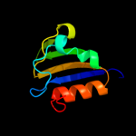
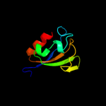
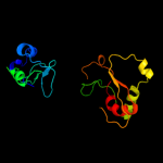
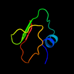
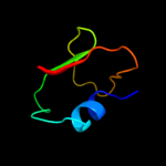
1 d1vcta2
99.5
23
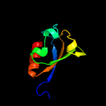
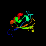
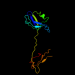
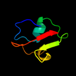
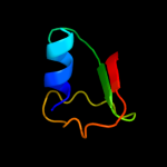
Fold: TrkA C-terminal domain-likeSuperfamily: TrkA C-terminal domain-likeFamily: TrkA C-terminal domain-like2 d2fy8a2
99.5
13
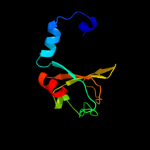
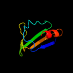
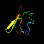
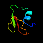
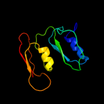
Fold: TrkA C-terminal domain-likeSuperfamily: TrkA C-terminal domain-likeFamily: TrkA C-terminal domain-like3 c2bknA_
99.5
18
PDB header: membrane proteinChain: A: PDB Molecule: hypothetical protein ph0236;PDBTitle: structure analysis of unknown function protein
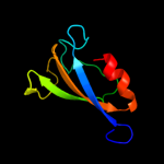
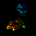
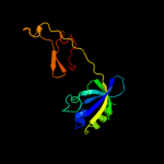
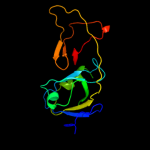
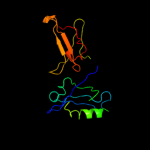
4 c3jxoB_
99.3
20
PDB header: transport proteinChain: B: PDB Molecule: trka-n domain protein;PDBTitle: crystal structure of an octomeric two-subunit trka k+ channel ring2 gating assembly, tm1088a:tm1088b, from thermotoga maritima
5 c2fy8A_
99.1
23
PDB header: transport proteinChain: A: PDB Molecule: calcium-gated potassium channel mthk;PDBTitle: crystal structure of mthk rck domain in its ligand-free gating-ring2 form
6 c1lnqC_
99.1
23
PDB header: metal transportChain: C: PDB Molecule: potassium channel related protein;PDBTitle: crystal structure of mthk at 3.3 a
7 c3l4bG_
98.5
20
PDB header: transport proteinChain: G: PDB Molecule: trka k+ channel protien tm1088b;PDBTitle: crystal structure of an octomeric two-subunit trka k+ channel ring2 gating assembly, tm1088a:tm1088b, from thermotoga maritima
8 c3mt5A_
97.7
10
PDB header: membrane protein, transport proteinChain: A: PDB Molecule: potassium large conductance calcium-activated channel,PDBTitle: crystal structure of the human bk gating apparatus
9 c3u6nC_
97.7
9
PDB header: transport proteinChain: C: PDB Molecule: high-conductance ca2+-activated k+ channel protein;PDBTitle: open structure of the bk channel gating ring
10 c2ka9A_
87.5
14
PDB header: cell adhesionChain: A: PDB Molecule: disks large homolog 4;PDBTitle: solution structure of psd-95 pdz12 complexed with cypin2 peptide
11 c3nafA_
85.1
14
PDB header: ion transportChain: A: PDB Molecule: calcium-activated potassium channel subunit alpha-1;PDBTitle: structure of the intracellular gating ring from the human high-2 conductance ca2+ gated k+ channel (bk channel)
12 c1u3bA_
77.7
15
PDB header: protein transportChain: A: PDB Molecule: amyloid beta a4 precursor protein-binding,PDBTitle: auto-inhibition mechanism of x11s/mints family scaffold2 proteins revealed by the closed conformation of the tandem3 pdz domains
13 d1zud21
76.7
20
Fold: beta-Grasp (ubiquitin-like)Superfamily: MoaD/ThiSFamily: ThiS14 d2cu3a1
76.0
19
Fold: beta-Grasp (ubiquitin-like)Superfamily: MoaD/ThiSFamily: ThiS15 c2kl0A_
72.0
13
PDB header: structural genomics, unknown functionChain: A: PDB Molecule: putative thiamin biosynthesis this;PDBTitle: solution nmr structure of rhodopseudomonas palustris rpa3574,2 northeast structural genomics consortium (nesg) target rpr325
16 c3r0hA_
70.3
18
PDB header: peptide binding proteinChain: A: PDB Molecule: inactivation-no-after-potential d protein;PDBTitle: structure of inad pdz45 in complex with ng2 peptide
17 c3cwiA_
64.6
16
PDB header: biosynthetic proteinChain: A: PDB Molecule: thiamine-biosynthesis protein this;PDBTitle: crystal structure of thiamine biosynthesis protein (this)2 from geobacter metallireducens. northeast structural3 genomics consortium target gmr137
18 d1tygb_
62.9
21
Fold: beta-Grasp (ubiquitin-like)Superfamily: MoaD/ThiSFamily: ThiS19 c2zc3F_
59.1
9
PDB header: biosynthetic proteinChain: F: PDB Molecule: penicillin-binding protein 2x;PDBTitle: penicillin-binding protein 2x (pbp 2x) acyl-enzyme complex2 (biapenem) from streptococcus pneumoniae
20 c1p1dA_
56.0
15
PDB header: protein bindingChain: A: PDB Molecule: glutamate receptor interacting protein;PDBTitle: structural insights into the inter-domain chaperoning of2 tandem pdz domains in glutamate receptor interacting3 proteins
21 c1tygG_
not modelled
53.0
20
PDB header: biosynthetic proteinChain: G: PDB Molecule: yjbs;PDBTitle: structure of the thiazole synthase/this complex
22 d1v62a_
not modelled
50.8
15
Fold: PDZ domain-likeSuperfamily: PDZ domain-likeFamily: PDZ domain23 c2jreA_
not modelled
48.1
14
PDB header: de novo proteinChain: A: PDB Molecule: c60-1 pdz domain peptide;PDBTitle: c60-1, a pdz domain designed using statistical coupling2 analysis
24 c2d8iA_
not modelled
44.8
17
PDB header: immune system, signaling proteinChain: A: PDB Molecule: t-cell lymphoma invasion and metastasis 1PDBTitle: solution structure of the pdz domain of t-cell lymphoma2 invasion and metastasis 1 varian
25 c2ejyA_
not modelled
43.1
24
PDB header: membrane proteinChain: A: PDB Molecule: 55 kda erythrocyte membrane protein;PDBTitle: solution structure of the p55 pdz t85c domain complexed2 with the glycophorin c f127c peptide
26 d1qlca_
not modelled
40.9
20
Fold: PDZ domain-likeSuperfamily: PDZ domain-likeFamily: PDZ domain27 c3ggeA_
not modelled
40.6
19
PDB header: protein bindingChain: A: PDB Molecule: pdz domain-containing protein gipc2;PDBTitle: crystal structure of the pdz domain of pdz domain-containing protein2 gipc2
28 c2qt5A_
not modelled
39.5
18
PDB header: peptide binding proteinChain: A: PDB Molecule: glutamate receptor-interacting protein 1;PDBTitle: crystal structure of grip1 pdz12 in complex with the fras12 peptide
29 d1wi2a_
not modelled
39.2
22
Fold: PDZ domain-likeSuperfamily: PDZ domain-likeFamily: PDZ domain30 d1uhpa_
not modelled
38.5
17
Fold: PDZ domain-likeSuperfamily: PDZ domain-likeFamily: PDZ domain31 d1i16a_
not modelled
38.3
16
Fold: PDZ domain-likeSuperfamily: PDZ domain-likeFamily: Interleukin 1632 d1d5ga_
not modelled
37.1
17
Fold: PDZ domain-likeSuperfamily: PDZ domain-likeFamily: PDZ domain33 d1um1a_
not modelled
35.6
23
Fold: PDZ domain-likeSuperfamily: PDZ domain-likeFamily: PDZ domain34 c2xkxB_
not modelled
34.9
16
PDB header: structural proteinChain: B: PDB Molecule: disks large homolog 4;PDBTitle: single particle analysis of psd-95 in negative stain
35 c2xrfA_
not modelled
34.9
26
PDB header: transferaseChain: A: PDB Molecule: uridine phosphorylase 2;PDBTitle: crystal structure of human uridine phosphorylase 2
36 d1wf8a1
not modelled
33.8
17
Fold: PDZ domain-likeSuperfamily: PDZ domain-likeFamily: PDZ domain37 c2fneB_
not modelled
31.8
15
PDB header: structural genomics, unknown functionChain: B: PDB Molecule: multiple pdz domain protein;PDBTitle: the crystal structure of the 13th pdz domain of mpdz
38 c4a8aI_
not modelled
30.7
19
PDB header: hydrolase/hydrolaseChain: I: PDB Molecule: periplasmic ph-dependent serine endoprotease degq;PDBTitle: asymmetric cryo-em reconstruction of e. coli degq 12-mer in complex2 with lysozyme
39 c2dazA_
not modelled
30.0
16
PDB header: protein bindingChain: A: PDB Molecule: inad-like protein;PDBTitle: solution structure of the 7th pdz domain of inad-like2 protein
40 c2d92A_
not modelled
28.9
16
PDB header: protein bindingChain: A: PDB Molecule: inad-like protein;PDBTitle: solution structure of the fifth pdz domain of inad-like2 protein
41 d2byga1
not modelled
28.6
19
Fold: PDZ domain-likeSuperfamily: PDZ domain-likeFamily: PDZ domain42 c2yt7A_
not modelled
28.6
18
PDB header: protein transportChain: A: PDB Molecule: amyloid beta a4 precursor protein-binding familyPDBTitle: solution structure of the pdz domain of amyloid beta a42 precursor protein-binding family a member 3
43 c2dluA_
not modelled
28.1
22
PDB header: protein bindingChain: A: PDB Molecule: inad-like protein;PDBTitle: solution structure of the second pdz domain of human inad-2 like protein
44 d2fe5a1
not modelled
27.9
23
Fold: PDZ domain-likeSuperfamily: PDZ domain-likeFamily: PDZ domain45 d1v6ba_
not modelled
27.8
12
Fold: PDZ domain-likeSuperfamily: PDZ domain-likeFamily: PDZ domain46 c3tl6B_
not modelled
27.8
21
PDB header: transferaseChain: B: PDB Molecule: purine nucleoside phosphorylase;PDBTitle: crystal structure of purine nucleoside phosphorylase from entamoeba2 histolytica
47 c2djtA_
not modelled
27.7
16
PDB header: signaling proteinChain: A: PDB Molecule: unnamed protein product;PDBTitle: solution structures of the pdz domain of human unnamed2 protein product
48 d1rp5a2
not modelled
27.0
9
Fold: Penicillin-binding protein 2x (pbp-2x), c-terminal domainSuperfamily: Penicillin-binding protein 2x (pbp-2x), c-terminal domainFamily: Penicillin-binding protein 2x (pbp-2x), c-terminal domain49 c2dm8A_
not modelled
26.6
13
PDB header: protein bindingChain: A: PDB Molecule: inad-like protein;PDBTitle: solution structure of the eighth pdz domain of human inad-2 like protein
50 d1uita_
not modelled
26.4
18
Fold: PDZ domain-likeSuperfamily: PDZ domain-likeFamily: PDZ domain51 c3kzeA_
not modelled
25.9
21
PDB header: signaling proteinChain: A: PDB Molecule: t-lymphoma invasion and metastasis-inducing protein 1;PDBTitle: crystal structure of t-cell lymphoma invasion and metastasis-1 pdz in2 complex with ssrkeyya peptide
52 d1pyya2
not modelled
25.4
9
Fold: Penicillin-binding protein 2x (pbp-2x), c-terminal domainSuperfamily: Penicillin-binding protein 2x (pbp-2x), c-terminal domainFamily: Penicillin-binding protein 2x (pbp-2x), c-terminal domain53 d1tp5a1
not modelled
24.7
22
Fold: PDZ domain-likeSuperfamily: PDZ domain-likeFamily: PDZ domain54 d1x6da1
not modelled
24.6
20
Fold: PDZ domain-likeSuperfamily: PDZ domain-likeFamily: Interleukin 1655 c2rqeA_
not modelled
24.4
15
PDB header: sugar binding proteinChain: A: PDB Molecule: beta-1,3-glucan-binding protein;PDBTitle: solution structure of the silkworm bgrp/gnbp3 n-terminal2 domain reveals the mechanism for b-1,3-glucan specific3 recognition
56 d1ufxa_
not modelled
24.3
27
Fold: PDZ domain-likeSuperfamily: PDZ domain-likeFamily: PDZ domain57 d1g9oa_
not modelled
24.0
23
Fold: PDZ domain-likeSuperfamily: PDZ domain-likeFamily: PDZ domain58 c2ogpA_
not modelled
23.3
17
PDB header: signaling proteinChain: A: PDB Molecule: partitioning-defective 3 homolog;PDBTitle: solution structure of the second pdz domain of par-3
59 c3krmB_
not modelled
23.1
12
PDB header: rna binding proteinChain: B: PDB Molecule: insulin-like growth factor 2 mrna-binding proteinPDBTitle: imp1 kh34
60 d1zoka1
not modelled
23.0
19
Fold: PDZ domain-likeSuperfamily: PDZ domain-likeFamily: PDZ domain61 c2opgB_
not modelled
22.7
13
PDB header: structural proteinChain: B: PDB Molecule: multiple pdz domain protein;PDBTitle: the crystal structure of the 10th pdz domain of mpdz
62 d1x5qa1
not modelled
22.5
16
Fold: PDZ domain-likeSuperfamily: PDZ domain-likeFamily: PDZ domain63 d1rzxa_
not modelled
22.4
25
Fold: PDZ domain-likeSuperfamily: PDZ domain-likeFamily: PDZ domain64 c2dmzA_
not modelled
21.8
12
PDB header: protein bindingChain: A: PDB Molecule: inad-like protein;PDBTitle: solution structure of the third pdz domain of human inad-2 like protein
65 d2i0ia1
not modelled
21.7
27
Fold: PDZ domain-likeSuperfamily: PDZ domain-likeFamily: PDZ domain66 d1va8a1
not modelled
21.7
18
Fold: PDZ domain-likeSuperfamily: PDZ domain-likeFamily: PDZ domain67 d1be9a_
not modelled
21.5
22
Fold: PDZ domain-likeSuperfamily: PDZ domain-likeFamily: PDZ domain68 d1r61a_
not modelled
21.0
20
Fold: The "swivelling" beta/beta/alpha domainSuperfamily: Putative cyclaseFamily: Putative cyclase69 c3npgD_
not modelled
20.3
17
PDB header: unknown functionChain: D: PDB Molecule: uncharacterized duf364 family protein;PDBTitle: crystal structure of a protein with unknown function from duf3642 family (ph1506) from pyrococcus horikoshii at 2.70 a resolution
70 d2fnea1
not modelled
20.1
17
Fold: PDZ domain-likeSuperfamily: PDZ domain-likeFamily: PDZ domain71 d1wgka_
not modelled
19.8
13
Fold: beta-Grasp (ubiquitin-like)Superfamily: MoaD/ThiSFamily: C9orf74 homolog72 c2vwrA_
not modelled
19.7
15
PDB header: protein-bindingChain: A: PDB Molecule: ligand of numb protein x 2;PDBTitle: crystal structure of the second pdz domain of numb-binding2 protein 2
73 c2hc8A_
not modelled
19.6
16
PDB header: transport proteinChain: A: PDB Molecule: cation-transporting atpase, p-type;PDBTitle: structure of the a. fulgidus copa a-domain
74 c2yubA_
not modelled
19.6
10
PDB header: transferaseChain: A: PDB Molecule: lim domain kinase 2;PDBTitle: solution structure of the pdz domain from mouse lim domain2 kinase
75 c2eehA_
not modelled
19.4
22
PDB header: metal binding proteinChain: A: PDB Molecule: pdz domain-containing protein 7;PDBTitle: solution structure of first pdz domain of pdz domain2 containing protein 7
76 d1n7ea_
not modelled
19.2
15
Fold: PDZ domain-likeSuperfamily: PDZ domain-likeFamily: PDZ domain77 c1mhsA_
not modelled
18.9
18
PDB header: membrane protein, proton transportChain: A: PDB Molecule: plasma membrane atpase;PDBTitle: model of neurospora crassa proton atpase
78 c3ixzA_
not modelled
18.8
15
PDB header: hydrolaseChain: A: PDB Molecule: potassium-transporting atpase alpha;PDBTitle: pig gastric h+/k+-atpase complexed with aluminium fluoride
79 c2k1zA_
not modelled
17.4
16
PDB header: signaling proteinChain: A: PDB Molecule: partitioning-defective 3 homolog;PDBTitle: solution structure of par-3 pdz3
80 c2egkC_
not modelled
17.1
18
PDB header: protein bindingChain: C: PDB Molecule: general receptor for phosphoinositides 1-PDBTitle: crystal structure of tamalin pdz-intrinsic ligand fusion2 protein
81 d1whaa_
not modelled
17.1
16
Fold: PDZ domain-likeSuperfamily: PDZ domain-likeFamily: PDZ domain82 c2jilA_
not modelled
16.4
20
PDB header: membrane proteinChain: A: PDB Molecule: glutamate receptor interacting protein-1;PDBTitle: crystal structure of 2nd pdz domain of glutamate receptor2 interacting protein-1 (grip1)
83 c2vspA_
not modelled
16.3
24
PDB header: transport proteinChain: A: PDB Molecule: pdz domain-containing protein 1;PDBTitle: crystal structure of the fourth pdz domain of pdz domain-2 containing protein 1
84 c2xlkB_
not modelled
16.2
16
PDB header: hydrolase/rnaChain: B: PDB Molecule: csy4 endoribonuclease;PDBTitle: crystal structure of the csy4-crrna complex, orthorhombic form
85 d1ozia_
not modelled
16.0
23
Fold: PDZ domain-likeSuperfamily: PDZ domain-likeFamily: PDZ domain86 d1wpga1
not modelled
15.9
19
Fold: Double-stranded beta-helixSuperfamily: Calcium ATPase, transduction domain AFamily: Calcium ATPase, transduction domain A87 d1ueqa_
not modelled
15.8
13
Fold: PDZ domain-likeSuperfamily: PDZ domain-likeFamily: PDZ domain88 c2q3gA_
not modelled
15.4
22
PDB header: structural genomicsChain: A: PDB Molecule: pdz and lim domain protein 7;PDBTitle: structure of the pdz domain of human pdlim7 bound to a c-2 terminal extension from human beta-tropomyosin
89 c2k9xA_
not modelled
15.2
26
PDB header: unknown functionChain: A: PDB Molecule: uncharacterized protein;PDBTitle: solution structure of urm1 from trypanosoma brucei
90 d2piaa1
not modelled
15.2
29
Fold: Reductase/isomerase/elongation factor common domainSuperfamily: Riboflavin synthase domain-likeFamily: Ferredoxin reductase FAD-binding domain-like91 d1qava_
not modelled
14.8
22
Fold: PDZ domain-likeSuperfamily: PDZ domain-likeFamily: PDZ domain92 d1wi4a1
not modelled
14.8
23
Fold: PDZ domain-likeSuperfamily: PDZ domain-likeFamily: PDZ domain93 c3kw0D_
not modelled
14.5
38
PDB header: hydrolaseChain: D: PDB Molecule: cysteine peptidase;PDBTitle: crystal structure of cysteine peptidase (np_982244.1) from bacillus2 cereus atcc 10987 at 2.50 a resolution
94 d1kwaa_
not modelled
14.5
24
Fold: PDZ domain-likeSuperfamily: PDZ domain-likeFamily: PDZ domain95 d1y7ma2
not modelled
14.4
10
Fold: LysM domainSuperfamily: LysM domainFamily: LysM domain96 c3pr9A_
not modelled
14.3
24
PDB header: chaperoneChain: A: PDB Molecule: fkbp-type peptidyl-prolyl cis-trans isomerase;PDBTitle: structural analysis of protein folding by the methanococcus jannaschii2 chaperone fkbp26
97 d2h1qa1
not modelled
14.1
15
Fold: PLP-dependent transferase-likeSuperfamily: Dhaf3308-likeFamily: Dhaf3308-like98 c2gzvA_
not modelled
14.1
31
PDB header: signaling proteinChain: A: PDB Molecule: prkca-binding protein;PDBTitle: the cystal structure of the pdz domain of human pick1 (casp target)
99 c3eggC_
not modelled
13.9
17
PDB header: hydrolaseChain: C: PDB Molecule: spinophilin;PDBTitle: crystal structure of a complex between protein phosphatase 1 alpha2 (pp1) and the pp1 binding and pdz domains of spinophilin


