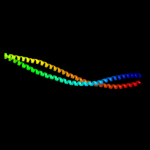
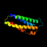
1 c1qu7A_
99.9
72
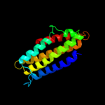
PDB header: signaling proteinChain: A: PDB Molecule: methyl-accepting chemotaxis protein i;PDBTitle: four helical-bundle structure of the cytoplasmic domain of a serine2 chemotaxis receptor
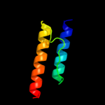
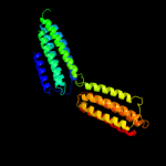
2 c2ch7A_
99.8
24
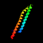
PDB header: chemotaxisChain: A: PDB Molecule: methyl-accepting chemotaxis protein;PDBTitle: crystal structure of the cytoplasmic domain of a bacterial2 chemoreceptor from thermotoga maritima
3 c3g67A_
99.7
23
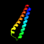
PDB header: signaling proteinChain: A: PDB Molecule: methyl-accepting chemotaxis protein;PDBTitle: crystal structure of a soluble chemoreceptor from thermotoga2 maritima
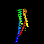
4 c3lnrA_
98.6
14
PDB header: signaling proteinChain: A: PDB Molecule: aerotaxis transducer aer2;PDBTitle: crystal structure of poly-hamp domains from the p. aeruginosa soluble2 receptor aer2
5 c2d4uA_
98.6
21
PDB header: signaling proteinChain: A: PDB Molecule: methyl-accepting chemotaxis protein i;PDBTitle: crystal structure of the ligand binding domain of the bacterial serine2 chemoreceptor tsr
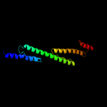
6 d2liga_
97.9
19
Fold: Four-helical up-and-down bundleSuperfamily: Aspartate receptor, ligand-binding domainFamily: Aspartate receptor, ligand-binding domain7 d2asxa1
97.8
23
Fold: HAMP domain-likeSuperfamily: HAMP domain-likeFamily: HAMP domain8 c2rm8A_
97.7
18
PDB header: signaling proteinChain: A: PDB Molecule: sensory rhodopsin ii transducer;PDBTitle: the solution structure of phototactic transducer protein2 htrii linker region from natronomonas pharaonis
9 c1sj8A_
97.4
11
PDB header: structural proteinChain: A: PDB Molecule: talin 1;PDBTitle: crystal structure of talin residues 482-789
10 c3zrwB_
97.2
18
PDB header: signaling proteinChain: B: PDB Molecule: af1503 protein, osmolarity sensor protein envz;PDBTitle: the structure of the dimeric hamp-dhp fusion a291v mutant
11 d2asra_
96.7
20
Fold: Four-helical up-and-down bundleSuperfamily: Aspartate receptor, ligand-binding domainFamily: Aspartate receptor, ligand-binding domain12 d1vlta_
96.4
20
Fold: Four-helical up-and-down bundleSuperfamily: Aspartate receptor, ligand-binding domainFamily: Aspartate receptor, ligand-binding domain13 c3dyjA_
95.4
11
PDB header: structural proteinChain: A: PDB Molecule: talin-1;PDBTitle: crystal structure a talin rod fragment
14 c2wpqA_
94.0
7
PDB header: membrane proteinChain: A: PDB Molecule: trimeric autotransporter adhesin fragment;PDBTitle: salmonella enterica sada 479-519 fused to gcn4 adaptors (2 sadak3, in-register fusion)
15 c3ojaB_
80.5
11
PDB header: protein bindingChain: B: PDB Molecule: anopheles plasmodium-responsive leucine-rich repeat proteinPDBTitle: crystal structure of lrim1/apl1c complex
16 c2kbbA_
77.6
11
PDB header: structural proteinChain: A: PDB Molecule: talin-1;PDBTitle: nmr structure of the talin rod domain, 1655-1822
17 c3gvmA_
72.9
11
PDB header: viral proteinChain: A: PDB Molecule: putative uncharacterized protein sag1039;PDBTitle: structure of the homodimeric wxg-100 family protein from streptococcus2 agalactiae
18 c1ei3E_
67.7
12
PDB header: PDB COMPND: 19 c2vs0B_
67.1
12
PDB header: cell invasionChain: B: PDB Molecule: virulence factor esxa;PDBTitle: structural analysis of homodimeric staphylococcal aureus2 virulence factor esxa
20 c1deqO_
66.6
12
PDB header: PDB COMPND: 21 c1ei3C_
not modelled
63.1
9
PDB header: PDB COMPND: 22 c1deqF_
not modelled
59.5
9
PDB header: PDB COMPND: 23 c2ieqC_
not modelled
55.0
13
PDB header: viral proteinChain: C: PDB Molecule: spike glycoprotein;PDBTitle: core structure of s2 from the human coronavirus nl63 spike2 glycoprotein
24 c2qihA_
not modelled
54.7
13
PDB header: cell adhesionChain: A: PDB Molecule: protein uspa1;PDBTitle: crystal structure of 527-665 fragment of uspa1 protein from2 moraxella catarrhalis
25 c2d4yA_
not modelled
53.8
14
PDB header: structural proteinChain: A: PDB Molecule: flagellar hook-associated protein 1;PDBTitle: crystal structure of a 49k fragment of hap1 (flgk)
26 c3ghgK_
not modelled
43.9
15
PDB header: blood clottingChain: K: PDB Molecule: fibrinogen beta chain;PDBTitle: crystal structure of human fibrinogen
27 c1urqA_
not modelled
42.8
21
PDB header: transport proteinChain: A: PDB Molecule: m-tomosyn isoform;PDBTitle: crystal structure of neuronal q-snares in complex with2 r-snare motif of tomosyn
28 c3ipdB_
not modelled
42.7
15
PDB header: exocytosisChain: B: PDB Molecule: syntaxin-1a;PDBTitle: helical extension of the neuronal snare complex into the2 membrane, spacegroup i 21 21 21
29 c1sfcJ_
not modelled
39.6
16
PDB header: transport proteinChain: J: PDB Molecule: protein (syntaxin 1a);PDBTitle: neuronal synaptic fusion complex
30 c3hd7A_
not modelled
39.1
22
PDB header: exocytosisChain: A: PDB Molecule: vesicle-associated membrane protein 2;PDBTitle: helical extension of the neuronal snare complex into the membrane,2 spacegroup c 1 2 1
31 c3b5nF_
not modelled
36.2
12
PDB header: membrane proteinChain: F: PDB Molecule: protein sso1;PDBTitle: structure of the yeast plasma membrane snare complex
32 c2npsB_
not modelled
30.3
16
PDB header: transport proteinChain: B: PDB Molecule: syntaxin 13;PDBTitle: crystal structure of the early endosomal snare complex
33 c2kseA_
not modelled
29.6
17
PDB header: transferaseChain: A: PDB Molecule: sensor protein qsec;PDBTitle: backbone structure of the membrane domain of e. coli2 histidine kinase receptor qsec, center for structures of3 membrane proteins (csmp) target 4311c
34 c1n7sA_
not modelled
29.6
22
PDB header: transport proteinChain: A: PDB Molecule: vesicle-associated membrane protein 2;PDBTitle: high resolution structure of a truncated neuronal snare2 complex
35 c2dnxA_
not modelled
29.3
12
PDB header: transport proteinChain: A: PDB Molecule: syntaxin-12;PDBTitle: solution structure of rsgi ruh-063, an n-terminal domain of2 syntaxin 12 from human cdna
36 c1i49A_
not modelled
29.2
10
PDB header: signaling proteinChain: A: PDB Molecule: arfaptin 2;PDBTitle: crystal structure analysis of arfaptin
37 c2npsD_
not modelled
28.8
12
PDB header: transport proteinChain: D: PDB Molecule: syntaxin-6;PDBTitle: crystal structure of the early endosomal snare complex
38 c3b5nE_
not modelled
28.3
15
PDB header: membrane proteinChain: E: PDB Molecule: synaptobrevin homolog 1;PDBTitle: structure of the yeast plasma membrane snare complex
39 c1n7sB_
not modelled
27.9
16
PDB header: transport proteinChain: B: PDB Molecule: syntaxin 1a;PDBTitle: high resolution structure of a truncated neuronal snare2 complex
40 c1gl2A_
not modelled
27.4
15
PDB header: membrane proteinChain: A: PDB Molecule: endobrevin;PDBTitle: crystal structure of an endosomal snare core complex
41 c1kmiZ_
not modelled
26.4
7
PDB header: signaling proteinChain: Z: PDB Molecule: chemotaxis protein chez;PDBTitle: crystal structure of an e.coli chemotaxis protein, chez
42 d1wa8a1
not modelled
25.2
11
Fold: Ferritin-likeSuperfamily: EsxAB dimer-likeFamily: ESAT-6 like43 c1sfcI_
not modelled
25.2
22
PDB header: transport proteinChain: I: PDB Molecule: protein (synaptobrevin 2);PDBTitle: neuronal synaptic fusion complex
44 d1ez3a_
not modelled
24.1
11
Fold: STAT-likeSuperfamily: t-snare proteinsFamily: t-snare proteins45 c2npsA_
not modelled
22.7
26
PDB header: transport proteinChain: A: PDB Molecule: vesicle-associated membrane protein 4;PDBTitle: crystal structure of the early endosomal snare complex
46 c1nafA_
not modelled
21.0
15
PDB header: signaling protein, membrane proteinChain: A: PDB Molecule: adp-ribosylation factor binding protein gga1;PDBTitle: crystal structure of the human gga1 gat domain
47 c1s94A_
not modelled
16.1
6
PDB header: endocytosis/exocytosisChain: A: PDB Molecule: s-syntaxin;PDBTitle: crystal structure of the habc domain of neuronal syntaxin from the2 squid loligo pealei
48 d1s94a_
not modelled
16.1
6
Fold: STAT-likeSuperfamily: t-snare proteinsFamily: t-snare proteins49 c2bezC_
not modelled
15.8
15
PDB header: viral proteinChain: C: PDB Molecule: e2 glycoprotein;PDBTitle: structure of a proteolitically resistant core from the2 severe acute respiratory syndrome coronavirus s2 fusion3 protein
50 c1l4aD_
not modelled
15.8
8
PDB header: endocytosis/exocytosisChain: D: PDB Molecule: s-snap25 fusion protein;PDBTitle: x-ray structure of the neuronal complexin/snare complex2 from the squid loligo pealei
51 c1sfcD_
not modelled
15.8
7
PDB header: transport proteinChain: D: PDB Molecule: protein (snap-25b);PDBTitle: neuronal synaptic fusion complex
52 c3c98B_
not modelled
15.7
11
PDB header: endocytosis/exocytosisChain: B: PDB Molecule: syntaxin-1a;PDBTitle: revised structure of the munc18a-syntaxin1 complex
53 c3ok8A_
not modelled
14.5
7
PDB header: protein bindingChain: A: PDB Molecule: brain-specific angiogenesis inhibitor 1-associated proteinPDBTitle: i-bar of pinkbar
54 c3arcl_
not modelled
14.3
30
PDB header: electron transport, photosynthesisChain: L: PDB Molecule: photosystem ii reaction center protein l;PDBTitle: crystal structure of oxygen-evolving photosystem ii at 1.9 angstrom2 resolution
55 c1l4aC_
not modelled
14.2
12
PDB header: endocytosis/exocytosisChain: C: PDB Molecule: s-snap25 fusion protein;PDBTitle: x-ray structure of the neuronal complexin/snare complex2 from the squid loligo pealei
56 c2dq3A_
not modelled
13.7
7
PDB header: ligaseChain: A: PDB Molecule: seryl-trna synthetase;PDBTitle: crystal structure of aq_298
57 d1oxza_
not modelled
13.6
14
Fold: Spectrin repeat-likeSuperfamily: GAT-like domainFamily: GAT domain58 c1oxzA_
not modelled
13.6
14
PDB header: membrane proteinChain: A: PDB Molecule: adp-ribosylation factor binding protein gga1;PDBTitle: crystal structure of the human gga1 gat domain
59 d1rkea1
not modelled
13.5
10
Fold: Four-helical up-and-down bundleSuperfamily: alpha-catenin/vinculin-likeFamily: alpha-catenin/vinculin60 d1wa8b1
not modelled
13.4
13
Fold: Ferritin-likeSuperfamily: EsxAB dimer-likeFamily: ESAT-6 like61 d1i4da_
not modelled
13.0
10
Fold: BAR/IMD domain-likeSuperfamily: BAR/IMD domain-likeFamily: Arfaptin, Rac-binding fragment62 c1urqC_
not modelled
12.9
15
PDB header: transport proteinChain: C: PDB Molecule: synaptosomal-associated protein 25;PDBTitle: crystal structure of neuronal q-snares in complex with2 r-snare motif of tomosyn
63 c3hnwB_
not modelled
12.7
11
PDB header: structural genomics, unknown functionChain: B: PDB Molecule: uncharacterized protein;PDBTitle: crystal structure of a basic coiled-coil protein of unknown function2 from eubacterium eligens atcc 27750
64 c2efrB_
not modelled
12.0
9
PDB header: contractile proteinChain: B: PDB Molecule: general control protein gcn4 and tropomyosin 1 alpha chain;PDBTitle: crystal structure of the c-terminal tropomyosin fragment with n- and2 c-terminal extensions of the leucine zipper at 1.8 angstroms3 resolution
65 d2obpa1
not modelled
11.2
35
Fold: DNA/RNA-binding 3-helical bundleSuperfamily: "Winged helix" DNA-binding domainFamily: ReutB4095-like66 c1nfoA_
not modelled
11.0
17
PDB header: lipid transportChain: A: PDB Molecule: apolipoprotein e2;PDBTitle: apolipoprotein e2 (apoe2, d154a mutation)
67 c3arcL_
not modelled
11.0
30
PDB header: electron transport, photosynthesisChain: L: PDB Molecule: photosystem ii reaction center protein l;PDBTitle: crystal structure of oxygen-evolving photosystem ii at 1.9 angstrom2 resolution
68 c3prqL_
not modelled
11.0
30
PDB header: photosynthesisChain: L: PDB Molecule: photosystem ii reaction center protein l;PDBTitle: crystal structure of cyanobacterial photosystem ii in complex with2 terbutryn (part 1 of 2). this file contains first monomer of psii3 dimer
69 c1s5ll_
not modelled
11.0
30
PDB header: photosynthesisChain: L: PDB Molecule: photosystem ii reaction center l protein;PDBTitle: architecture of the photosynthetic oxygen evolving center
70 c3bz2L_
not modelled
11.0
30
PDB header: electron transportChain: L: PDB Molecule: photosystem ii reaction center protein l;PDBTitle: crystal structure of cyanobacterial photosystem ii (part 22 of 2). this file contains second monomer of psii dimer
71 c3a0bl_
not modelled
11.0
30
PDB header: electron transportChain: L: PDB Molecule: photosystem ii reaction center protein l;PDBTitle: crystal structure of br-substituted photosystem ii complex
72 c3a0hl_
not modelled
11.0
30
PDB header: electron transportChain: L: PDB Molecule: photosystem ii reaction center protein l;PDBTitle: crystal structure of i-substituted photosystem ii complex
73 c3a0hL_
not modelled
11.0
30
PDB header: electron transportChain: L: PDB Molecule: photosystem ii reaction center protein l;PDBTitle: crystal structure of i-substituted photosystem ii complex
74 c3a0bL_
not modelled
11.0
30
PDB header: electron transportChain: L: PDB Molecule: photosystem ii reaction center protein l;PDBTitle: crystal structure of br-substituted photosystem ii complex
75 c2axtl_
not modelled
11.0
30
PDB header: electron transportChain: L: PDB Molecule: photosystem ii reaction center l protein;PDBTitle: crystal structure of photosystem ii from thermosynechococcus elongatus
76 c1s5lL_
not modelled
11.0
30
PDB header: photosynthesisChain: L: PDB Molecule: photosystem ii reaction center l protein;PDBTitle: architecture of the photosynthetic oxygen evolving center
77 c3bz1L_
not modelled
11.0
30
PDB header: electron transportChain: L: PDB Molecule: photosystem ii reaction center protein l;PDBTitle: crystal structure of cyanobacterial photosystem ii (part 12 of 2). this file contains first monomer of psii dimer
78 d2axtl1
not modelled
11.0
30
Fold: Single transmembrane helixSuperfamily: Photosystem II reaction center protein L, PsbLFamily: PsbL-like79 c2axtL_
not modelled
11.0
30
PDB header: electron transportChain: L: PDB Molecule: photosystem ii reaction center l protein;PDBTitle: crystal structure of photosystem ii from thermosynechococcus elongatus
80 c3kziL_
not modelled
11.0
30
PDB header: electron transportChain: L: PDB Molecule: photosystem ii reaction center protein l;PDBTitle: crystal structure of monomeric form of cyanobacterial photosystem ii
81 c3prrL_
not modelled
11.0
30
PDB header: photosynthesisChain: L: PDB Molecule: photosystem ii reaction center protein l;PDBTitle: crystal structure of cyanobacterial photosystem ii in complex with2 terbutryn (part 2 of 2). this file contains second monomer of psii3 dimer
82 c2js5B_
not modelled
10.9
6
PDB header: structural genomics, unknown functionChain: B: PDB Molecule: uncharacterized protein;PDBTitle: nmr structure of protein q60c73_metca. northeast structural2 genomics consortium target mcr1
83 c3b5nL_
not modelled
10.9
11
PDB header: membrane proteinChain: L: PDB Molecule: protein transport protein sec9;PDBTitle: structure of the yeast plasma membrane snare complex
84 c3u59C_
not modelled
10.8
12
PDB header: contractile proteinChain: C: PDB Molecule: tropomyosin beta chain;PDBTitle: n-terminal 98-aa fragment of smooth muscle tropomyosin beta
85 c2d3eD_
not modelled
10.6
6
PDB header: contractile proteinChain: D: PDB Molecule: general control protein gcn4 and tropomyosin 1PDBTitle: crystal structure of the c-terminal fragment of rabbit2 skeletal alpha-tropomyosin
86 c1zvaA_
not modelled
9.9
13
PDB header: viral proteinChain: A: PDB Molecule: e2 glycoprotein;PDBTitle: a structure-based mechanism of sars virus membrane fusion
87 c1gl2D_
not modelled
9.8
25
PDB header: membrane proteinChain: D: PDB Molecule: syntaxin 8;PDBTitle: crystal structure of an endosomal snare core complex
88 c3csxA_
not modelled
8.9
15
PDB header: metal binding protein,unknown functionChain: A: PDB Molecule: putative uncharacterized protein;PDBTitle: structural characterization of a protein in the duf6832 family- crystal structure of cce_0567 from the3 cyanobacterium cyanothece 51142.
89 d1o3xa_
not modelled
8.8
15
Fold: Spectrin repeat-likeSuperfamily: GAT-like domainFamily: GAT domain90 c1zv8I_
not modelled
8.6
11
PDB header: viral proteinChain: I: PDB Molecule: e2 glycoprotein;PDBTitle: a structure-based mechanism of sars virus membrane fusion
91 d2h8pc1
not modelled
8.3
0
Fold: Voltage-gated potassium channelsSuperfamily: Voltage-gated potassium channelsFamily: Voltage-gated potassium channels92 c2l9uA_
not modelled
8.3
21
PDB header: membrane proteinChain: A: PDB Molecule: receptor tyrosine-protein kinase erbb-3;PDBTitle: spatial structure of dimeric erbb3 transmembrane domain
93 c2qrxA_
not modelled
8.3
11
PDB header: dna binding proteinChain: A: PDB Molecule: gm27569p;PDBTitle: crystal structure of drosophila melanogaster translin2 protein
94 c2ks1B_
not modelled
8.2
21
PDB header: transferaseChain: B: PDB Molecule: epidermal growth factor receptor;PDBTitle: heterodimeric association of transmembrane domains of erbb1 and erbb22 receptors enabling kinase activation
95 c3fxeA_
not modelled
8.2
46
PDB header: unknown functionChain: A: PDB Molecule: protein icmq;PDBTitle: crystal structure of interacting domains of icmr and icmq (seleno-2 derivative)
96 d1pzxa_
not modelled
8.2
7
Fold: DAK1/DegV-likeSuperfamily: DAK1/DegV-likeFamily: DegV-like97 c1wyyB_
not modelled
8.2
16
PDB header: viral proteinChain: B: PDB Molecule: e2 glycoprotein;PDBTitle: post-fusion hairpin conformation of the sars coronavirus spike2 glycoprotein
98 c1wdfA_
not modelled
8.2
13
PDB header: viral proteinChain: A: PDB Molecule: e2 glycoprotein;PDBTitle: crystal structure of mhv spike protein fusion core
99 d2b0ha1
not modelled
8.2
11
Fold: Four-helical up-and-down bundleSuperfamily: alpha-catenin/vinculin-likeFamily: VBS domain