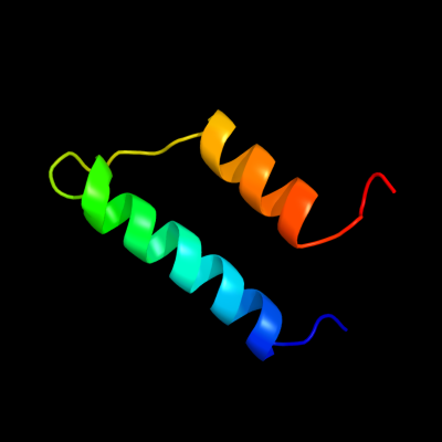
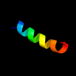
1 c2dmwA_
37.4
20
PDB header: membrane proteinChain: A: PDB Molecule: synaptobrevin-like 1 variant;PDBTitle: solution structure of the longin domain of synaptobrevin-2 like protein 1
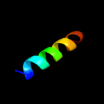
2 c3aqpB_
23.9
11
PDB header: membrane proteinChain: B: PDB Molecule: probable secdf protein-export membrane protein;PDBTitle: crystal structure of secdf, a translocon-associated membrane protein,2 from thermus thrmophilus
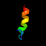
3 d1ljra1
21.6
13
Fold: GST C-terminal domain-likeSuperfamily: GST C-terminal domain-likeFamily: Glutathione S-transferase (GST), C-terminal domain4 d1iyxa2
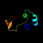
19.2
24
Fold: Enolase N-terminal domain-likeSuperfamily: Enolase N-terminal domain-likeFamily: Enolase N-terminal domain-like5 c2jttD_
17.2
13
PDB header: calcium binding protein/antitumor proteiChain: D: PDB Molecule: calcyclin-binding protein;PDBTitle: solution structure of calcium loaded s100a6 bound to c-2 terminal siah-1 interacting protein
6 c1zroB_
16.6
12
PDB header: cell invasionChain: B: PDB Molecule: erythrocyte binding antigen region ii;PDBTitle: crystal structure of eba-175 region ii (rii) crystallized2 in the presence of (alpha)2,3-sialyllactose
7 c3htkB_
15.8
13
PDB header: recombination/replication/ligaseChain: B: PDB Molecule: structural maintenance of chromosomes protein 5;PDBTitle: crystal structure of mms21 and smc5 complex
8 d1v97a1
14.8
20
Fold: CO dehydrogenase ISP C-domain likeSuperfamily: CO dehydrogenase ISP C-domain likeFamily: CO dehydrogenase ISP C-domain like9 d1oaia_
13.9
16
Fold: RuvA C-terminal domain-likeSuperfamily: UBA-likeFamily: TAP-C domain-like10 d1axda1
13.2
16
Fold: GST C-terminal domain-likeSuperfamily: GST C-terminal domain-likeFamily: Glutathione S-transferase (GST), C-terminal domain11 d1hh4e_
13.1
26
Fold: Immunoglobulin-like beta-sandwichSuperfamily: E set domainsFamily: RhoGDI-like12 c1yx5A_
12.6
29
PDB header: hydrolaseChain: A: PDB Molecule: 26s proteasome non-atpase regulatory subunit 4;PDBTitle: solution structure of s5a uim-1/ubiquitin complex
13 c1yhuW_
11.2
8
PDB header: oxygen storage/transportChain: W: PDB Molecule: hemoglobin b1a chain;PDBTitle: crystal structure of riftia pachyptila c1 hemoglobin reveals novel2 assembly of 24 subunits.
14 d2i4ra1
10.9
35
Fold: AtpF-likeSuperfamily: AtpF-likeFamily: AtpF-like15 d1ds6b_
10.9
29
Fold: Immunoglobulin-like beta-sandwichSuperfamily: E set domainsFamily: RhoGDI-like16 c3bd0D_
10.8
11
PDB header: peptide binding proteinChain: D: PDB Molecule: protein memo1;PDBTitle: crystal structure of memo, form ii
17 d3cx5d2
10.3
17
Fold: Single transmembrane helixSuperfamily: Cytochrome c1 subunit of cytochrome bc1 complex (Ubiquinol-cytochrome c reductase), transmembrane anchorFamily: Cytochrome c1 subunit of cytochrome bc1 complex (Ubiquinol-cytochrome c reductase), transmembrane anchor18 d1ppjd2
10.2
28
Fold: Single transmembrane helixSuperfamily: Cytochrome c1 subunit of cytochrome bc1 complex (Ubiquinol-cytochrome c reductase), transmembrane anchorFamily: Cytochrome c1 subunit of cytochrome bc1 complex (Ubiquinol-cytochrome c reductase), transmembrane anchor19 c2j96B_
10.0
22
PDB header: photosynthesisChain: B: PDB Molecule: phycoerythrocyanin alpha chain;PDBTitle: the e-configuration of alfa-phycoerythrocyanin
20 c3c9pA_
9.9
24
PDB header: structural genomics, unknown functionChain: A: PDB Molecule: uncharacterized protein sp1917;PDBTitle: crystal structure of uncharacterized protein sp1917
21 c1l2aD_
not modelled
9.7
16
PDB header: hydrolaseChain: D: PDB Molecule: cellobiohydrolase;PDBTitle: the crystal structure and catalytic mechanism of2 cellobiohydrolase cels, the major enzymatic component of3 the clostridium thermocellum cellulosome
22 d1l1ya_
not modelled
9.7
16
Fold: alpha/alpha toroidSuperfamily: Six-hairpin glycosidasesFamily: Cellulases catalytic domain23 d1go5a_
not modelled
9.7
20
Fold: RuvA C-terminal domain-likeSuperfamily: UBA-likeFamily: TAP-C domain-like24 d1vmja_
not modelled
9.4
23
Fold: YjbQ-likeSuperfamily: YjbQ-likeFamily: YjbQ-like25 d1k3ra2
not modelled
9.3
19
Fold: alpha/beta knotSuperfamily: alpha/beta knotFamily: Hypothetical protein MTH1 (MT0001), dimerisation domain26 c2kdtA_
not modelled
9.3
50
PDB header: protein transportChain: A: PDB Molecule: neuroendocrine convertase 1;PDBTitle: pc1/3 dcsg sorting domain structure in dpc
27 d1t3qa1
not modelled
9.1
11
Fold: CO dehydrogenase ISP C-domain likeSuperfamily: CO dehydrogenase ISP C-domain likeFamily: CO dehydrogenase ISP C-domain like28 d2ebfx1
not modelled
8.9
18
Fold: PMT central region-likeSuperfamily: PMT central region-likeFamily: PMT central region-like29 d1xeqa1
not modelled
8.7
50
Fold: S15/NS1 RNA-binding domainSuperfamily: S15/NS1 RNA-binding domainFamily: N-terminal, RNA-binding domain of nonstructural protein NS130 c2vh3B_
not modelled
8.6
29
PDB header: unknown functionChain: B: PDB Molecule: ranasmurfin;PDBTitle: ranasmurfin
31 c3r66A_
not modelled
8.5
50
PDB header: viral protein/antiviral proteinChain: A: PDB Molecule: non-structural protein 1;PDBTitle: crystal structure of human isg15 in complex with ns1 n-terminal region2 from influenza virus b, northeast structural genomics consortium3 target ids hx6481, hr2873, and or2
32 d1t33a1
not modelled
8.3
14
Fold: DNA/RNA-binding 3-helical bundleSuperfamily: Homeodomain-likeFamily: Tetracyclin repressor-like, N-terminal domain33 c3cloC_
not modelled
8.3
14
PDB header: transcription regulatorChain: C: PDB Molecule: transcriptional regulator;PDBTitle: crystal structure of putative transcriptional regulator containing a2 luxr dna binding domain (np_811094.1) from bacteroides3 thetaiotaomicron vpi-5482 at 2.04 a resolution
34 c2qr4B_
not modelled
8.2
8
PDB header: hydrolaseChain: B: PDB Molecule: peptidase m3b, oligoendopeptidase f;PDBTitle: crystal structure of oligoendopeptidase-f from enterococcus faecium
35 c2jp7A_
not modelled
8.2
21
PDB header: translationChain: A: PDB Molecule: mrna export factor mex67;PDBTitle: nmr structure of the mex67 uba domain
36 d1f1sa1
not modelled
8.1
21
Fold: alpha/alpha toroidSuperfamily: Chondroitin AC/alginate lyaseFamily: Hyaluronate lyase-like catalytic, N-terminal domain37 d1s29a_
not modelled
8.1
27
Fold: DNA/RNA-binding 3-helical bundleSuperfamily: "Winged helix" DNA-binding domainFamily: La domain38 d1doab_
not modelled
8.0
26
Fold: Immunoglobulin-like beta-sandwichSuperfamily: E set domainsFamily: RhoGDI-like39 d1r2za1
not modelled
7.9
36
Fold: S13-like H2TH domainSuperfamily: S13-like H2TH domainFamily: Middle domain of MutM-like DNA repair proteins40 d1q33a_
not modelled
7.9
12
Fold: NudixSuperfamily: NudixFamily: MutT-like41 c1bm4A_
not modelled
7.8
38
PDB header: viral proteinChain: A: PDB Molecule: protein (moloney murine leukemia virus capsid);PDBTitle: momlv capsid protein major homology region peptide analog
42 d1vi0a2
not modelled
7.6
15
Fold: Tetracyclin repressor-like, C-terminal domainSuperfamily: Tetracyclin repressor-like, C-terminal domainFamily: Tetracyclin repressor-like, C-terminal domain43 d1u7ka_
not modelled
7.4
20
Fold: Retrovirus capsid protein, N-terminal core domainSuperfamily: Retrovirus capsid protein, N-terminal core domainFamily: Retrovirus capsid protein, N-terminal core domain44 d1ee8a1
not modelled
7.4
43
Fold: S13-like H2TH domainSuperfamily: S13-like H2TH domainFamily: Middle domain of MutM-like DNA repair proteins45 d1j2za_
not modelled
7.3
19
Fold: Single-stranded left-handed beta-helixSuperfamily: Trimeric LpxA-like enzymesFamily: UDP N-acetylglucosamine acyltransferase46 d1g9ga_
not modelled
7.2
16
Fold: alpha/alpha toroidSuperfamily: Six-hairpin glycosidasesFamily: Cellulases catalytic domain47 c2rrfA_
not modelled
7.2
35
PDB header: unknown functionChain: A: PDB Molecule: zinc finger fyve domain-containing protein 21;PDBTitle: the solution structure of the c-terminal region of zinc finger fyve2 domain-containing protein 21
48 c1wyoA_
not modelled
7.1
17
PDB header: structural proteinChain: A: PDB Molecule: microtubule-associated protein rp/eb familyPDBTitle: solution structure of the ch domain of human microtubule-2 associated protein rp/eb family member 3
49 d1utga_
not modelled
7.1
18
Fold: Uteroglobin-likeSuperfamily: Uteroglobin-likeFamily: Uteroglobin-like50 d1rm6c1
not modelled
7.0
15
Fold: CO dehydrogenase ISP C-domain likeSuperfamily: CO dehydrogenase ISP C-domain likeFamily: CO dehydrogenase ISP C-domain like51 d2cqka1
not modelled
7.0
33
Fold: DNA/RNA-binding 3-helical bundleSuperfamily: "Winged helix" DNA-binding domainFamily: La domain52 c2r8uA_
not modelled
6.9
20
PDB header: cell cycleChain: A: PDB Molecule: microtubule-associated protein rp/eb familyPDBTitle: structure of fragment of human end-binding protein 1 (eb1)2 containing the n-terminal domain at 1.35 a resolution
53 c2p6cB_
not modelled
6.8
19
PDB header: structural genomics, unknown functionChain: B: PDB Molecule: aq_2013 protein;PDBTitle: crystal structure of hypothetical protein aq_2013 from aquifex2 aeolicus vf5.
54 d1i3da_
not modelled
6.6
14
Fold: Globin-likeSuperfamily: Globin-likeFamily: Globins55 d1cb8a1
not modelled
6.5
12
Fold: alpha/alpha toroidSuperfamily: Chondroitin AC/alginate lyaseFamily: Hyaluronate lyase-like catalytic, N-terminal domain56 c2veqA_
not modelled
6.4
40
PDB header: cell cycleChain: A: PDB Molecule: centromere dna-binding protein complex cbf3PDBTitle: insights into kinetochore-dna interactions from the2 structure of cep3p
57 c1x1jA_
not modelled
6.3
23
PDB header: lyaseChain: A: PDB Molecule: xanthan lyase;PDBTitle: crystal structure of xanthan lyase (n194a) with a substrate.
58 d1w6ta2
not modelled
6.1
24
Fold: Enolase N-terminal domain-likeSuperfamily: Enolase N-terminal domain-likeFamily: Enolase N-terminal domain-like59 c3n8bB_
not modelled
6.0
13
PDB header: nucleic acid binding proteinChain: B: PDB Molecule: uncharacterized protein;PDBTitle: crystal structure of borrelia burgdorferi pur-alpha
60 c1rm6F_
not modelled
6.0
16
PDB header: oxidoreductaseChain: F: PDB Molecule: 4-hydroxybenzoyl-coa reductase gamma subunit;PDBTitle: structure of 4-hydroxybenzoyl-coa reductase from thauera2 aromatica
61 d1l2la_
not modelled
5.9
36
Fold: Ribokinase-likeSuperfamily: Ribokinase-likeFamily: ADP-specific Phosphofructokinase/Glucokinase62 d1jroa1
not modelled
5.9
16
Fold: CO dehydrogenase ISP C-domain likeSuperfamily: CO dehydrogenase ISP C-domain likeFamily: CO dehydrogenase ISP C-domain like63 d2h8aa1
not modelled
5.8
15
Fold: MAPEG domain-likeSuperfamily: MAPEG domain-likeFamily: MAPEG domain64 c3zvrA_
not modelled
5.8
17
PDB header: hydrolaseChain: A: PDB Molecule: dynamin-1;PDBTitle: crystal structure of dynamin
65 d1m22a_
not modelled
5.8
25
Fold: Amidase signature (AS) enzymesSuperfamily: Amidase signature (AS) enzymesFamily: Amidase signature (AS) enzymes66 d1alla_
not modelled
5.7
18
Fold: Globin-likeSuperfamily: Globin-likeFamily: Phycocyanin-like phycobilisome proteins67 d1tw9a1
not modelled
5.7
12
Fold: GST C-terminal domain-likeSuperfamily: GST C-terminal domain-likeFamily: Glutathione S-transferase (GST), C-terminal domain68 c1cb8A_
not modelled
5.6
5
PDB header: lyaseChain: A: PDB Molecule: protein (chondroitinase ac);PDBTitle: chondroitinase ac lyase from flavobacterium heparinum
69 c3nm7D_
not modelled
5.6
13
PDB header: nucleic acid binding proteinChain: D: PDB Molecule: uncharacterized protein;PDBTitle: crystal structure of borrelia burgdorferi pur-alpha
70 c2rddB_
not modelled
5.5
11
PDB header: membrane protein/transport proteinChain: B: PDB Molecule: upf0092 membrane protein yajc;PDBTitle: x-ray crystal structure of acrb in complex with a novel2 transmembrane helix.
71 c1q0wA_
not modelled
5.5
40
PDB header: transport proteinChain: A: PDB Molecule: vacuolar protein sorting-associated proteinPDBTitle: solution structure of vps27 amino-terminal uim-ubiquitin2 complex
72 d1n9wa2
not modelled
5.4
21
Fold: Class II aaRS and biotin synthetasesSuperfamily: Class II aaRS and biotin synthetasesFamily: Class II aminoacyl-tRNA synthetase (aaRS)-like, catalytic domain73 d1ccda_
not modelled
5.4
15
Fold: Uteroglobin-likeSuperfamily: Uteroglobin-likeFamily: Uteroglobin-like74 c3snhA_
not modelled
5.3
17
PDB header: endocytosisChain: A: PDB Molecule: dynamin-1;PDBTitle: crystal structure of nucleotide-free human dynamin1
75 c1k3rA_
not modelled
5.3
19
PDB header: structural genomics, unknown functionChain: A: PDB Molecule: conserved protein mt0001;PDBTitle: crystal structure of the methyltransferase with a knot from2 methanobacterium thermoautotrophicum
76 d1ffva1
not modelled
5.2
9
Fold: CO dehydrogenase ISP C-domain likeSuperfamily: CO dehydrogenase ISP C-domain likeFamily: CO dehydrogenase ISP C-domain like77 d1k1xa1
not modelled
5.1
26
Fold: immunoglobulin/albumin-binding domain-likeSuperfamily: Families 57/38 glycoside transferase middle domainFamily: 4-alpha-glucanotransferase, domain 278 c3b9yA_
not modelled
5.1
10
PDB header: transport proteinChain: A: PDB Molecule: ammonium transporter family rh-like protein;PDBTitle: crystal structure of the nitrosomonas europaea rh protein