1 c2jbuB_
100.0
14
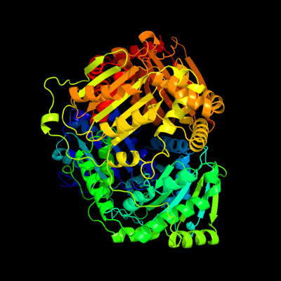
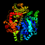
PDB header: hydrolaseChain: B: PDB Molecule: insulin-degrading enzyme;PDBTitle: crystal structure of human insulin degrading enzyme2 complexed with co-purified peptides.
2 c2wk3A_
100.0
14
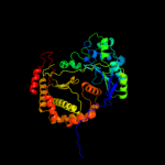
PDB header: hydrolaseChain: A: PDB Molecule: insulin degrading enzyme;PDBTitle: crystal structure of human insulin-degrading enzyme in2 complex with amyloid-beta (1-42)
3 c1q2lA_
100.0
13
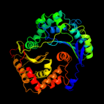
PDB header: hydrolaseChain: A: PDB Molecule: protease iii;PDBTitle: crystal structure of pitrilysin
4 c2fgeA_
100.0
10
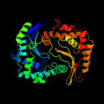
PDB header: hydrolase, plant proteinChain: A: PDB Molecule: zinc metalloprotease (insulinase family);PDBTitle: crystal structure of presequence protease prep from2 arabidopsis thaliana
5 c3go9A_
100.0
20
PDB header: hydrolaseChain: A: PDB Molecule: insulinase family protease;PDBTitle: predicted insulinase family protease from yersinia pestis
6 c1sqpA_
100.0
18
PDB header: oxidoreductaseChain: A: PDB Molecule: ubiquinol-cytochrome-c reductase complex core protein i,PDBTitle: crystal structure analysis of bovine bc1 with myxothiazol
7 c1hr9D_
100.0
17
PDB header: hydrolaseChain: D: PDB Molecule: mitochondrial processing peptidase beta subunit;PDBTitle: yeast mitochondrial processing peptidase beta-e73q mutant2 complexed with malate dehydrogenase signal peptide
8 c1nu1A_
100.0
18
PDB header: oxidoreductaseChain: A: PDB Molecule: ubiquinol-cytochrome c reductase complex core protein i,PDBTitle: crystal structure of mitochondrial cytochrome bc1 complexed with 2-2 nonyl-4-hydroxyquinoline n-oxide (nqno)
9 c1hr6C_
100.0
13
PDB header: hydrolaseChain: C: PDB Molecule: mitochondrial processing peptidase alpha subunit;PDBTitle: yeast mitochondrial processing peptidase
10 c3amiB_
100.0
20
PDB header: hydrolaseChain: B: PDB Molecule: zinc peptidase;PDBTitle: the crystal structure of the m16b metallopeptidase subunit from2 sphingomonas sp. a1
11 c3eoqB_
100.0
20
PDB header: hydrolaseChain: B: PDB Molecule: putative zinc protease;PDBTitle: the crystal structure of putative zinc protease beta-2 subunit from thermus thermophilus hb8
12 c3hdiA_
100.0
22
PDB header: hydrolaseChain: A: PDB Molecule: processing protease;PDBTitle: crystal structure of bacillus halodurans metallo peptidase
13 c1l0lB_
100.0
15
PDB header: oxidoreductaseChain: B: PDB Molecule: ubiquinol-cytochrome c reductase complex core protein 2;PDBTitle: structure of bovine mitochondrial cytochrome bc1 complex with a bound2 fungicide famoxadone
14 c3gwbA_
100.0
16
PDB header: hydrolaseChain: A: PDB Molecule: peptidase m16 inactive domain family protein;PDBTitle: crystal structure of peptidase m16 inactive domain from pseudomonas2 fluorescens. northeast structural genomics target plr293l
15 c3amjB_
100.0
15
PDB header: hydrolaseChain: B: PDB Molecule: zinc peptidase inactive subunit;PDBTitle: the crystal structure of the heterodimer of m16b peptidase from2 sphingomonas sp. a1
16 c3cx5L_
100.0
16
PDB header: oxidoreductaseChain: L: PDB Molecule: cytochrome b-c1 complex subunit 1, mitochondrial;PDBTitle: structure of complex iii with bound cytochrome c in reduced2 state and definition of a minimal core interface for3 electron transfer.
17 c3d3yA_
100.0
16
PDB header: structural genomics, unknown functionChain: A: PDB Molecule: uncharacterized protein;PDBTitle: crystal structure of a conserved protein from enterococcus faecalis2 v583
18 d2fgea4
100.0
16
Fold: LuxS/MPP-like metallohydrolaseSuperfamily: LuxS/MPP-like metallohydrolaseFamily: MPP-like19 d1ppja1
100.0
24
Fold: LuxS/MPP-like metallohydrolaseSuperfamily: LuxS/MPP-like metallohydrolaseFamily: MPP-like20 c3cxhM_
100.0
15
PDB header: oxidoreductaseChain: M: PDB Molecule: cytochrome b-c1 complex subunit 2, mitochondrial;PDBTitle: structure of yeast complex iii with isoform-2 cytochrome c2 bound and definition of a minimal core interface for3 electron transfer.
21 d1hr6b1
not modelled
100.0
21
Fold: LuxS/MPP-like metallohydrolaseSuperfamily: LuxS/MPP-like metallohydrolaseFamily: MPP-like22 d1bcca1
not modelled
100.0
22
Fold: LuxS/MPP-like metallohydrolaseSuperfamily: LuxS/MPP-like metallohydrolaseFamily: MPP-like23 d1hr6a1
not modelled
100.0
19
Fold: LuxS/MPP-like metallohydrolaseSuperfamily: LuxS/MPP-like metallohydrolaseFamily: MPP-like24 d1q2la4
not modelled
100.0
17
Fold: LuxS/MPP-like metallohydrolaseSuperfamily: LuxS/MPP-like metallohydrolaseFamily: MPP-like25 d1ppjb1
not modelled
100.0
18
Fold: LuxS/MPP-like metallohydrolaseSuperfamily: LuxS/MPP-like metallohydrolaseFamily: MPP-like26 d1bccb1
not modelled
100.0
16
Fold: LuxS/MPP-like metallohydrolaseSuperfamily: LuxS/MPP-like metallohydrolaseFamily: MPP-like27 d3cx5b1
not modelled
100.0
17
Fold: LuxS/MPP-like metallohydrolaseSuperfamily: LuxS/MPP-like metallohydrolaseFamily: MPP-like28 d3cx5a1
not modelled
100.0
14
Fold: LuxS/MPP-like metallohydrolaseSuperfamily: LuxS/MPP-like metallohydrolaseFamily: MPP-like29 d1q2la1
not modelled
99.9
15
Fold: LuxS/MPP-like metallohydrolaseSuperfamily: LuxS/MPP-like metallohydrolaseFamily: MPP-like30 d1hr6b2
not modelled
99.9
10
Fold: LuxS/MPP-like metallohydrolaseSuperfamily: LuxS/MPP-like metallohydrolaseFamily: MPP-like31 d1ppjb2
not modelled
99.9
7
Fold: LuxS/MPP-like metallohydrolaseSuperfamily: LuxS/MPP-like metallohydrolaseFamily: MPP-like32 d1q2la2
not modelled
99.9
11
Fold: LuxS/MPP-like metallohydrolaseSuperfamily: LuxS/MPP-like metallohydrolaseFamily: MPP-like33 c3ivlA_
not modelled
99.9
13
PDB header: hydrolaseChain: A: PDB Molecule: putative zinc protease;PDBTitle: the crystal structure of the inactive peptidase domain of a putative2 zinc protease from bordetella parapertussis to 2.2a
34 d1bcca2
not modelled
99.9
12
Fold: LuxS/MPP-like metallohydrolaseSuperfamily: LuxS/MPP-like metallohydrolaseFamily: MPP-like35 d1hr6a2
not modelled
99.9
9
Fold: LuxS/MPP-like metallohydrolaseSuperfamily: LuxS/MPP-like metallohydrolaseFamily: MPP-like36 d1ppja2
not modelled
99.8
12
Fold: LuxS/MPP-like metallohydrolaseSuperfamily: LuxS/MPP-like metallohydrolaseFamily: MPP-like37 d2fgea2
not modelled
99.8
6
Fold: LuxS/MPP-like metallohydrolaseSuperfamily: LuxS/MPP-like metallohydrolaseFamily: MPP-like38 d1bccb2
not modelled
99.8
9
Fold: LuxS/MPP-like metallohydrolaseSuperfamily: LuxS/MPP-like metallohydrolaseFamily: MPP-like39 d3cx5a2
not modelled
99.8
11
Fold: LuxS/MPP-like metallohydrolaseSuperfamily: LuxS/MPP-like metallohydrolaseFamily: MPP-like40 d2fgea1
not modelled
99.5
11
Fold: LuxS/MPP-like metallohydrolaseSuperfamily: LuxS/MPP-like metallohydrolaseFamily: MPP-like41 d2fgea3
not modelled
99.4
11
Fold: LuxS/MPP-like metallohydrolaseSuperfamily: LuxS/MPP-like metallohydrolaseFamily: MPP-like42 d1q2la3
not modelled
99.2
9
Fold: LuxS/MPP-like metallohydrolaseSuperfamily: LuxS/MPP-like metallohydrolaseFamily: MPP-like43 d1nsha_
not modelled
39.5
16
Fold: EF Hand-likeSuperfamily: EF-handFamily: S100 proteins44 c2rgiA_
not modelled
35.8
16
PDB header: metal binding proteinChain: A: PDB Molecule: protein s100-a2;PDBTitle: crystal structure of ca2+-free s100a2 at 1.6 a resolution
45 d1a4pa_
not modelled
35.2
9
Fold: EF Hand-likeSuperfamily: EF-handFamily: S100 proteins46 d1qlsa_
not modelled
33.8
12
Fold: EF Hand-likeSuperfamily: EF-handFamily: S100 proteins47 d1g5ta_
not modelled
23.2
17
Fold: P-loop containing nucleoside triphosphate hydrolasesSuperfamily: P-loop containing nucleoside triphosphate hydrolasesFamily: RecA protein-like (ATPase-domain)48 c1ko7B_
not modelled
22.8
15
PDB header: transferase,hydrolaseChain: B: PDB Molecule: hpr kinase/phosphatase;PDBTitle: x-ray structure of the hpr kinase/phosphatase from2 staphylococcus xylosus at 1.95 a resolution
49 c1g6uB_
not modelled
22.2
37
PDB header: de novo proteinChain: B: PDB Molecule: domain swapped dimer;PDBTitle: crystal structure of a domain swapped dimer
50 d1ko7a1
not modelled
20.8
15
Fold: MurF and HprK N-domain-likeSuperfamily: HprK N-terminal domain-likeFamily: HPr kinase/phoshatase HprK N-terminal domain51 c2kfvA_
not modelled
20.5
23
PDB header: isomeraseChain: A: PDB Molecule: fk506-binding protein 3;PDBTitle: structure of the amino-terminal domain of human fk506-2 binding protein 3 / northeast structural genomics3 consortium target ht99a
52 d1e8aa_
not modelled
20.3
12
Fold: EF Hand-likeSuperfamily: EF-handFamily: S100 proteins53 d2rdea1
not modelled
19.5
25
Fold: Split barrel-likeSuperfamily: PilZ domain-likeFamily: PilZ domain54 c2y5iF_
not modelled
19.2
16
PDB header: metal-binding proteinChain: F: PDB Molecule: s100 calcium binding protein z;PDBTitle: s100z from zebrafish in complex with calcium
55 c1qpoA_
not modelled
17.9
10
PDB header: transferaseChain: A: PDB Molecule: quinolinate acid phosphoribosyl transferase;PDBTitle: quinolinate phosphoribosyl transferase (qaprtase) apo-enzyme from2 mycobacterium tuberculosis
56 c3l7vA_
not modelled
17.4
13
PDB header: transcriptionChain: A: PDB Molecule: putative uncharacterized protein smu.1377c;PDBTitle: crystal structure of a hypothetical protein smu.1377c from2 streptococcus mutans ua159
57 d1ksoa_
not modelled
15.7
10
Fold: EF Hand-likeSuperfamily: EF-handFamily: S100 proteins58 c2uxsA_
not modelled
15.2
13
PDB header: hydrolaseChain: A: PDB Molecule: inorganic pyrophosphatase;PDBTitle: 2.7a crystal structure of inorganic pyrophosphatase (rv3628)2 from mycobacterium tuberculosis at ph 7.5
59 d1knxa1
not modelled
14.2
14
Fold: MurF and HprK N-domain-likeSuperfamily: HprK N-terminal domain-likeFamily: HPr kinase/phoshatase HprK N-terminal domain60 d1g64b_
not modelled
13.5
17
Fold: P-loop containing nucleoside triphosphate hydrolasesSuperfamily: P-loop containing nucleoside triphosphate hydrolasesFamily: RecA protein-like (ATPase-domain)61 c3llkA_
not modelled
12.7
14
PDB header: oxidoreductaseChain: A: PDB Molecule: sulfhydryl oxidase 1;PDBTitle: sulfhydryl oxidase fragment of human qsox1
62 d3c1va1
not modelled
12.3
14
Fold: EF Hand-likeSuperfamily: EF-handFamily: S100 proteins63 d3cr5x1
not modelled
11.8
12
Fold: EF Hand-likeSuperfamily: EF-handFamily: S100 proteins64 d1zfsa1
not modelled
11.3
15
Fold: EF Hand-likeSuperfamily: EF-handFamily: S100 proteins65 c3fhkF_
not modelled
11.2
17
PDB header: structural genomics, unknown functionChain: F: PDB Molecule: upf0403 protein yphp;PDBTitle: crystal structure of apc1446, b.subtilis yphp disulfide2 isomerase
66 d1qlka_
not modelled
10.6
10
Fold: EF Hand-likeSuperfamily: EF-handFamily: S100 proteins67 c2pbrB_
not modelled
10.6
12
PDB header: transferaseChain: B: PDB Molecule: thymidylate kinase;PDBTitle: crystal structure of thymidylate kinase (aq_969) from aquifex aeolicus2 vf5
68 d2prda_
not modelled
10.6
17
Fold: OB-foldSuperfamily: Inorganic pyrophosphataseFamily: Inorganic pyrophosphatase69 d3c7ba2
not modelled
10.5
12
Fold: Ferredoxin-likeSuperfamily: Nitrite/Sulfite reductase N-terminal domain-likeFamily: DsrA/DsrB N-terminal-domain-like70 c3ostA_
not modelled
10.1
8
PDB header: lipid binding proteinChain: A: PDB Molecule: serine/threonine-protein kinase kcc4;PDBTitle: structure of the kinase associated-1 (ka1) from kcc4p
71 c3lv8A_
not modelled
9.6
12
PDB header: transferaseChain: A: PDB Molecule: thymidylate kinase;PDBTitle: 1.8 angstrom resolution crystal structure of a thymidylate kinase2 (tmk) from vibrio cholerae o1 biovar eltor str. n16961 in complex3 with tmp, thymidine-5'-diphosphate and adp
72 d2ikba1
not modelled
9.4
17
Fold: Lysozyme-likeSuperfamily: Lysozyme-likeFamily: NMB1012-like73 d1zavz1
not modelled
9.2
21
Fold: Ribosomal protein L7/12, oligomerisation (N-terminal) domainSuperfamily: Ribosomal protein L7/12, oligomerisation (N-terminal) domainFamily: Ribosomal protein L7/12, oligomerisation (N-terminal) domain74 c1zawZ_
not modelled
9.2
21
PDB header: structural proteinChain: Z: PDB Molecule: 50s ribosomal protein l7/l12;PDBTitle: ribosomal protein l10-l12(ntd) complex, space group p212121,2 form a
75 c1zavZ_
not modelled
9.2
21
PDB header: structural proteinChain: Z: PDB Molecule: 50s ribosomal protein l7/l12;PDBTitle: ribosomal protein l10-l12(ntd) complex, space group p21
76 d1l7ba_
not modelled
9.0
22
Fold: BRCT domainSuperfamily: BRCT domainFamily: DNA ligase77 c1zawY_
not modelled
8.9
21
PDB header: structural proteinChain: Y: PDB Molecule: 50s ribosomal protein l7/l12;PDBTitle: ribosomal protein l10-l12(ntd) complex, space group p212121,2 form a
78 c1zawX_
not modelled
8.9
21
PDB header: structural proteinChain: X: PDB Molecule: 50s ribosomal protein l7/l12;PDBTitle: ribosomal protein l10-l12(ntd) complex, space group p212121,2 form a
79 d1pc2a_
not modelled
8.9
14
Fold: alpha-alpha superhelixSuperfamily: TPR-likeFamily: Tetratricopeptide repeat (TPR)80 c3pajA_
not modelled
8.8
16
PDB header: transferaseChain: A: PDB Molecule: nicotinate-nucleotide pyrophosphorylase, carboxylating;PDBTitle: 2.00 angstrom resolution crystal structure of a quinolinate2 phosphoribosyltransferase from vibrio cholerae o1 biovar eltor str.3 n16961
81 d1qpoa1
not modelled
8.7
10
Fold: TIM beta/alpha-barrelSuperfamily: Nicotinate/Quinolinate PRTase C-terminal domain-likeFamily: NadC C-terminal domain-like82 d1udea_
not modelled
8.6
15
Fold: OB-foldSuperfamily: Inorganic pyrophosphataseFamily: Inorganic pyrophosphatase83 d1v33a_
not modelled
8.6
17
Fold: Prim-pol domainSuperfamily: Prim-pol domainFamily: PriA-like84 c1knxF_
not modelled
8.6
12
PDB header: transferase/hydrolaseChain: F: PDB Molecule: probable hpr(ser) kinase/phosphatase;PDBTitle: hpr kinase/phosphatase from mycoplasma pneumoniae
85 d1tuza_
not modelled
8.2
6
Fold: EF Hand-likeSuperfamily: EF-handFamily: EF-hand modules in multidomain proteins86 c2yw3E_
not modelled
8.2
11
PDB header: lyaseChain: E: PDB Molecule: 4-hydroxy-2-oxoglutarate aldolase/2-deydro-3-PDBTitle: crystal structure analysis of the 4-hydroxy-2-oxoglutarate aldolase/2-2 deydro-3-deoxyphosphogluconate aldolase from tthb1
87 c3qf2B_
not modelled
8.0
11
PDB header: apoptosisChain: B: PDB Molecule: nacht, lrr and pyd domains-containing protein 3;PDBTitle: crystal structure of nalp3 pyd
88 d1u1qa_
not modelled
8.0
7
Fold: Ferredoxin-likeSuperfamily: RNA-binding domain, RBDFamily: Canonical RBD89 c3dzzB_
not modelled
8.0
13
PDB header: transferaseChain: B: PDB Molecule: putative pyridoxal 5'-phosphate-dependent c-s lyase;PDBTitle: crystal structure of a putative plp-dependent aminotransferase2 (lbul_1103) from lactobacillus delbrueckii subsp. at 1.61 a3 resolution
90 c3gnnA_
not modelled
7.9
21
PDB header: transferaseChain: A: PDB Molecule: nicotinate-nucleotide pyrophosphorylase;PDBTitle: crystal structure of nicotinate-nucleotide2 pyrophosphorylase from burkholderi pseudomallei
91 d1kyqa2
not modelled
7.9
11
Fold: Siroheme synthase middle domains-likeSuperfamily: Siroheme synthase middle domains-likeFamily: Siroheme synthase middle domains-like92 c2ebuA_
not modelled
7.8
17
PDB header: replicationChain: A: PDB Molecule: replication factor c subunit 1;PDBTitle: solution structure of the brct domain from human2 replication factor c large subunit 1
93 c1zawU_
not modelled
7.7
21
PDB header: structural proteinChain: U: PDB Molecule: 50s ribosomal protein l7/l12;PDBTitle: ribosomal protein l10-l12(ntd) complex, space group p212121,2 form a
94 c1zawW_
not modelled
7.7
21
PDB header: structural proteinChain: W: PDB Molecule: 50s ribosomal protein l7/l12;PDBTitle: ribosomal protein l10-l12(ntd) complex, space group p212121,2 form a
95 c1zawV_
not modelled
7.7
21
PDB header: structural proteinChain: V: PDB Molecule: 50s ribosomal protein l7/l12;PDBTitle: ribosomal protein l10-l12(ntd) complex, space group p212121,2 form a
96 c2b7pA_
not modelled
7.7
7
PDB header: transferaseChain: A: PDB Molecule: probable nicotinate-nucleotide pyrophosphorylase;PDBTitle: crystal structure of quinolinic acid phosphoribosyltransferase from2 helicobacter pylori
97 c2ld7A_
not modelled
7.6
7
PDB header: transcriptionChain: A: PDB Molecule: histone deacetylase complex subunit sap30;PDBTitle: solution structure of the msin3a pah3-sap30 sid complex
98 c1zaxZ_
not modelled
7.6
21
PDB header: structural proteinChain: Z: PDB Molecule: 50s ribosomal protein l7/l12;PDBTitle: ribosomal protein l10-l12(ntd) complex, space group p212121,2 form b
99 c1zaxV_
not modelled
7.5
21
PDB header: structural proteinChain: V: PDB Molecule: 50s ribosomal protein l7/l12;PDBTitle: ribosomal protein l10-l12(ntd) complex, space group p212121,2 form b