1 c2l26A_
100.0
36
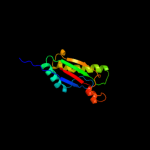
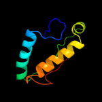
PDB header: membrane proteinChain: A: PDB Molecule: uncharacterized protein rv0899/mt0922;PDBTitle: rv0899 from mycobacterium tuberculosis contains two separated domains
2 c2kgwA_
100.0
31
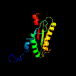
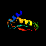
PDB header: membrane proteinChain: A: PDB Molecule: outer membrane protein a;PDBTitle: solution structure of the carboxy-terminal domain of ompatb, a pore2 forming protein from mycobacterium tuberculosis
3 c3td4D_
100.0
38
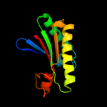
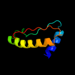
PDB header: membrane protein,peptide binding proteinChain: D: PDB Molecule: outer membrane protein omp38;PDBTitle: crystal structure of ompa-like domain from acinetobacter baumannii in2 complex with diaminopimelate
4 d2aizp1
100.0
23
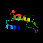
Fold: Bacillus chorismate mutase-likeSuperfamily: OmpA-likeFamily: OmpA-like5 c2k1sA_
100.0
32
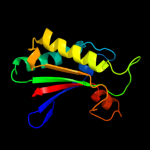
PDB header: lipoproteinChain: A: PDB Molecule: inner membrane lipoprotein yiad;PDBTitle: solution nmr structure of the folded c-terminal fragment of yiad from2 escherichia coli. northeast structural genomics consortium target3 er553.
6 c3khnB_
100.0
26
PDB header: structural genomics, unknown functionChain: B: PDB Molecule: motb protein, putative;PDBTitle: crystal structure of putative motb like protein dvu_22282 from desulfovibrio vulgaris.
7 d2hqsc1
100.0
24
Fold: Bacillus chorismate mutase-likeSuperfamily: OmpA-likeFamily: OmpA-like8 c1r1mA_
100.0
36
PDB header: membrane proteinChain: A: PDB Molecule: outer membrane protein class 4;PDBTitle: structure of the ompa-like domain of rmpm from neisseria2 meningitidis
9 d1r1ma_
100.0
36
Fold: Bacillus chorismate mutase-likeSuperfamily: OmpA-likeFamily: OmpA-like10 c3cyqM_
100.0
22
PDB header: membrane proteinChain: M: PDB Molecule: chemotaxis protein motb;PDBTitle: the crystal structure of the complex of the c-terminal domain of2 helicobacter pylori motb (residues 125-256) with n-acetylmuramic acid
11 c2zvyB_
100.0
19
PDB header: membrane proteinChain: B: PDB Molecule: chemotaxis protein motb;PDBTitle: structure of the periplasmic domain of motb from salmonella2 (crystal form ii)
12 c3ldtA_
100.0
21
PDB header: membrane proteinChain: A: PDB Molecule: outer membrane protein, ompa family protein;PDBTitle: crystal structure of an outer membrane protein(ompa)from2 legionella pneumophila
13 c3oonA_
99.9
25
PDB header: membrane proteinChain: A: PDB Molecule: outer membrane protein (tpn50);PDBTitle: the structure of an outer membrance protein from borrelia burgdorferi2 b31
14 c2zovA_
99.9
23
PDB header: membrane proteinChain: A: PDB Molecule: chemotaxis protein motb;PDBTitle: structure of the periplasmic domain of motb from salmonella2 (crystal form i)
15 c2zf8A_
99.8
18
PDB header: structural proteinChain: A: PDB Molecule: component of sodium-driven polar flagellar motor;PDBTitle: crystal structure of moty
16 d1tiba_
56.0
17
Fold: alpha/beta-HydrolasesSuperfamily: alpha/beta-HydrolasesFamily: Fungal lipases17 d1tiaa_
49.5
19
Fold: alpha/beta-HydrolasesSuperfamily: alpha/beta-HydrolasesFamily: Fungal lipases18 c3ngmB_
45.5
23
PDB header: hydrolaseChain: B: PDB Molecule: extracellular lipase;PDBTitle: crystal structure of lipase from gibberella zeae
19 c3o0dF_
41.7
17
PDB header: hydrolaseChain: F: PDB Molecule: triacylglycerol lipase;PDBTitle: crystal structure of lip2 lipase from yarrowia lipolytica at 1.7 a2 resolution
20 d1uwca_
31.8
17
Fold: alpha/beta-HydrolasesSuperfamily: alpha/beta-HydrolasesFamily: Fungal lipases21 c3g7nA_
not modelled
31.0
22
PDB header: hydrolaseChain: A: PDB Molecule: lipase;PDBTitle: crystal structure of a triacylglycerol lipase from2 penicillium expansum at 1.3
22 d1qy7a_
not modelled
29.3
11
Fold: Ferredoxin-likeSuperfamily: GlnB-likeFamily: Prokaryotic signal transducing protein23 c3ds8A_
not modelled
24.8
18
PDB header: structural genomics, unknown functionChain: A: PDB Molecule: lin2722 protein;PDBTitle: the crysatl structure of the gene lin2722 products from listeria2 innocua
24 d1uzhc1
not modelled
24.2
12
Fold: RuBisCO, small subunitSuperfamily: RuBisCO, small subunitFamily: RuBisCO, small subunit25 d1rbli_
not modelled
23.9
19
Fold: RuBisCO, small subunitSuperfamily: RuBisCO, small subunitFamily: RuBisCO, small subunit26 c1ir6A_
not modelled
22.3
17
PDB header: hydrolaseChain: A: PDB Molecule: exonuclease recj;PDBTitle: crystal structure of exonuclease recj bound to manganese
27 d1ir6a_
not modelled
22.3
17
Fold: DHH phosphoesterasesSuperfamily: DHH phosphoesterasesFamily: Exonuclease RecJ28 c2rd5D_
not modelled
22.2
17
PDB header: protein bindingChain: D: PDB Molecule: pii protein;PDBTitle: structural basis for the regulation of n-acetylglutamate kinase by pii2 in arabidopsis thaliana
29 d1svdm1
not modelled
22.0
4
Fold: RuBisCO, small subunitSuperfamily: RuBisCO, small subunitFamily: RuBisCO, small subunit30 c2r6hC_
not modelled
19.4
33
PDB header: oxidoreductaseChain: C: PDB Molecule: nadh:ubiquinone oxidoreductase, na translocating, fPDBTitle: crystal structure of the domain comprising the nad binding and the fad2 binding regions of the nadh:ubiquinone oxidoreductase, na3 translocating, f subunit from porphyromonas gingivalis
31 d8ruci_
not modelled
18.9
12
Fold: RuBisCO, small subunitSuperfamily: RuBisCO, small subunitFamily: RuBisCO, small subunit32 c2oghA_
not modelled
18.3
13
PDB header: translationChain: A: PDB Molecule: eukaryotic translation initiation factor eif-1;PDBTitle: solution structure of yeast eif1
33 d1ej7s_
not modelled
18.3
12
Fold: RuBisCO, small subunitSuperfamily: RuBisCO, small subunitFamily: RuBisCO, small subunit34 d3tgla_
not modelled
17.7
18
Fold: alpha/beta-HydrolasesSuperfamily: alpha/beta-HydrolasesFamily: Fungal lipases35 d1ir1s_
not modelled
17.1
12
Fold: RuBisCO, small subunitSuperfamily: RuBisCO, small subunitFamily: RuBisCO, small subunit36 d2if1a_
not modelled
17.0
13
Fold: eIF1-likeSuperfamily: eIF1-likeFamily: eIF1-like37 d1wdds_
not modelled
16.8
12
Fold: RuBisCO, small subunitSuperfamily: RuBisCO, small subunitFamily: RuBisCO, small subunit38 d1bwvs_
not modelled
16.6
9
Fold: RuBisCO, small subunitSuperfamily: RuBisCO, small subunitFamily: RuBisCO, small subunit39 d1lgya_
not modelled
16.0
16
Fold: alpha/beta-HydrolasesSuperfamily: alpha/beta-HydrolasesFamily: Fungal lipases40 d2v6ai1
not modelled
14.7
12
Fold: RuBisCO, small subunitSuperfamily: RuBisCO, small subunitFamily: RuBisCO, small subunit41 d1bxni_
not modelled
12.7
3
Fold: RuBisCO, small subunitSuperfamily: RuBisCO, small subunitFamily: RuBisCO, small subunit42 d1sxra_
not modelled
12.7
11
Fold: N-acetylmuramoyl-L-alanine amidase-likeSuperfamily: N-acetylmuramoyl-L-alanine amidase-likeFamily: N-acetylmuramoyl-L-alanine amidase-like43 c1nauA_
not modelled
12.7
18
PDB header: hormone/growth factorChain: A: PDB Molecule: glucagon;PDBTitle: nmr solution structure of the glucagon antagonist [deshis1,2 desphe6, glu9]glucagon amide in the presence of3 perdeuterated dodecylphosphocholine micelles
44 d2ckca1
not modelled
11.7
10
Fold: GYF/BRK domain-likeSuperfamily: BRK domain-likeFamily: BRK domain-like45 c2ckcA_
not modelled
11.7
10
PDB header: hydrolaseChain: A: PDB Molecule: chromodomain-helicase-dna-binding protein 7;PDBTitle: solution structures of the brk domains of the human chromo2 helicase domain 7 and 8, reveals structural similarity3 with gyf domain suggesting a role in protein interaction
46 d1gvha3
not modelled
11.1
15
Fold: Ferredoxin reductase-like, C-terminal NADP-linked domainSuperfamily: Ferredoxin reductase-like, C-terminal NADP-linked domainFamily: Flavohemoglobin, C-terminal domain47 d1gk8i_
not modelled
11.0
10
Fold: RuBisCO, small subunitSuperfamily: RuBisCO, small subunitFamily: RuBisCO, small subunit48 d2piia_
not modelled
10.7
17
Fold: Ferredoxin-likeSuperfamily: GlnB-likeFamily: Prokaryotic signal transducing protein49 d1uzdc1
not modelled
10.3
10
Fold: RuBisCO, small subunitSuperfamily: RuBisCO, small subunitFamily: RuBisCO, small subunit50 c1s2jA_
not modelled
10.2
10
PDB header: hydrolaseChain: A: PDB Molecule: peptidoglycan recognition protein sa cg11709-pa;PDBTitle: crystal structure of the drosophila pattern-recognition2 receptor pgrp-sa
51 d1d4oa_
not modelled
9.8
19
Fold: DHS-like NAD/FAD-binding domainSuperfamily: DHS-like NAD/FAD-binding domainFamily: Transhydrogenase domain III (dIII)52 d2v0ea1
not modelled
8.9
8
Fold: GYF/BRK domain-likeSuperfamily: BRK domain-likeFamily: BRK domain-like53 c1pt9B_
not modelled
8.9
19
PDB header: oxidoreductaseChain: B: PDB Molecule: nad(p) transhydrogenase, mitochondrial;PDBTitle: crystal structure analysis of the diii component of transhydrogenase2 with a thio-nicotinamide nucleotide analogue
54 d1tvca2
not modelled
8.3
11
Fold: Ferredoxin reductase-like, C-terminal NADP-linked domainSuperfamily: Ferredoxin reductase-like, C-terminal NADP-linked domainFamily: Aromatic dioxygenase reductase-like55 c3b9eA_
not modelled
8.1
9
PDB header: hydrolaseChain: A: PDB Molecule: chitinase a;PDBTitle: crystal structure of inactive mutant e315m chitinase a from2 vibrio harveyi
56 c3bzqA_
not modelled
7.2
15
PDB header: signaling protein/transcriptionChain: A: PDB Molecule: nitrogen regulatory protein p-ii;PDBTitle: high resolution crystal structure of nitrogen regulatory protein2 (rv2919c) of mycobacterium tuberculosis
57 c3rxyA_
not modelled
6.9
25
PDB header: structural genomics, unknown functionChain: A: PDB Molecule: nif3 protein;PDBTitle: crystal structure of nif3 superfamily protein from sphaerobacter2 thermophilus
58 d2gova1
not modelled
6.6
10
Fold: Probable bacterial effector-binding domainSuperfamily: Probable bacterial effector-binding domainFamily: SOUL heme-binding protein59 c1gvhA_
not modelled
6.6
14
PDB header: oxidoreductaseChain: A: PDB Molecule: flavohemoprotein;PDBTitle: the x-ray structure of ferric escherichia coli2 flavohemoglobin reveals an unespected geometry of the3 distal heme pocket
60 c3devB_
not modelled
6.5
4
PDB header: structural genomics, unknown functionChain: B: PDB Molecule: sh1221;PDBTitle: crystal structure of sh1221 protein from staphylococcus haemolyticus,2 northeast structural genomics consortium target shr87
61 c3mhyC_
not modelled
6.2
13
PDB header: signaling proteinChain: C: PDB Molecule: pii-like protein pz;PDBTitle: a new pii protein structure
62 c2ckaA_
not modelled
6.0
8
PDB header: hydrolaseChain: A: PDB Molecule: chromodomain-helicase-dna-binding protein 8;PDBTitle: solution structures of the brk domains of the human chromo2 helicase domain 7 and 8, reveals structural similarity3 with gyf domain suggesting a role in protein interaction
63 d2ckaa1
not modelled
6.0
8
Fold: GYF/BRK domain-likeSuperfamily: BRK domain-likeFamily: BRK domain-like64 d1zpda1
not modelled
6.0
13
Fold: DHS-like NAD/FAD-binding domainSuperfamily: DHS-like NAD/FAD-binding domainFamily: Pyruvate oxidase and decarboxylase, middle domain65 d1e32a3
not modelled
5.8
19
Fold: Cdc48 domain 2-likeSuperfamily: Cdc48 domain 2-likeFamily: Cdc48 domain 2-like66 c3dmaA_
not modelled
5.7
11
PDB header: structural genomics, unknown functionChain: A: PDB Molecule: exopolyphosphatase-related protein;PDBTitle: crystal structure of an exopolyphosphatase-related protein2 from bacteroides fragilis. northeast structural genomics3 target bfr192
67 d1cqxa3
not modelled
5.7
17
Fold: Ferredoxin reductase-like, C-terminal NADP-linked domainSuperfamily: Ferredoxin reductase-like, C-terminal NADP-linked domainFamily: Flavohemoglobin, C-terminal domain