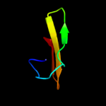
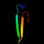
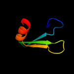
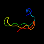
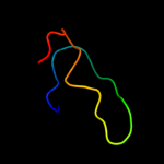
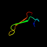
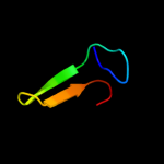
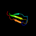
1 c1nuiA_
68.9
32
PDB header: replicationChain: A: PDB Molecule: dna primase/helicase;PDBTitle: crystal structure of the primase fragment of bacteriophage t7 primase-2 helicase protein
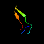
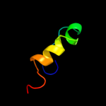
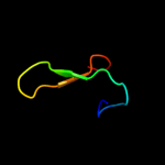
2 d1wiia_
54.8
19
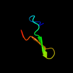
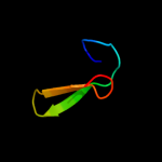
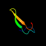
Fold: Rubredoxin-likeSuperfamily: Zinc beta-ribbonFamily: Putative zinc binding domain3 c2dcuB_
46.2
31
PDB header: translationChain: B: PDB Molecule: translation initiation factor 2 beta subunit;PDBTitle: crystal structure of translation initiation factor aif2betagamma2 heterodimer with gdp
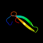
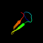
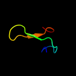
4 d2drpa2
45.7
86
Fold: beta-beta-alpha zinc fingersSuperfamily: beta-beta-alpha zinc fingersFamily: Classic zinc finger, C2H25 d1k81a_
45.2
25
Fold: Zinc-binding domain of translation initiation factor 2 betaSuperfamily: Zinc-binding domain of translation initiation factor 2 betaFamily: Zinc-binding domain of translation initiation factor 2 beta6 c2au3A_
44.6
33
PDB header: transferaseChain: A: PDB Molecule: dna primase;PDBTitle: crystal structure of the aquifex aeolicus primase (zinc binding and2 rna polymerase domains)
7 c1neeA_
42.4
21
PDB header: translationChain: A: PDB Molecule: probable translation initiation factor 2 betaPDBTitle: structure of archaeal translation factor aif2beta from2 methanobacterium thermoautrophicum
8 c3cw2M_
39.9
21
PDB header: translationChain: M: PDB Molecule: translation initiation factor 2 subunit beta;PDBTitle: crystal structure of the intact archaeal translation2 initiation factor 2 from sulfolobus solfataricus .
9 d1d0qa_
39.6
42
Fold: Rubredoxin-likeSuperfamily: Zinc beta-ribbonFamily: DNA primase zinc finger10 c4a17Y_
36.4
35
PDB header: ribosomeChain: Y: PDB Molecule: rpl37a;PDBTitle: t.thermophila 60s ribosomal subunit in complex with2 initiation factor 6. this file contains 5s rrna,3 5.8s rrna and proteins of molecule 2.
11 d1vqoz1
36.2
25
Fold: Rubredoxin-likeSuperfamily: Zn-binding ribosomal proteinsFamily: Ribosomal protein L37ae12 d2fiya1
35.5
29
Fold: FdhE-likeSuperfamily: FdhE-likeFamily: FdhE-like13 c1yshD_
35.0
33
PDB header: structural protein/rnaChain: D: PDB Molecule: ribosomal protein l37a;PDBTitle: localization and dynamic behavior of ribosomal protein l30e
14 c2yrmA_
34.5
37
PDB header: gene regulationChain: A: PDB Molecule: b-cell lymphoma 6 protein;PDBTitle: solution structure of the 1st zf-c2h2 domain from human b-2 cell lymphoma 6 protein
15 c2zkrz_
34.4
25
PDB header: ribosomal protein/rnaChain: Z: PDB Molecule: e site t-rna;PDBTitle: structure of a mammalian ribosomal 60s subunit within an2 80s complex obtained by docking homology models of the rna3 and proteins into an 8.7 a cryo-em map
16 d1jj2y_
33.8
22
Fold: Rubredoxin-likeSuperfamily: Zn-binding ribosomal proteinsFamily: Ribosomal protein L37ae17 c2e9hA_
33.3
25
PDB header: translationChain: A: PDB Molecule: eukaryotic translation initiation factor 5;PDBTitle: solution structure of the eif-5_eif-2b domain from human2 eukaryotic translation initiation factor 5
18 c2qa4Z_
33.1
25
PDB header: ribosomeChain: Z: PDB Molecule: 50s ribosomal protein l37ae;PDBTitle: a more complete structure of the the l7/l12 stalk of the2 haloarcula marismortui 50s large ribosomal subunit
19 c1nnjA_
31.9
21
PDB header: hydrolaseChain: A: PDB Molecule: formamidopyrimidine-dna glycosylase;PDBTitle: crystal structure complex between the lactococcus lactis fpg and an2 abasic site containing dna
20 d1ffkw_
31.9
29
Fold: Rubredoxin-likeSuperfamily: Zn-binding ribosomal proteinsFamily: Ribosomal protein L37ae21 c3cc4Z_
not modelled
29.6
25
PDB header: ribosomeChain: Z: PDB Molecule: 50s ribosomal protein l37ae;PDBTitle: co-crystal structure of anisomycin bound to the 50s ribosomal subunit
22 c1s1i9_
not modelled
28.2
29
PDB header: ribosomeChain: 9: PDB Molecule: 60s ribosomal protein l43;PDBTitle: structure of the ribosomal 80s-eef2-sordarin complex from2 yeast obtained by docking atomic models for rna and protein3 components into a 11.7 a cryo-em map. this file, 1s1i,4 contains 60s subunit. the 40s ribosomal subunit is in file5 1s1h.
23 c3jyw9_
not modelled
27.7
29
PDB header: ribosomeChain: 9: PDB Molecule: 60s ribosomal protein l43;PDBTitle: structure of the 60s proteins for eukaryotic ribosome based on cryo-em2 map of thermomyces lanuginosus ribosome at 8.9a resolution
24 c3hi2C_
not modelled
26.2
57
PDB header: dna binding protein/toxinChain: C: PDB Molecule: hth-type transcriptional regulator mqsa(ygit);PDBTitle: structure of the n-terminal domain of the e. coli antitoxin mqsa2 (ygit/b3021) in complex with the e. coli toxin mqsr (ygiu/b3022)
25 c1x31D_
not modelled
26.0
67
PDB header: oxidoreductaseChain: D: PDB Molecule: sarcosine oxidase delta subunit;PDBTitle: crystal structure of heterotetrameric sarcosine oxidase from2 corynebacterium sp. u-96
26 c3eswA_
not modelled
25.9
27
PDB header: hydrolaseChain: A: PDB Molecule: peptide-n(4)-(n-acetyl-beta-glucosaminyl)asparaginePDBTitle: complex of yeast pngase with glcnac2-iac.
27 d1wd2a_
not modelled
25.2
45
Fold: RING/U-boxSuperfamily: RING/U-boxFamily: RING finger domain, C3HC428 c3k7aM_
not modelled
24.1
19
PDB header: transcriptionChain: M: PDB Molecule: transcription initiation factor iib;PDBTitle: crystal structure of an rna polymerase ii-tfiib complex
29 d1g6ma_
not modelled
23.7
100
Fold: Snake toxin-likeSuperfamily: Snake toxin-likeFamily: Snake venom toxins30 c2kdxA_
not modelled
22.3
21
PDB header: metal-binding proteinChain: A: PDB Molecule: hydrogenase/urease nickel incorporation proteinPDBTitle: solution structure of hypa protein
31 d1v6pa_
not modelled
21.7
100
Fold: Snake toxin-likeSuperfamily: Snake toxin-likeFamily: Snake venom toxins32 d1x3za1
not modelled
21.2
27
Fold: Cysteine proteinasesSuperfamily: Cysteine proteinasesFamily: Transglutaminase core33 c1k82D_
not modelled
21.2
33
PDB header: hydrolase/dnaChain: D: PDB Molecule: formamidopyrimidine-dna glycosylase;PDBTitle: crystal structure of e.coli formamidopyrimidine-dna2 glycosylase (fpg) covalently trapped with dna
34 d1pfta_
not modelled
20.3
22
Fold: Rubredoxin-likeSuperfamily: Zinc beta-ribbonFamily: Transcriptional factor domain35 c2gb5B_
not modelled
18.6
26
PDB header: hydrolaseChain: B: PDB Molecule: nadh pyrophosphatase;PDBTitle: crystal structure of nadh pyrophosphatase (ec 3.6.1.22) (1790429) from2 escherichia coli k12 at 2.30 a resolution
36 c2kvgA_
not modelled
18.5
78
PDB header: transcriptionChain: A: PDB Molecule: zinc finger and btb domain-containing protein 32;PDBTitle: structure of the three-cys2his2 domain of mouse testis zinc2 finger protein
37 d1i4pa1
not modelled
17.7
30
Fold: OB-foldSuperfamily: Bacterial enterotoxinsFamily: Superantigen toxins, N-terminal domain38 c3izbP_
not modelled
17.0
44
PDB header: ribosomeChain: P: PDB Molecule: 40s ribosomal protein rps11 (s17p);PDBTitle: localization of the small subunit ribosomal proteins into a 6.1 a2 cryo-em map of saccharomyces cerevisiae translating 80s ribosome
39 d1dx8a_
not modelled
17.0
32
Fold: Rubredoxin-likeSuperfamily: Rubredoxin-likeFamily: Rubredoxin40 c2xqyA_
not modelled
16.9
26
PDB header: immune system/viral proteinChain: A: PDB Molecule: envelope glycoprotein h;PDBTitle: crystal structure of pseudorabies core fragment of2 glycoprotein h in complex with fab d6.3
41 d2ds5a1
not modelled
16.7
40
Fold: Glucocorticoid receptor-like (DNA-binding domain)Superfamily: Glucocorticoid receptor-like (DNA-binding domain)Family: ClpX chaperone zinc binding domain42 c1ovxB_
not modelled
16.3
40
PDB header: metal binding proteinChain: B: PDB Molecule: atp-dependent clp protease atp-binding subunit clpx;PDBTitle: nmr structure of the e. coli clpx chaperone zinc binding domain dimer
43 d1ubdc4
not modelled
16.0
71
Fold: beta-beta-alpha zinc fingersSuperfamily: beta-beta-alpha zinc fingersFamily: Classic zinc finger, C2H244 c1hk8A_
not modelled
15.4
26
PDB header: oxidoreductaseChain: A: PDB Molecule: anaerobic ribonucleotide-triphosphate reductase;PDBTitle: structural basis for allosteric substrate specificity2 regulation in class iii ribonucleotide reductases:3 nrdd in complex with dgtp
45 d1hk8a_
not modelled
15.4
26
Fold: PFL-like glycyl radical enzymesSuperfamily: PFL-like glycyl radical enzymesFamily: Class III anaerobic ribonucleotide reductase NRDD subunit46 c3iz6P_
not modelled
15.1
44
PDB header: ribosomeChain: P: PDB Molecule: 40s ribosomal protein s11 (s17p);PDBTitle: localization of the small subunit ribosomal proteins into a 5.5 a2 cryo-em map of triticum aestivum translating 80s ribosome
47 c2ds8A_
not modelled
15.1
40
PDB header: metal binding protein, protein bindingChain: A: PDB Molecule: atp-dependent clp protease atp-binding subunitPDBTitle: structure of the zbd-xb complex
48 d1qm7a_
not modelled
13.8
83
Fold: Snake toxin-likeSuperfamily: Snake toxin-likeFamily: Snake venom toxins49 d2g6ta1
not modelled
13.3
41
Fold: CAC2185-likeSuperfamily: CAC2185-likeFamily: CAC2185-like50 d1igtb3
not modelled
13.2
43
Fold: Immunoglobulin-like beta-sandwichSuperfamily: ImmunoglobulinFamily: C1 set domains (antibody constant domain-like)51 d2f4ma1
not modelled
12.6
22
Fold: Cysteine proteinasesSuperfamily: Cysteine proteinasesFamily: Transglutaminase core52 d1vdda_
not modelled
12.5
23
Fold: Recombination protein RecRSuperfamily: Recombination protein RecRFamily: Recombination protein RecR53 d1l1ta3
not modelled
12.3
36
Fold: Glucocorticoid receptor-like (DNA-binding domain)Superfamily: Glucocorticoid receptor-like (DNA-binding domain)Family: C-terminal, Zn-finger domain of MutM-like DNA repair proteins54 d1tfsa_
not modelled
12.1
67
Fold: Snake toxin-likeSuperfamily: Snake toxin-likeFamily: Snake venom toxins55 c1vddC_
not modelled
12.0
23
PDB header: recombinationChain: C: PDB Molecule: recombination protein recr;PDBTitle: crystal structure of recombinational repair protein recr
56 c3ov5A_
not modelled
10.9
44
PDB header: protein transportChain: A: PDB Molecule: uncharacterized protein;PDBTitle: atomic structure of the xanthomonas citri virb7 globular domain.
57 d1iq9a_
not modelled
10.8
83
Fold: Snake toxin-likeSuperfamily: Snake toxin-likeFamily: Snake venom toxins58 d1tdza3
not modelled
10.4
24
Fold: Glucocorticoid receptor-like (DNA-binding domain)Superfamily: Glucocorticoid receptor-like (DNA-binding domain)Family: C-terminal, Zn-finger domain of MutM-like DNA repair proteins59 d1ntxa_
not modelled
10.3
83
Fold: Snake toxin-likeSuperfamily: Snake toxin-likeFamily: Snake venom toxins60 d3ebxa_
not modelled
10.2
83
Fold: Snake toxin-likeSuperfamily: Snake toxin-likeFamily: Snake venom toxins61 d1lkoa2
not modelled
9.9
22
Fold: Rubredoxin-likeSuperfamily: Rubredoxin-likeFamily: Rubredoxin62 c2ja1A_
not modelled
9.1
44
PDB header: transferaseChain: A: PDB Molecule: thymidine kinase;PDBTitle: thymidine kinase from b. cereus with ttp bound as phosphate2 donor.
63 c2jvnA_
not modelled
8.9
33
PDB header: transferaseChain: A: PDB Molecule: poly [adp-ribose] polymerase 1;PDBTitle: domain c of human parp-1
64 c3hh7A_
not modelled
8.9
45
PDB header: toxinChain: A: PDB Molecule: muscarinic toxin-like protein 3 homolog;PDBTitle: structural and functional characterization of a novel2 homodimeric three-finger neurotoxin from the venom of3 ophiophagus hannah (king cobra)
65 d1k3xa3
not modelled
8.7
29
Fold: Glucocorticoid receptor-like (DNA-binding domain)Superfamily: Glucocorticoid receptor-like (DNA-binding domain)Family: C-terminal, Zn-finger domain of MutM-like DNA repair proteins66 c2riqA_
not modelled
8.5
33
PDB header: transferaseChain: A: PDB Molecule: poly [adp-ribose] polymerase 1;PDBTitle: crystal structure of the third zinc-binding domain of human parp-1
67 d1vb0a_
not modelled
8.2
83
Fold: Snake toxin-likeSuperfamily: Snake toxin-likeFamily: Snake venom toxins68 c1iclA_
not modelled
8.2
89
PDB header: de novo proteinChain: A: PDB Molecule: th1ox;PDBTitle: solution structure of designed beta-sheet mini-protein th1ox
69 c3htkC_
not modelled
8.1
50
PDB header: recombination/replication/ligaseChain: C: PDB Molecule: e3 sumo-protein ligase mms21;PDBTitle: crystal structure of mms21 and smc5 complex
70 c2f5qA_
not modelled
8.1
30
PDB header: hydrolase/dnaChain: A: PDB Molecule: formamidopyrimidine-dna glycosidase;PDBTitle: catalytically inactive (e3q) mutm crosslinked to oxog:c2 containing dna cc2
71 c2elpA_
not modelled
8.0
60
PDB header: transcriptionChain: A: PDB Molecule: zinc finger protein 406;PDBTitle: solution structure of the 13th c2h2 zinc finger of human2 zinc finger protein 406
72 d1r2za3
not modelled
7.8
36
Fold: Glucocorticoid receptor-like (DNA-binding domain)Superfamily: Glucocorticoid receptor-like (DNA-binding domain)Family: C-terminal, Zn-finger domain of MutM-like DNA repair proteins73 c3mv2A_
not modelled
7.7
19
PDB header: protein transportChain: A: PDB Molecule: coatomer subunit alpha;PDBTitle: crystal structure of a-cop in complex with e-cop
74 d1k82a3
not modelled
7.6
50
Fold: Glucocorticoid receptor-like (DNA-binding domain)Superfamily: Glucocorticoid receptor-like (DNA-binding domain)Family: C-terminal, Zn-finger domain of MutM-like DNA repair proteins75 d1ee8a3
not modelled
7.3
38
Fold: Glucocorticoid receptor-like (DNA-binding domain)Superfamily: Glucocorticoid receptor-like (DNA-binding domain)Family: C-terminal, Zn-finger domain of MutM-like DNA repair proteins76 c2l4wA_
not modelled
7.2
44
PDB header: protein transportChain: A: PDB Molecule: uncharacterized protein;PDBTitle: nmr structure of the xanthomonas virb7
77 c2qq0B_
not modelled
7.0
48
PDB header: transferaseChain: B: PDB Molecule: thymidine kinase;PDBTitle: thymidine kinase from thermotoga maritima in complex with2 thymidine + appnhp
78 c2d88A_
not modelled
6.9
33
PDB header: signaling protein, protein bindingChain: A: PDB Molecule: protein mical-3;PDBTitle: solution structure of the ch domain from human mical-32 protein
79 c3cngC_
not modelled
6.4
23
PDB header: hydrolaseChain: C: PDB Molecule: nudix hydrolase;PDBTitle: crystal structure of nudix hydrolase from nitrosomonas europaea
80 c2opfA_
not modelled
6.3
30
PDB header: hydrolase/dnaChain: A: PDB Molecule: endonuclease viii;PDBTitle: crystal structure of the dna repair enzyme endonuclease-viii (nei)2 from e. coli (r252a) in complex with ap-site containing dna substrate
81 d1z84a1
not modelled
6.2
67
Fold: HIT-likeSuperfamily: HIT-likeFamily: Hexose-1-phosphate uridylyltransferase82 c2qgpA_
not modelled
5.8
43
PDB header: hydrolaseChain: A: PDB Molecule: hnh endonuclease;PDBTitle: x-ray structure of the nhn endonuclease from geobacter2 metallireducens. northeast structural genomics consortium3 target gmr87.
83 c2eoyA_
not modelled
5.8
83
PDB header: transcriptionChain: A: PDB Molecule: zinc finger protein 473;PDBTitle: solution structure of the c2h2 type zinc finger (region 557-2 589) of human zinc finger protein 473
84 c2k5cA_
not modelled
5.8
71
PDB header: metal binding proteinChain: A: PDB Molecule: uncharacterized protein pf0385;PDBTitle: nmr structure for pf0385
85 d1yuza2
not modelled
5.7
50
Fold: Rubredoxin-likeSuperfamily: Rubredoxin-likeFamily: Rubredoxin86 d1qxfa_
not modelled
5.6
33
Fold: Rubredoxin-likeSuperfamily: Zn-binding ribosomal proteinsFamily: Ribosomal protein S27e87 c1dvbA_
not modelled
5.5
20
PDB header: electron transportChain: A: PDB Molecule: rubrerythrin;PDBTitle: rubrerythrin
88 d2j0151
not modelled
5.5
28
Fold: Rubredoxin-likeSuperfamily: Zn-binding ribosomal proteinsFamily: Ribosomal protein L32p89 d1drsa_
not modelled
5.3
35
Fold: Snake toxin-likeSuperfamily: Snake toxin-likeFamily: Dendroaspin90 d1wwra1
not modelled
5.3
40
Fold: Cytidine deaminase-likeSuperfamily: Cytidine deaminase-likeFamily: Deoxycytidylate deaminase-like91 c3a9fA_
not modelled
5.3
43
PDB header: electron transportChain: A: PDB Molecule: cytochrome c;PDBTitle: crystal structure of the c-terminal domain of cytochrome cz2 from chlorobium tepidum
92 c2hr5B_
not modelled
5.1
63
PDB header: metal binding proteinChain: B: PDB Molecule: rubrerythrin;PDBTitle: pf1283- rubrerythrin from pyrococcus furiosus iron bound form
93 c2xznQ_
not modelled
5.1
42
PDB header: ribosomeChain: Q: PDB Molecule: ribosomal protein s17 containing protein;PDBTitle: crystal structure of the eukaryotic 40s ribosomal2 subunit in complex with initiation factor 1. this file3 contains the 40s subunit and initiation factor for4 molecule 2


