|
| ||||||
| Protein Homology/analogY Recognition Engine V 2.2 | |||||||
|
|
|
| ||||||
| Protein Homology/analogY Recognition Engine V 2.2 | |||||||
|
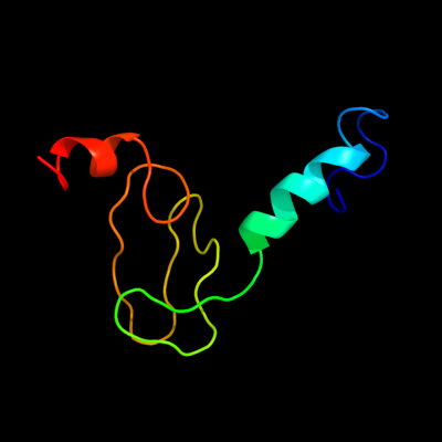
| Fold library id | PDB Header | Molecule | Title |
|---|---|---|---|
| c3jyw9_ | PDB header: ribosome | Chain: 9: PDB Molecule: 60s ribosomal protein l43; | PDBTitle: structure of the 60s proteins for eukaryotic ribosome based on cryo-em2 map of thermomyces lanuginosus ribosome at 8.9a resolution |
| Added to library: Sat Aug 28 20:53:08 2010 | |
| Links to external resources | |
|---|---|
| 1 | . | . | . | . | . | . | . | . | 10 | . | . | . | . | . | . | . | . | . | 20 | . | . | . | . | . | . | . | . | . | 30 | . | . | . | . | . | . | . | . | . | 40 | . | . | . | . | . | . | . | . | . | 50 | . | . | . | . | . | . | . | . | . | 60 | . | . | . | . | . | . | . | . | . | 70 | |
| Sequence | T | G | K | Y | G | V | R | Y | G | S | S | L | R | R | Q | V | K | K | L | E | I | Q | Q | H | A | R | Y | D | C | S | F | C | G | K | K | T | V | K | R | G | A | A | G | I | W | T | C | S | C | C | K | K | T | V | A | G | G | A | Y | T | V | S | T | A | A | A | A | T | V | R |
| Predicted secondary structure |  | | | | | | | | | | | | | | |  | | | | | | | | | | |  | | | | | | | | | | | | ||||||||||||||||||||||||||||||||
| SS confidence | ||||||||||||||||||||||||||||||||||||||||||||||||||||||||||||||||||||||
| Known secondary structure (DSSP) | T | T | T | T | T | S | S | | | | | T | T | T | | | | | | | | | S | B | S | S | S | S | B | S | B | S | S | S | B | S | S | S | S | S | S | S | S | | | | | | | T | . | . | ||||||||||||||||||
| Sequence | S | T | ||||||||||||||||||||||||||||||||||||||||||||||||||||||||||||||||||||
| Predicted secondary structure | ||||||||||||||||||||||||||||||||||||||||||||||||||||||||||||||||||||||
| SS confidence | ||||||||||||||||||||||||||||||||||||||||||||||||||||||||||||||||||||||
| Known secondary structure (DSSP) | T |
| Download: | PDB structure | FASTA sequence |
Phyre is now FREE for commercial users! All images and data generated by Phyre2 are free to use in any publication with acknowledgement Accessibility Statement
| ||||||||||||||||||||||||