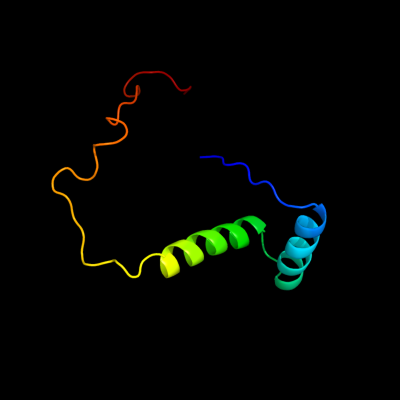
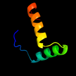
| 1 | c2k5jB_
|
|
 |
100.0 |
100 |
PDB header:structural genomics, unknown function
Chain: B: PDB Molecule:uncharacterized protein yiif;
PDBTitle: solution structure of protein yiif from shigella flexneri2 serotype 5b (strain 8401) . northeast structural genomics3 consortium target sft1
|
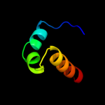
| 2 | c2bj3D_
|
|
 |
98.1 |
11 |
PDB header:transcription
Chain: D: PDB Molecule:nickel responsive regulator;
PDBTitle: nikr-apo
|
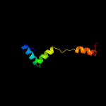
| 3 | d2bj7a1
|
|
 |
98.1 |
12 |
Fold:Ribbon-helix-helix
Superfamily:Ribbon-helix-helix
Family:CopG-like |
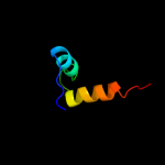
| 4 | c1q5vB_
|
|
 |
98.0 |
21 |
PDB header:transcription
Chain: B: PDB Molecule:nickel responsive regulator;
PDBTitle: apo-nikr
|
| 5 | c2ca9B_
|
|
 |
98.0 |
18 |
PDB header:transcriptional regulation
Chain: B: PDB Molecule:putative nickel-responsive regulator;
PDBTitle: apo-nikr from helicobacter pylori in closed trans-2 conformation
|
| 6 | d2hzaa1
|
|
 |
97.3 |
23 |
Fold:Ribbon-helix-helix
Superfamily:Ribbon-helix-helix
Family:CopG-like |
| 7 | d2hzab1
|
|
 |
97.1 |
23 |
Fold:Ribbon-helix-helix
Superfamily:Ribbon-helix-helix
Family:CopG-like |
| 8 | d2cpga_
|
|
 |
95.0 |
23 |
Fold:Ribbon-helix-helix
Superfamily:Ribbon-helix-helix
Family:CopG-like |
| 9 | c2rbfB_
|
|
 |
94.4 |
13 |
PDB header:oxidoreductase/dna
Chain: B: PDB Molecule:bifunctional protein puta;
PDBTitle: structure of the ribbon-helix-helix domain of escherichia coli puta2 (puta52) complexed with operator dna (o2)
|
| 10 | c2k9iB_
|
|
 |
93.5 |
24 |
PDB header:dna binding protein
Chain: B: PDB Molecule:plasmid prn1, complete sequence;
PDBTitle: nmr structure of plasmid copy control protein orf56 from2 sulfolobus islandicus
|
| 11 | c3h87D_
|
|
 |
91.5 |
20 |
PDB header:toxin/antitoxin
Chain: D: PDB Molecule:putative uncharacterized protein;
PDBTitle: rv0301 rv0300 toxin antitoxin complex from mycobacterium tuberculosis
|
| 12 | c2an7A_
|
|
 |
88.7 |
19 |
PDB header:dna binding protein
Chain: A: PDB Molecule:protein pard;
PDBTitle: solution structure of the bacterial antidote pard
|
| 13 | c2kelB_
|
|
 |
86.3 |
19 |
PDB header:transcription repressor
Chain: B: PDB Molecule:uncharacterized protein 56b;
PDBTitle: structure of the transcription regulator svtr from the2 hyperthermophilic archaeal virus sirv1
|
| 14 | d2ay0a1
|
|
 |
79.0 |
13 |
Fold:Ribbon-helix-helix
Superfamily:Ribbon-helix-helix
Family:PutA pre-N-terminal region-like |
| 15 | d1p94a_
|
|
 |
78.0 |
15 |
Fold:Ribbon-helix-helix
Superfamily:Ribbon-helix-helix
Family:CopG-like |
| 16 | c2k29A_
|
|
 |
76.5 |
18 |
PDB header:transcription
Chain: A: PDB Molecule:antitoxin relb;
PDBTitle: structure of the dbd domain of e. coli antitoxin relb
|
| 17 | c3kxeD_
|
|
 |
66.5 |
11 |
PDB header:protein binding
Chain: D: PDB Molecule:antitoxin protein pard-1;
PDBTitle: a conserved mode of protein recognition and binding in a2 pard-pare toxin-antitoxin complex
|
| 18 | d2bsqe1
|
|
 |
64.5 |
14 |
Fold:Ribbon-helix-helix
Superfamily:Ribbon-helix-helix
Family:Trafficking protein A-like |
| 19 | d1mnta_
|
|
 |
48.7 |
17 |
Fold:Ribbon-helix-helix
Superfamily:Ribbon-helix-helix
Family:Arc/Mnt-like phage repressors |
| 20 | c2q2kA_
|
|
 |
32.9 |
26 |
PDB header:dna binding protein/dna
Chain: A: PDB Molecule:hypothetical protein;
PDBTitle: structure of nucleic-acid binding protein
|
| 21 | c1kcfB_ |
|
not modelled |
28.1 |
33 |
PDB header:hydrolase
Chain: B: PDB Molecule:hypothetical 30.2 kd protein c25g10.02 in
PDBTitle: crystal structure of the yeast mitochondrial holliday2 junction resolvase, ydc2
|
| 22 | d1y9ba1 |
|
not modelled |
24.8 |
12 |
Fold:Ribbon-helix-helix
Superfamily:Ribbon-helix-helix
Family:VCA0319-like |
| 23 | c3c5yD_ |
|
not modelled |
21.3 |
9 |
PDB header:isomerase
Chain: D: PDB Molecule:ribose/galactose isomerase;
PDBTitle: crystal structure of a putative ribose 5-phosphate isomerase2 (saro_3514) from novosphingobium aromaticivorans dsm at 1.81 a3 resolution
|
| 24 | c2qhoF_ |
|
not modelled |
19.9 |
28 |
PDB header:protein binding/ligase
Chain: F: PDB Molecule:e3 ubiquitin-protein ligase edd1;
PDBTitle: crystal structure of the uba domain from edd ubiquitin2 ligase in complex with ubiquitin
|
| 25 | c2hwyB_ |
|
not modelled |
18.6 |
27 |
PDB header:rna binding protein
Chain: B: PDB Molecule:protein smg5;
PDBTitle: structure of pin domain of human smg5.
|
| 26 | c2kkeA_ |
|
not modelled |
18.3 |
17 |
PDB header:structural genomics, unknown function
Chain: A: PDB Molecule:uncharacterized protein;
PDBTitle: solution nmr structure of a dimeric protein of unknown2 function from methanobacterium thermoautotrophicum,3 northeast structural genomics consortium target tr5
|
| 27 | c2ppwA_ |
|
not modelled |
15.6 |
8 |
PDB header:isomerase
Chain: A: PDB Molecule:conserved domain protein;
PDBTitle: the crystal structure of uncharacterized ribose 5-phosphate isomerase2 rpib from streptococcus pneumoniae
|
| 28 | c1ny9A_ |
|
not modelled |
14.8 |
13 |
PDB header:transcription
Chain: A: PDB Molecule:transcriptional activator tipa-s;
PDBTitle: antibiotic binding domain of a tipa-class multidrug2 resistance transcriptional regulator
|
| 29 | d1ny9a_ |
|
not modelled |
14.8 |
13 |
Fold:Antibiotic binding domain of TipA-like multidrug resistance regulators
Superfamily:Antibiotic binding domain of TipA-like multidrug resistance regulators
Family:Antibiotic binding domain of TipA-like multidrug resistance regulators |
| 30 | c1yxeA_ |
|
not modelled |
14.2 |
10 |
PDB header:immune system
Chain: A: PDB Molecule:apical membrane antigen 1;
PDBTitle: structure and inter-domain interactions of domain ii from the blood2 stage malarial protein, apical membrane antigen 1
|
| 31 | c1u9pA_ |
|
not modelled |
12.8 |
10 |
PDB header:unknown function
Chain: A: PDB Molecule:parc;
PDBTitle: permuted single-chain arc
|
| 32 | c2xv9A_ |
|
not modelled |
11.3 |
17 |
PDB header:lipid binding protein
Chain: A: PDB Molecule:aba-1a1 repeat unit;
PDBTitle: the solution structure of aba-1a saturated with oleic acid
|
| 33 | d2proc1 |
|
not modelled |
10.7 |
42 |
Fold:Alpha-lytic protease prodomain-like
Superfamily:Alpha-lytic protease prodomain
Family:Alpha-lytic protease prodomain |
| 34 | c3iwfA_ |
|
not modelled |
10.7 |
12 |
PDB header:transcription regulator
Chain: A: PDB Molecule:transcription regulator rpir family;
PDBTitle: the crystal structure of the n-terminal domain of a rpir2 transcriptional regulator from staphylococcus epidermidis to 1.4a
|
| 35 | c3onoA_ |
|
not modelled |
10.3 |
5 |
PDB header:isomerase
Chain: A: PDB Molecule:ribose/galactose isomerase;
PDBTitle: crystal structure of ribose-5-phosphate isomerase lacab_rpib from2 vibrio parahaemolyticus
|
| 36 | d2o3fa1 |
|
not modelled |
9.9 |
18 |
Fold:DNA/RNA-binding 3-helical bundle
Superfamily:Homeodomain-like
Family:RpiR-like |
| 37 | c2o3fC_ |
|
not modelled |
9.9 |
18 |
PDB header:transcription
Chain: C: PDB Molecule:putative hth-type transcriptional regulator ybbh;
PDBTitle: structural genomics, the crystal structure of the n-2 terminal domain of the putative transcriptional regulator3 ybbh from bacillus subtilis subsp. subtilis str. 168.
|
| 38 | c3oeoD_ |
|
not modelled |
9.8 |
12 |
PDB header:signaling protein
Chain: D: PDB Molecule:spheroplast protein y;
PDBTitle: the crystal structure e. coli spy
|
| 39 | c3kk4B_ |
|
not modelled |
9.5 |
10 |
PDB header:structural genomics, unknown function
Chain: B: PDB Molecule:uncharacterized protein bp1543;
PDBTitle: uncharacterized protein bp1543 from bordetella pertussis tohama i
|
| 40 | d2je6b2 |
|
not modelled |
9.1 |
25 |
Fold:Ribonuclease PH domain 2-like
Superfamily:Ribonuclease PH domain 2-like
Family:Ribonuclease PH domain 2-like |
| 41 | c3o39A_ |
|
not modelled |
9.0 |
12 |
PDB header:chaperone
Chain: A: PDB Molecule:periplasmic protein related to spheroblast formation;
PDBTitle: crystal structure of spy
|
| 42 | d1uxca_ |
|
not modelled |
8.9 |
18 |
Fold:lambda repressor-like DNA-binding domains
Superfamily:lambda repressor-like DNA-binding domains
Family:GalR/LacI-like bacterial regulator |
| 43 | d2phcb1 |
|
not modelled |
8.8 |
12 |
Fold:Cyclophilin-like
Superfamily:Cyclophilin-like
Family:PH0987 C-terminal domain-like |
| 44 | d2qklb1 |
|
not modelled |
8.5 |
28 |
Fold:Dcp2 domain-like
Superfamily:Dcp2 domain-like
Family:Dcp2 box A domain |
| 45 | d1znda1 |
|
not modelled |
8.4 |
23 |
Fold:Lipocalins
Superfamily:Lipocalins
Family:Retinol binding protein-like |
| 46 | d1to0a_ |
|
not modelled |
8.3 |
10 |
Fold:alpha/beta knot
Superfamily:alpha/beta knot
Family:YbeA-like |
| 47 | d1gm6a_ |
|
not modelled |
8.0 |
13 |
Fold:Lipocalins
Superfamily:Lipocalins
Family:Retinol binding protein-like |
| 48 | d1l5aa2 |
|
not modelled |
7.5 |
13 |
Fold:CoA-dependent acyltransferases
Superfamily:CoA-dependent acyltransferases
Family:NRPS condensation domain (amide synthase) |
| 49 | c2l8nA_ |
|
not modelled |
7.5 |
18 |
PDB header:transcription regulator
Chain: A: PDB Molecule:transcriptional repressor cytr;
PDBTitle: nmr structure of the cytidine repressor dna binding domain in presence2 of operator half-site dna
|
| 50 | d2c2vv1 |
|
not modelled |
7.3 |
17 |
Fold:RING/U-box
Superfamily:RING/U-box
Family:U-box |
| 51 | d1q9ja2 |
|
not modelled |
7.2 |
16 |
Fold:CoA-dependent acyltransferases
Superfamily:CoA-dependent acyltransferases
Family:NRPS condensation domain (amide synthase) |
| 52 | c2lcvA_ |
|
not modelled |
7.0 |
18 |
PDB header:transcription regulator
Chain: A: PDB Molecule:hth-type transcriptional repressor cytr;
PDBTitle: structure of the cytidine repressor dna-binding domain; an alternate2 calculation
|
| 53 | c2zp2B_ |
|
not modelled |
6.9 |
12 |
PDB header:transferase inhibitor
Chain: B: PDB Molecule:kinase a inhibitor;
PDBTitle: c-terminal domain of kipi from bacillus subtilis
|
| 54 | c2e4jA_ |
|
not modelled |
6.9 |
16 |
PDB header:isomerase
Chain: A: PDB Molecule:prostaglandin-h2 d-isomerase;
PDBTitle: solution structure of mouse lipocalin-type prostaglandin d2 synthase
|
| 55 | c3epvB_ |
|
not modelled |
6.9 |
13 |
PDB header:metal binding protein
Chain: B: PDB Molecule:nickel and cobalt resistance protein cnrr;
PDBTitle: x-ray structure of the metal-sensor cnrx in both the apo- and copper-2 bound forms
|
| 56 | d1cjya2 |
|
not modelled |
6.9 |
23 |
Fold:FabD/lysophospholipase-like
Superfamily:FabD/lysophospholipase-like
Family:Lysophospholipase |
| 57 | c2wteB_ |
|
not modelled |
6.8 |
26 |
PDB header:antiviral protein
Chain: B: PDB Molecule:csa3;
PDBTitle: the structure of the crispr-associated protein, csa3, from2 sulfolobus solfataricus at 1.8 angstrom resolution.
|
| 58 | c3qzcA_ |
|
not modelled |
6.8 |
14 |
PDB header:signaling protein
Chain: A: PDB Molecule:periplasmic protein cpxp;
PDBTitle: structure of the periplasmic stress response protein cpxp
|
| 59 | c4a1qB_ |
|
not modelled |
6.4 |
26 |
PDB header:viral protein
Chain: B: PDB Molecule:orf e73;
PDBTitle: solution structure of e73 protein from sulfolobus spindle-2 shaped virus ragged hills, a hyperthermophilic3 crenarchaeal virus from yellowstone national park
|
| 60 | c2qkmF_ |
|
not modelled |
6.4 |
23 |
PDB header:hydrolase
Chain: F: PDB Molecule:spac19a8.12 protein;
PDBTitle: the crystal structure of fission yeast mrna decapping enzyme dcp1-dcp22 complex
|
| 61 | d2obpa1 |
|
not modelled |
5.9 |
29 |
Fold:DNA/RNA-binding 3-helical bundle
Superfamily:"Winged helix" DNA-binding domain
Family:ReutB4095-like |
| 62 | d1y0ya2 |
|
not modelled |
5.9 |
12 |
Fold:Phosphorylase/hydrolase-like
Superfamily:Zn-dependent exopeptidases
Family:Bacterial dinuclear zinc exopeptidases |
| 63 | c2kp6A_ |
|
not modelled |
5.8 |
8 |
PDB header:structural genomics, unknown function
Chain: A: PDB Molecule:uncharacterized protein;
PDBTitle: solution nmr structure of protein cv0237 from2 chromobacterium violaceum. northeast structural genomics3 consortium (nesg) target cvt1
|
| 64 | d1ns5a_ |
|
not modelled |
5.8 |
17 |
Fold:alpha/beta knot
Superfamily:alpha/beta knot
Family:YbeA-like |
| 65 | d1lcda_ |
|
not modelled |
5.8 |
18 |
Fold:lambda repressor-like DNA-binding domains
Superfamily:lambda repressor-like DNA-binding domains
Family:GalR/LacI-like bacterial regulator |
| 66 | d1uxda_ |
|
not modelled |
5.7 |
18 |
Fold:lambda repressor-like DNA-binding domains
Superfamily:lambda repressor-like DNA-binding domains
Family:GalR/LacI-like bacterial regulator |
| 67 | d2hsga1 |
|
not modelled |
5.7 |
18 |
Fold:lambda repressor-like DNA-binding domains
Superfamily:lambda repressor-like DNA-binding domains
Family:GalR/LacI-like bacterial regulator |
| 68 | d1qpza1 |
|
not modelled |
5.7 |
12 |
Fold:lambda repressor-like DNA-binding domains
Superfamily:lambda repressor-like DNA-binding domains
Family:GalR/LacI-like bacterial regulator |
| 69 | d2nn6d2 |
|
not modelled |
5.5 |
13 |
Fold:Ribonuclease PH domain 2-like
Superfamily:Ribonuclease PH domain 2-like
Family:Ribonuclease PH domain 2-like |
| 70 | c2xstA_ |
|
not modelled |
5.5 |
23 |
PDB header:transport protein
Chain: A: PDB Molecule:lipocalin 15;
PDBTitle: crystal structure of the human lipocalin 15
|
| 71 | c3by0B_ |
|
not modelled |
5.4 |
16 |
PDB header:ligand binding protein
Chain: B: PDB Molecule:neutrophil gelatinase-associated lipocalin;
PDBTitle: crystal structure of siderocalin (ngal, lipocalin 2) w79a-r81a2 complexed with ferric enterobactin
|
| 72 | c3p3vB_ |
|
not modelled |
5.2 |
10 |
PDB header:transferase
Chain: B: PDB Molecule:pts system, n-acetylgalactosamine-specific iib component;
PDBTitle: crystal structure of a pts dependent n-acetyl-galactosamine-iib2 component (agav, spy_0631) from streptococcus pyogenes at 1.65 a3 resolution
|
| 73 | d1t1ha_ |
|
not modelled |
5.2 |
8 |
Fold:RING/U-box
Superfamily:RING/U-box
Family:U-box |
| 74 | c3i8oA_ |
|
not modelled |
5.1 |
10 |
PDB header:rna binding protein
Chain: A: PDB Molecule:kh domain-containing protein mj1533;
PDBTitle: a domain of a functionally unknown protein from2 methanocaldococcus jannaschii dsm 2661.
|
| 75 | d1cuka1 |
|
not modelled |
5.1 |
18 |
Fold:RuvA C-terminal domain-like
Superfamily:DNA helicase RuvA subunit, C-terminal domain
Family:DNA helicase RuvA subunit, C-terminal domain |