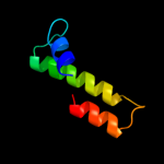
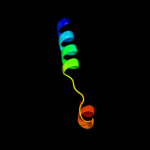
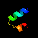
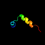
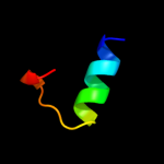
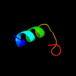
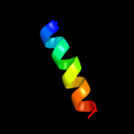
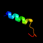
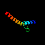
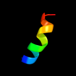
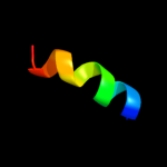
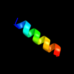
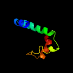
1 d2gsva1
52.5
22
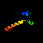
Fold: Open three-helical up-and-down bundleSuperfamily: YvfG-likeFamily: YvfG-like2 c3kheB_
21.0
19
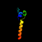
PDB header: metal binding proteinChain: B: PDB Molecule: calmodulin-like domain protein kinase isoform 3;PDBTitle: crystal structure of the calcium-loaded calmodulin-like domain of the2 cdpk, 541.m00134 from toxoplasma gondii
3 c2fvhB_
20.8
14
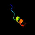
PDB header: hydrolaseChain: B: PDB Molecule: urease gamma subunit;PDBTitle: crystal structure of rv1848, a urease gamma subunit urea (urea2 amidohydrolase), from mycobacterium tuberculosis
4 d1e7la2
20.2
27
Fold: His-Me finger endonucleasesSuperfamily: His-Me finger endonucleasesFamily: Recombination endonuclease VII, N-terminal domain5 d4ubpa_
19.6
11
Fold: Urease, gamma-subunitSuperfamily: Urease, gamma-subunitFamily: Urease, gamma-subunit6 d1ejxa_
19.0
21
Fold: Urease, gamma-subunitSuperfamily: Urease, gamma-subunitFamily: Urease, gamma-subunit7 c1ztaA_
16.2
41
PDB header: dna-binding motifChain: A: PDB Molecule: leucine zipper monomer;PDBTitle: the solution structure of a leucine-zipper motif peptide
8 c2f8mB_
15.8
18
PDB header: isomeraseChain: B: PDB Molecule: ribose 5-phosphate isomerase;PDBTitle: ribose 5-phosphate isomerase from plasmodium falciparum
9 c1zdbA_
15.8
28
PDB header: igg binding domainChain: A: PDB Molecule: mini protein a domain, z38;PDBTitle: phage-selected mini protein a domain, z38, nmr, minimized2 mean structure
10 d1r8ja1
14.1
20
Fold: KaiA/RbsU domainSuperfamily: KaiA/RbsU domainFamily: Circadian clock protein KaiA, C-terminal domain11 d1sv1a_
13.7
20
Fold: KaiA/RbsU domainSuperfamily: KaiA/RbsU domainFamily: Circadian clock protein KaiA, C-terminal domain12 c3l19B_
13.1
29
PDB header: transferaseChain: B: PDB Molecule: calcium/calmodulin dependent protein kinase with a kinasePDBTitle: crystal structure of calcium binding domain of cpcdpk3, cgd5_820
13 c2vz4A_
12.4
8
PDB header: transcriptionChain: A: PDB Molecule: hth-type transcriptional activator tipa;PDBTitle: the n-terminal domain of merr-like protein tipal bound to2 promoter dna
14 c1y2iC_
12.0
18
PDB header: structural genomics, unknown functionChain: C: PDB Molecule: hypothetical protein s0862;PDBTitle: crystal structure of mcsg target apc27401 from shigella2 flexneri
15 d1y2ia_
12.0
18
Fold: Dodecin subunit-likeSuperfamily: YbjQ-likeFamily: YbjQ-like16 d1oeyj_
11.9
33
Fold: beta-Grasp (ubiquitin-like)Superfamily: CAD & PB1 domainsFamily: PB1 domain17 c1nvpB_
11.8
21
PDB header: transcription/dnaChain: B: PDB Molecule: transcription initiation factor iia alpha chain;PDBTitle: human tfiia/tbp/dna complex
18 d1nvpb_
11.8
21
Fold: Transcription factor IIA (TFIIA), alpha-helical domainSuperfamily: Transcription factor IIA (TFIIA), alpha-helical domainFamily: Transcription factor IIA (TFIIA), alpha-helical domain19 c1ce0B_
11.0
29
PDB header: hiv-1 envelope proteinChain: B: PDB Molecule: protein (leucine zipper model h38-p1);PDBTitle: trimerization specificity in hiv-1 gp41: analysis with a2 gcn4 leucine zipper model
20 c3mejA_
10.8
18
PDB header: transcriptional regulatorChain: A: PDB Molecule: transcriptional regulator ywtf;PDBTitle: crystal structure of putative transcriptional regulator ywtf from2 bacillus subtilis, northeast structural genomics consortium target3 sr736
21 c2oqqB_
not modelled
10.7
50
PDB header: transcriptionChain: B: PDB Molecule: transcription factor hy5;PDBTitle: crystal structure of hy5 leucine zipper homodimer from2 arabidopsis thaliana
22 d2gr7a1
not modelled
10.6
23
Fold: Pili subunitsSuperfamily: Pili subunitsFamily: YadA C-terminal domain-like23 c2gr7C_
not modelled
10.6
23
PDB header: membrane proteinChain: C: PDB Molecule: adhesin;PDBTitle: hia 992-1098
24 c3hh0C_
not modelled
10.5
23
PDB header: transcription regulatorChain: C: PDB Molecule: transcriptional regulator, merr family;PDBTitle: crystal strucure of a transcriptional regulator, merr family2 from bacillus cereus
25 d1v8ga1
not modelled
10.5
19
Fold: Methionine synthase domain-likeSuperfamily: Nucleoside phosphorylase/phosphoribosyltransferase N-terminal domainFamily: Nucleoside phosphorylase/phosphoribosyltransferase N-terminal domain26 d1nh2b_
not modelled
10.2
14
Fold: Transcription factor IIA (TFIIA), alpha-helical domainSuperfamily: Transcription factor IIA (TFIIA), alpha-helical domainFamily: Transcription factor IIA (TFIIA), alpha-helical domain27 c2jd3B_
not modelled
10.1
14
PDB header: dna binding proteinChain: B: PDB Molecule: stbb protein;PDBTitle: parr from plasmid pb171
28 d1edla_
not modelled
10.0
33
Fold: immunoglobulin/albumin-binding domain-likeSuperfamily: Bacterial immunoglobulin/albumin-binding domainsFamily: Immunoglobulin-binding protein A modules29 c3kltB_
not modelled
9.9
13
PDB header: structural proteinChain: B: PDB Molecule: vimentin;PDBTitle: crystal structure of a vimentin fragment
30 d1u2ca2
not modelled
8.9
17
Fold: Dystroglycan, domain 2Superfamily: Dystroglycan, domain 2Family: Dystroglycan, domain 231 c2bcxB_
not modelled
8.6
63
PDB header: calcium binding proteinChain: B: PDB Molecule: ryanodine receptor 1;PDBTitle: crystal structure of calmodulin in complex with a ryanodine2 receptor peptide
32 c2o7hF_
not modelled
8.5
35
PDB header: transcriptionChain: F: PDB Molecule: general control protein gcn4;PDBTitle: crystal structure of trimeric coiled coil gcn4 leucine zipper
33 c1ij2C_
not modelled
8.4
35
PDB header: transcriptionChain: C: PDB Molecule: general control protein gcn4;PDBTitle: gcn4-pvtl coiled-coil trimer with threonine at the a(16)2 position
34 c3k7zB_
not modelled
8.4
35
PDB header: dna binding proteinChain: B: PDB Molecule: general control protein gcn4;PDBTitle: gcn4-leucine zipper core mutant as n16a trigonal automatic2 solution
35 c3k7zA_
not modelled
8.4
35
PDB header: dna binding proteinChain: A: PDB Molecule: general control protein gcn4;PDBTitle: gcn4-leucine zipper core mutant as n16a trigonal automatic2 solution
36 c1rb6C_
not modelled
8.4
35
PDB header: dna binding proteinChain: C: PDB Molecule: general control protein gcn4;PDBTitle: antiparallel trimer of gcn4-leucine zipper core mutant as2 n16a tetragonal form
37 c1swiA_
not modelled
8.4
35
PDB header: leucine zipperChain: A: PDB Molecule: gcn4p1;PDBTitle: gcn4-leucine zipper core mutant as n16a complexed with2 benzene
38 c1rb1A_
not modelled
8.4
35
PDB header: dna binding proteinChain: A: PDB Molecule: general control protein gcn4;PDBTitle: gcn4-leucine zipper core mutant as n16a trigonal automatic2 solution
39 c1rb1B_
not modelled
8.4
35
PDB header: dna binding proteinChain: B: PDB Molecule: general control protein gcn4;PDBTitle: gcn4-leucine zipper core mutant as n16a trigonal automatic2 solution
40 c1ij3B_
not modelled
8.4
35
PDB header: transcriptionChain: B: PDB Molecule: general control protein gcn4;PDBTitle: gcn4-pvsl coiled-coil trimer with serine at the a(16)2 position
41 c1ij3C_
not modelled
8.4
35
PDB header: transcriptionChain: C: PDB Molecule: general control protein gcn4;PDBTitle: gcn4-pvsl coiled-coil trimer with serine at the a(16)2 position
42 c3cvfA_
not modelled
8.2
32
PDB header: signaling proteinChain: A: PDB Molecule: homer protein homolog 3;PDBTitle: crystal structure of the carboxy terminus of homer3
43 c3qfiA_
not modelled
8.0
13
PDB header: transcription regulatorChain: A: PDB Molecule: transcriptional regulator;PDBTitle: x-ray crystal structure of transcriptional regulator (ef0465) from2 enterococcus faecalis, northeast structural genomics consortium3 target efr190
44 d1fc2c_
not modelled
7.6
39
Fold: immunoglobulin/albumin-binding domain-likeSuperfamily: Bacterial immunoglobulin/albumin-binding domainsFamily: Immunoglobulin-binding protein A modules45 c2pxgA_
not modelled
7.5
13
PDB header: membrane proteinChain: A: PDB Molecule: outer membrane protein;PDBTitle: nmr solution structure of omla
46 c1ij2B_
not modelled
7.4
35
PDB header: transcriptionChain: B: PDB Molecule: general control protein gcn4;PDBTitle: gcn4-pvtl coiled-coil trimer with threonine at the a(16)2 position
47 d1vr4a1
not modelled
7.3
20
Fold: Dodecin subunit-likeSuperfamily: YbjQ-likeFamily: YbjQ-like48 c1uixA_
not modelled
7.3
37
PDB header: transferaseChain: A: PDB Molecule: rho-associated kinase;PDBTitle: coiled-coil structure of the rhoa-binding domain in rho-2 kinase
49 c1xtzA_
not modelled
7.2
9
PDB header: isomeraseChain: A: PDB Molecule: ribose-5-phosphate isomerase;PDBTitle: crystal structure of the s. cerevisiae d-ribose-5-phosphate isomerase:2 comparison with the archeal and bacterial enzymes
50 d1lp1a_
not modelled
7.1
22
Fold: immunoglobulin/albumin-binding domain-likeSuperfamily: Bacterial immunoglobulin/albumin-binding domainsFamily: Immunoglobulin-binding protein A modules51 c2a45J_
not modelled
7.1
20
PDB header: hydrolase/hydrolase inhibitorChain: J: PDB Molecule: fibrinogen alpha chain;PDBTitle: crystal structure of the complex between thrombin and the central "e"2 region of fibrin
52 c1b6aA_
not modelled
6.9
25
PDB header: angiogenesis inhibitorChain: A: PDB Molecule: methionine aminopeptidase;PDBTitle: human methionine aminopeptidase 2 complexed with tnp-470
53 c1e9zA_
not modelled
6.8
11
PDB header: hydrolaseChain: A: PDB Molecule: urease subunit alpha;PDBTitle: crystal structure of helicobacter pylori urease
54 d1e9ya2
not modelled
6.8
11
Fold: Urease, gamma-subunitSuperfamily: Urease, gamma-subunitFamily: Urease, gamma-subunit55 c3okzB_
not modelled
6.7
21
PDB header: structural genomics, unknown functionChain: B: PDB Molecule: putative uncharacterized protein gbs0355;PDBTitle: crystal structure of protein gbs0355 from streptococcus agalactiae,2 northeast structural genomics consortium target sar127
56 d2etda1
not modelled
6.6
27
Fold: Bromodomain-likeSuperfamily: LemA-likeFamily: LemA-like57 d1deeg_
not modelled
6.3
33
Fold: immunoglobulin/albumin-binding domain-likeSuperfamily: Bacterial immunoglobulin/albumin-binding domainsFamily: Immunoglobulin-binding protein A modules58 c2juiA_
not modelled
6.0
46
PDB header: toxinChain: A: PDB Molecule: plne;PDBTitle: three-dimensional structure of the two peptides that2 constitute the two-peptide bacteriocin plantaracin ef
59 d1jfib_
not modelled
5.9
24
Fold: Histone-foldSuperfamily: Histone-foldFamily: TBP-associated factors, TAFs60 c1jfiB_
not modelled
5.9
24
PDB header: transcription/dnaChain: B: PDB Molecule: transcription regulator nc2 beta chain;PDBTitle: crystal structure of the nc2-tbp-dna ternary complex
61 d2jwda1
not modelled
5.9
39
Fold: immunoglobulin/albumin-binding domain-likeSuperfamily: Bacterial immunoglobulin/albumin-binding domainsFamily: Immunoglobulin-binding protein A modules62 d1lp1b_
not modelled
5.5
33
Fold: immunoglobulin/albumin-binding domain-likeSuperfamily: Bacterial immunoglobulin/albumin-binding domainsFamily: Immunoglobulin-binding protein A modules63 c1m25A_
not modelled
5.5
100
PDB header: unknown functionChain: A: PDB Molecule: major prion protein;PDBTitle: structure of synthetic 26-mer peptide containing 145-1692 sheep prion protein segment and c-terminal cysteine in tfe3 solution
64 c1r8jB_
not modelled
5.5
20
PDB header: circadian clock proteinChain: B: PDB Molecule: kaia;PDBTitle: crystal structure of circadian clock protein kaia from2 synechococcus elongatus
65 c3cveC_
not modelled
5.4
18
PDB header: signaling proteinChain: C: PDB Molecule: homer protein homolog 1;PDBTitle: crystal structure of the carboxy terminus of homer1
66 c3qgaD_
not modelled
5.4
16
PDB header: hydrolaseChain: D: PDB Molecule: fusion of urease beta and gamma subunits;PDBTitle: 3.0 a model of iron containing urease urea2b2 from helicobacter2 mustelae
67 d2a3qa1
not modelled
5.2
20
Fold: all-alpha NTP pyrophosphatasesSuperfamily: all-alpha NTP pyrophosphatasesFamily: MazG-like68 c2q4pA_
not modelled
5.2
20
PDB header: structural genomics, unknown functionChain: A: PDB Molecule: protein rs21-c6;PDBTitle: ensemble refinement of the crystal structure of protein from mus2 musculus mm.29898
69 d1v2za_
not modelled
5.2
20
Fold: KaiA/RbsU domainSuperfamily: KaiA/RbsU domainFamily: Circadian clock protein KaiA, C-terminal domain70 c3jzaB_
not modelled
5.2
37
PDB header: transport proteinChain: B: PDB Molecule: uncharacterized protein drra;PDBTitle: crystal structure of human rab1b in complex with the gef domain of2 drra/sidm from legionella pneumophila
71 c1vtpA_
not modelled
5.0
40
PDB header: targeting peptideChain: A: PDB Molecule: vacuolar targeting peptide;PDBTitle: vacuolar targeting peptide from na-propi


