Phyre Home Retrieve Phyre Job Id
Emaill.a.kelley@imperial.ac.uk
DescriptionP76506
DateThu Jan 5 12:23:43 GMT 2012
Unique Job IDcb51189c03d9ed96

Summary 

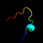
Top model
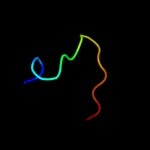
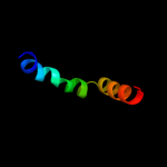
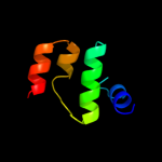
Image coloured by rainbow N → C terminus
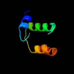
Model (left) based on template c3kuzA_
Top template information
PDB header:signaling protein
Chain: A: PDB Molecule:plexin-c1;
PDBTitle: crystal structure of the ubiquitin like domain of plxnc1
Confidence and coverage
Confidence: 38.4% Coverage: 6%
15 residues ( 6% of your sequence) have been modelled with 38.4% confidence by the single highest scoring template.
You may wish to submit your sequence to Phyrealarm. This will automatically scan your sequence every week for new potential templates as they appear in the Phyre2 library.
Please note: You must be registered and logged in to use Phyrealarm.
3D viewing
Interactive 3D view in Jmol

Sequence analysis 

Secondary structure and disorder prediction 

   1........10.........20.........30.........40.........50.........60
Sequence  MKLRLSALALGTTLLVGCASSGTDQQGRSDPLEGFNRTMYNFNFNVLDPYIVRPVAVAWR
Secondary structure 


SS confidence 


Disorder  ???


???????????


Disorder confidence 


   .........70.........80.........90.........100.........110.........120
Sequence  DYVPQPARNGLSNFTGNLEEPAVMVNYFLQGDPYQGMVHFTRFFLNTILGMGGFIDVAGM
Secondary structure 


SS confidence 


Disorder 


Disorder confidence 


   .........130.........140.........150.........160.........170.........180
Sequence  ANPKLQRTEPHRFGSTLGHYGVGYGPYVQLPFYGSFTLRDDGGDMADGFYPVLSWLTWPM
Secondary structure 


SS confidence 


Disorder  ???????????


Disorder confidence 


   .........190.........200.........210.........220.........230.........240
Sequence  SVGKWTLEGIETRAQLLDSDGLLRQSSDPYIMVREAYFQRHDFIANGGELKPQENPNAQA
Secondary structure 


SS confidence 


Disorder 


??


????????????????
Disorder confidence 


   .........250.
Sequence  IQDDLKDIDSE
Secondary structure 


SS confidence 


Disorder  ??


??????
Disorder confidence 


Confidence Key
High(9)                    Low (0)
?Disordered
Alpha helix
Beta strand

Domain analysis 

Hover over an aligned region to see model and summary info

Please note, only up to the top 20 hits are modelled to reduce computer load

RankAligned region

PDB 3kuz chain A

3D model

Region: 136 - 150
Aligned: 15
Modelled: 15
Confidence: 38.4%
Identity: 33%
PDB header:signaling protein
Chain: A: PDB Molecule:plexin-c1;
PDBTitle: crystal structure of the ubiquitin like domain of plxnc1

Phyre2

PDB 2jph chain A

3D model

Region: 136 - 150
Aligned: 15
Modelled: 15
Confidence: 38.0%
Identity: 53%
PDB header:signaling protein, protein binding
Chain: A: PDB Molecule:plexin-b1;
PDBTitle: nmr solution structure of the rho gtpase binding domain of2 human plexin-b1

Phyre2

PDB 1wjn chain A

3D model

Region: 135 - 157
Aligned: 23
Modelled: 23
Confidence: 34.3%
Identity: 22%
Fold: beta-Grasp (ubiquitin-like)
Superfamily: Ubiquitin-like
Family: Ubiquitin-related

Phyre2

PDB 3h6n chain A

3D model

Region: 136 - 150
Aligned: 15
Modelled: 14
Confidence: 34.2%
Identity: 33%
PDB header:signaling protein
Chain: A: PDB Molecule:plexin-d1;
PDBTitle: crystal structure of the ubiquitin-like domain of plexin d1

Phyre2

PDB 3q3j chain A

3D model

Region: 136 - 150
Aligned: 15
Modelled: 15
Confidence: 33.3%
Identity: 47%
PDB header:membrane protein/protein binding
Chain: A: PDB Molecule:plexin-a2;
PDBTitle: crystal structure of plexin a2 rbd in complex with rnd1

Phyre2

PDB 1z2m chain A domain 1

3D model

Region: 134 - 150
Aligned: 17
Modelled: 17
Confidence: 21.7%
Identity: 35%
Fold: beta-Grasp (ubiquitin-like)
Superfamily: Ubiquitin-like
Family: Ubiquitin-related

Phyre2

PDB 2odg chain C domain 1

3D model

Region: 129 - 146
Aligned: 18
Modelled: 18
Confidence: 17.2%
Identity: 22%
Fold: LEM/SAP HeH motif
Superfamily: LEM domain
Family: LEM domain

Phyre2

PDB 1we7 chain A

3D model

Region: 134 - 157
Aligned: 24
Modelled: 24
Confidence: 16.2%
Identity: 21%
Fold: beta-Grasp (ubiquitin-like)
Superfamily: Ubiquitin-like
Family: Ubiquitin-related

Phyre2

PDB 2wx3 chain A

3D model

Region: 187 - 220
Aligned: 34
Modelled: 34
Confidence: 14.2%
Identity: 29%
PDB header:structural protein
Chain: A: PDB Molecule:mrna-decapping enzyme 1a;
PDBTitle: asymmetric trimer of the human dcp1a c-terminal domain

Phyre2

PDB 1we6 chain A

3D model

Region: 134 - 157
Aligned: 24
Modelled: 24
Confidence: 14.1%
Identity: 33%
Fold: beta-Grasp (ubiquitin-like)
Superfamily: Ubiquitin-like
Family: Ubiquitin-related

Phyre2

PDB 1wzi chain A

3D model

Region: 138 - 154
Aligned: 17
Modelled: 17
Confidence: 13.6%
Identity: 29%
PDB header:oxidoreductase
Chain: A: PDB Molecule:malate dehydrogenase;
PDBTitle: structural basis for alteration of cofactor specificity of2 malate dehydrogenase from thermus flavus

Phyre2

PDB 3su8 chain X

3D model

Region: 136 - 150
Aligned: 15
Modelled: 15
Confidence: 13.3%
Identity: 53%
PDB header:apoptosis/signaling protein
Chain: X: PDB Molecule:plexin-b1;
PDBTitle: crystal structure of a truncated intracellular domain of plexin-b1 in2 complex with rac1

Phyre2

PDB 157l chain A

3D model

Region: 49 - 102
Aligned: 54
Modelled: 54
Confidence: 12.9%
Identity: 17%
Fold: Lysozyme-like
Superfamily: Lysozyme-like
Family: Phage lysozyme

Phyre2

PDB 2aga chain A

3D model

Region: 82 - 98
Aligned: 17
Modelled: 17
Confidence: 12.8%
Identity: 24%
PDB header:transcription
Chain: A: PDB Molecule:machado-joseph disease protein 1;
PDBTitle: de-ubiquitinating function of ataxin-3: insights from the2 solution structure of the josephin domain

Phyre2

PDB 1ogw chain A

3D model

Region: 134 - 154
Aligned: 21
Modelled: 21
Confidence: 12.1%
Identity: 29%
Fold: beta-Grasp (ubiquitin-like)
Superfamily: Ubiquitin-like
Family: Ubiquitin-related

Phyre2

PDB 3ig3 chain A

3D model

Region: 136 - 151
Aligned: 16
Modelled: 16
Confidence: 10.1%
Identity: 44%
PDB header:signaling protein, membrane protein
Chain: A: PDB Molecule:plxna3 protein;
PDBTitle: crystal strucure of mouse plexin a3 intracellular domain

Phyre2

PDB 1zkh chain A domain 1

3D model

Region: 134 - 150
Aligned: 17
Modelled: 17
Confidence: 9.7%
Identity: 24%
Fold: beta-Grasp (ubiquitin-like)
Superfamily: Ubiquitin-like
Family: Ubiquitin-related

Phyre2

PDB 1p37 chain A

3D model

Region: 49 - 102
Aligned: 54
Modelled: 54
Confidence: 9.7%
Identity: 20%
Fold: Lysozyme-like
Superfamily: Lysozyme-like
Family: Phage lysozyme

Phyre2

PDB 1y7t chain A domain 2

3D model

Region: 138 - 153
Aligned: 16
Modelled: 16
Confidence: 9.4%
Identity: 31%
Fold: LDH C-terminal domain-like
Superfamily: LDH C-terminal domain-like
Family: Lactate & malate dehydrogenases, C-terminal domain

Phyre2

PDB 1i0z chain A domain 2

3D model

Region: 138 - 153
Aligned: 16
Modelled: 16
Confidence: 9.4%
Identity: 31%
Fold: LDH C-terminal domain-like
Superfamily: LDH C-terminal domain-like
Family: Lactate & malate dehydrogenases, C-terminal domain

Phyre2
1

c3kuzA_
2

c2jphA_
3

d1wjna_
4

c3h6nA_
5

c3q3jA_
6

d1z2ma1
7

d2odgc1
8

d1we7a_
9

c2wx3A_
10

d1we6a_
11

c1wziA_
12

c3su8X_
13

d157la_
14

c2agaA_
15

d1ogwa_
16

c3ig3A_
17

d1zkha1
18

d1p37a_
19

d1y7ta2
20

d1i0za2
21


22


23


24


25


26


27


28


29


30


31


32


33


34


35


36


37


38


39


40


41


42


43


44


45


46


47


48


49


50


51


52


53


54


Detailed template information 

#
Template Alignment Coverage3D Model Confidence
% i.d. Template Information
1c3kuzA_


38.4 33 PDB header:signaling protein
Chain: A: PDB Molecule:plexin-c1;
PDBTitle: crystal structure of the ubiquitin like domain of plxnc1
2c2jphA_


38.0 53 PDB header:signaling protein, protein binding
Chain: A: PDB Molecule:plexin-b1;
PDBTitle: nmr solution structure of the rho gtpase binding domain of2 human plexin-b1
3d1wjna_


34.3 22 Fold:beta-Grasp (ubiquitin-like)
Superfamily:Ubiquitin-like
Family:Ubiquitin-related
4c3h6nA_


34.2 33 PDB header:signaling protein
Chain: A: PDB Molecule:plexin-d1;
PDBTitle: crystal structure of the ubiquitin-like domain of plexin d1
5c3q3jA_


33.3 47 PDB header:membrane protein/protein binding
Chain: A: PDB Molecule:plexin-a2;
PDBTitle: crystal structure of plexin a2 rbd in complex with rnd1
6d1z2ma1


21.7 35 Fold:beta-Grasp (ubiquitin-like)
Superfamily:Ubiquitin-like
Family:Ubiquitin-related
7d2odgc1


17.2 22 Fold:LEM/SAP HeH motif
Superfamily:LEM domain
Family:LEM domain
8d1we7a_


16.2 21 Fold:beta-Grasp (ubiquitin-like)
Superfamily:Ubiquitin-like
Family:Ubiquitin-related
9c2wx3A_


14.2 29 PDB header:structural protein
Chain: A: PDB Molecule:mrna-decapping enzyme 1a;
PDBTitle: asymmetric trimer of the human dcp1a c-terminal domain
10d1we6a_


14.1 33 Fold:beta-Grasp (ubiquitin-like)
Superfamily:Ubiquitin-like
Family:Ubiquitin-related
11c1wziA_


13.6 29 PDB header:oxidoreductase
Chain: A: PDB Molecule:malate dehydrogenase;
PDBTitle: structural basis for alteration of cofactor specificity of2 malate dehydrogenase from thermus flavus
12c3su8X_


13.3 53 PDB header:apoptosis/signaling protein
Chain: X: PDB Molecule:plexin-b1;
PDBTitle: crystal structure of a truncated intracellular domain of plexin-b1 in2 complex with rac1
13d157la_


12.9 17 Fold:Lysozyme-like
Superfamily:Lysozyme-like
Family:Phage lysozyme
14c2agaA_


12.8 24 PDB header:transcription
Chain: A: PDB Molecule:machado-joseph disease protein 1;
PDBTitle: de-ubiquitinating function of ataxin-3: insights from the2 solution structure of the josephin domain
15d1ogwa_


12.1 29 Fold:beta-Grasp (ubiquitin-like)
Superfamily:Ubiquitin-like
Family:Ubiquitin-related
16c3ig3A_


10.1 44 PDB header:signaling protein, membrane protein
Chain: A: PDB Molecule:plxna3 protein;
PDBTitle: crystal strucure of mouse plexin a3 intracellular domain
17d1zkha1


9.7 24 Fold:beta-Grasp (ubiquitin-like)
Superfamily:Ubiquitin-like
Family:Ubiquitin-related
18d1p37a_


9.7 20 Fold:Lysozyme-like
Superfamily:Lysozyme-like
Family:Phage lysozyme
19d1y7ta2


9.4 31 Fold:LDH C-terminal domain-like
Superfamily:LDH C-terminal domain-like
Family:Lactate & malate dehydrogenases, C-terminal domain
20d1i0za2


9.4 31 Fold:LDH C-terminal domain-like
Superfamily:LDH C-terminal domain-like
Family:Lactate & malate dehydrogenases, C-terminal domain
21c3m62B_


not modelled 9.2 25 PDB header:ligase/protein binding
Chain: B: PDB Molecule:uv excision repair protein rad23;
PDBTitle: crystal structure of ufd2 in complex with the ubiquitin-like (ubl)2 domain of rad23
22d2f2qa1


not modelled 9.1 19 Fold:Lysozyme-like
Superfamily:Lysozyme-like
Family:Phage lysozyme
23c3hm6X_


not modelled 9.0 50 PDB header:signaling protein
Chain: X: PDB Molecule:plexin-b1;
PDBTitle: crystal structure of the cytoplasmic domain of human plexin b1
24d1v5oa_


not modelled 8.3 44 Fold:beta-Grasp (ubiquitin-like)
Superfamily:Ubiquitin-like
Family:Ubiquitin-related
25d1j8ca_


not modelled 8.3 35 Fold:beta-Grasp (ubiquitin-like)
Superfamily:Ubiquitin-like
Family:Ubiquitin-related
26d1b8pa2


not modelled 8.0 31 Fold:LDH C-terminal domain-like
Superfamily:LDH C-terminal domain-like
Family:Lactate & malate dehydrogenases, C-terminal domain
27c3jrnA_


not modelled 7.6 27 PDB header:plant protein
Chain: A: PDB Molecule:at1g72930 protein;
PDBTitle: crystal structure of tir domain from arabidopsis thaliana
28d1oqya4


not modelled 7.3 13 Fold:beta-Grasp (ubiquitin-like)
Superfamily:Ubiquitin-like
Family:Ubiquitin-related
29d9ldta2


not modelled 7.0 25 Fold:LDH C-terminal domain-like
Superfamily:LDH C-terminal domain-like
Family:Lactate & malate dehydrogenases, C-terminal domain
30d2c5lc1


not modelled 6.8 13 Fold:beta-Grasp (ubiquitin-like)
Superfamily:Ubiquitin-like
Family:Ras-binding domain, RBD
31c2o4wA_


not modelled 6.7 20 PDB header:hydrolase
Chain: A: PDB Molecule:lysozyme;
PDBTitle: t4 lysozyme circular permutant
32d1wxva1


not modelled 6.7 31 Fold:beta-Grasp (ubiquitin-like)
Superfamily:Ubiquitin-like
Family:Ubiquitin-related
33d1v5ta_


not modelled 6.7 12 Fold:beta-Grasp (ubiquitin-like)
Superfamily:Ubiquitin-like
Family:Ubiquitin-related
34d1icha_


not modelled 6.4 29 Fold:DEATH domain
Superfamily:DEATH domain
Family:DEATH domain, DD
35c1ichA_


not modelled 6.4 29 PDB header:apoptosis
Chain: A: PDB Molecule:tumor necrosis factor receptor-1;
PDBTitle: solution structure of the tumor necrosis factor receptor-12 death domain
36c3pbpL_


not modelled 6.1 25 PDB header:transport protein,structural protein
Chain: L: PDB Molecule:nucleoporin nup159;
PDBTitle: structure of the yeast heterotrimeric nup82-nup159-nup116 nucleoporin2 complex
37d1z2ma2


not modelled 6.1 29 Fold:beta-Grasp (ubiquitin-like)
Superfamily:Ubiquitin-like
Family:Ubiquitin-related
38d1sifa_


not modelled 6.1 29 Fold:beta-Grasp (ubiquitin-like)
Superfamily:Ubiquitin-like
Family:Ubiquitin-related
39d2ihoa2


not modelled 6.0 50 Fold:Cysteine proteinases
Superfamily:Cysteine proteinases
Family:MOA C-terminal domain-like
40d1uela_


not modelled 6.0 16 Fold:beta-Grasp (ubiquitin-like)
Superfamily:Ubiquitin-like
Family:Ubiquitin-related
41c2kj6A_


not modelled 5.9 19 PDB header:chaperone
Chain: A: PDB Molecule:tubulin folding cofactor b;
PDBTitle: nmr solution structure of a tubulin folding cofactor b2 obtained from arabidopsis thaliana: northeast structural3 genomics consortium target ar3436a
42c2k25A_


not modelled 5.9 29 PDB header:unknown function
Chain: A: PDB Molecule:ubb;
PDBTitle: automated nmr structure of the ubb by fapsy
43d1l64a_


not modelled 5.7 16 Fold:Lysozyme-like
Superfamily:Lysozyme-like
Family:Phage lysozyme
44c2l7rA_


not modelled 5.7 29 PDB header:protein binding
Chain: A: PDB Molecule:ubiquitin-like protein fubi;
PDBTitle: solution nmr structure of n-terminal ubiquitin-like domain of fubi, a2 ribosomal protein s30 precursor from homo sapiens. northeast3 structural genomics consortium (nesg) target hr6166
45d1ldma2


not modelled 5.7 25 Fold:LDH C-terminal domain-like
Superfamily:LDH C-terminal domain-like
Family:Lactate & malate dehydrogenases, C-terminal domain
46d2bh1a1


not modelled 5.6 25 Fold:Ribonuclease H-like motif
Superfamily:Actin-like ATPase domain
Family:Cyto-EpsL domain
47c2kanA_


not modelled 5.6 13 PDB header:structural genomics, unknown function
Chain: A: PDB Molecule:uncharacterized protein ar3433a;
PDBTitle: solution nmr structure of ubiquitin-like domain of2 arabidopsis thaliana protein at2g32350. northeast3 structural genomics consortium target ar3433a
48d1wx8a1


not modelled 5.6 24 Fold:beta-Grasp (ubiquitin-like)
Superfamily:Ubiquitin-like
Family:Ubiquitin-related
49d2ldxa2


not modelled 5.4 19 Fold:LDH C-terminal domain-like
Superfamily:LDH C-terminal domain-like
Family:Lactate & malate dehydrogenases, C-terminal domain
50d1jtma_


not modelled 5.4 18 Fold:Lysozyme-like
Superfamily:Lysozyme-like
Family:Phage lysozyme
51c1yx5B_


not modelled 5.3 29 PDB header:hydrolase
Chain: B: PDB Molecule:ubiquitin;
PDBTitle: solution structure of s5a uim-1/ubiquitin complex
52d1qbaa2


not modelled 5.3 12 Fold:Common fold of diphtheria toxin/transcription factors/cytochrome f
Superfamily:Carbohydrate-binding domain
Family:Bacterial chitobiase, n-terminal domain
53d2g7ja1


not modelled 5.1 30 Fold:Secretion chaperone-like
Superfamily:YgaC/TfoX-N like
Family:YgaC-like
54d1bt0a_


not modelled 5.1 24 Fold:beta-Grasp (ubiquitin-like)
Superfamily:Ubiquitin-like
Family:Ubiquitin-related

Binding site prediction 

Due to computational demand, binding site predictions are not run for batch jobs

If you want to predict binding sites, please manually submit your model of choice to 3DLigandSite


Phyre is for academic use only

Please cite: Protein structure prediction on the web: a case study using the Phyre server
Kelley LA and Sternberg MJE. Nature Protocols 4, 363 - 371 (2009) [pdf] [Import into BibTeX]
 
If you use the binding site predictions from 3DLigandSite, please also cite:
3DLigandSite: predicting ligand-binding sites using similar structures.
Wass MN, Kelley LA and Sternberg MJ Nucleic Acids Research 38, W469-73 (2010) [PubMed]
 
© Structural Bioinformatics Group
Imperial College London
Lawrence Kelley, Benjamin Jefferys
Disclaimer
Terms and Conditions
Component software
Template detection: HHpred 1.51
Secondary structure prediction: Psi-pred 2.5
Disorder prediction: Disopred 2.4
Transmembrane prediction: Memsat_SVM
Multi-template modelling and ab initio: Poing 1.0