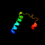
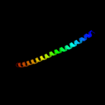
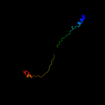
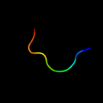
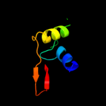
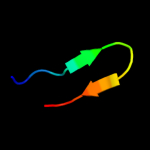
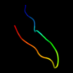
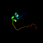
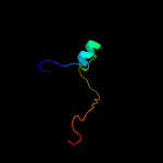
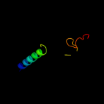
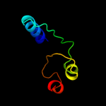
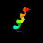
1 d1jmxa1
55.4
12
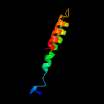
Fold: Cytochrome cSuperfamily: Cytochrome cFamily: Quinohemoprotein amine dehydrogenase A chain, domains 1 and 22 c1eboE_
51.9
13
PDB header: viral proteinChain: E: PDB Molecule: ebola virus envelope protein chimera consistingPDBTitle: crystal structure of the ebola virus membrane-fusion2 subunit, gp2, from the envelope glycoprotein ectodomain
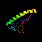
3 d1pbya1
51.6
24
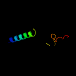
Fold: Cytochrome cSuperfamily: Cytochrome cFamily: Quinohemoprotein amine dehydrogenase A chain, domains 1 and 24 c3ifqD_
50.0
28
PDB header: cell adhesionChain: D: PDB Molecule: e-cadherin;PDBTitle: interction of plakoglobin and beta-catenin with desmosomal2 cadherins
5 d2q09a1
43.3
89
Fold: Composite domain of metallo-dependent hydrolasesSuperfamily: Composite domain of metallo-dependent hydrolasesFamily: Imidazolonepropionase-like6 c3k5bE_
42.0
19
PDB header: hydrolaseChain: E: PDB Molecule: v-type atp synthase subunit e;PDBTitle: crystal structure of the peripheral stalk of thermus thermophilus h+-2 atpase/synthase
7 c2jo1A_
41.8
28
PDB header: hydrolase regulatorChain: A: PDB Molecule: phospholemman;PDBTitle: structure of the na,k-atpase regulatory protein fxyd1 in2 micelles
8 d2isba1
31.2
18
Fold: The "swivelling" beta/beta/alpha domainSuperfamily: FumA C-terminal domain-likeFamily: FumA C-terminal domain-like9 c3mk7F_
31.0
17
PDB header: oxidoreductaseChain: F: PDB Molecule: cytochrome c oxidase, cbb3-type, subunit p;PDBTitle: the structure of cbb3 cytochrome oxidase
10 d1y0na_
28.2
30
Fold: YehU-likeSuperfamily: YehU-likeFamily: YehU-like11 c3c6eC_
24.2
33
PDB header: viral proteinChain: C: PDB Molecule: prm;PDBTitle: crystal structure of the precursor membrane protein- envelope protein2 heterodimer from the dengue 2 virus at neutral ph
12 c3c6rE_
24.1
33
PDB header: virusChain: E: PDB Molecule: peptide pr;PDBTitle: low ph immature dengue virus
13 d1ny722
23.1
16
Fold: Nucleoplasmin-like/VP (viral coat and capsid proteins)Superfamily: Positive stranded ssRNA virusesFamily: Comoviridae-like VP14 d1pgl22
23.1
24
Fold: Nucleoplasmin-like/VP (viral coat and capsid proteins)Superfamily: Positive stranded ssRNA virusesFamily: Comoviridae-like VP15 c3rfrI_
22.7
24
PDB header: oxidoreductaseChain: I: PDB Molecule: pmob;PDBTitle: crystal structure of particulate methane monooxygenase (pmmo) from2 methylocystis sp. strain m
16 c3i24B_
21.7
19
PDB header: hydrolaseChain: B: PDB Molecule: hit family hydrolase;PDBTitle: crystal structure of a hit family hydrolase protein from2 vibrio fischeri. northeast structural genomics consortium3 target id vfr176
17 d1v54k_
21.6
17
Fold: Single transmembrane helixSuperfamily: Mitochondrial cytochrome c oxidase subunit VIIbFamily: Mitochondrial cytochrome c oxidase subunit VIIb18 c2y69X_
21.6
17
PDB header: electron transportChain: X: PDB Molecule: cytochrome c oxidase polypeptide 7b;PDBTitle: bovine heart cytochrome c oxidase re-refined with molecular2 oxygen
19 c3nohA_
20.2
26
PDB header: peptide binding proteinChain: A: PDB Molecule: putative peptide binding protein;PDBTitle: crystal structure of a putative peptide binding protein (rumgna_00914)2 from ruminococcus gnavus atcc 29149 at 1.60 a resolution
20 c2bpbB_
19.4
18
PDB header: oxidoreductaseChain: B: PDB Molecule: sulfite\:cytochrome c oxidoreductase subunit b;PDBTitle: sulfite dehydrogenase from starkeya novella
21 d1u61a_
not modelled
18.9
24
Fold: RNase III domain-likeSuperfamily: RNase III domain-likeFamily: RNase III catalytic domain-like22 c3heiF_
not modelled
18.6
25
PDB header: transferase/signaling proteinChain: F: PDB Molecule: ephrin-a1;PDBTitle: ligand recognition by a-class eph receptors: crystal structures of the2 epha2 ligand-binding domain and the epha2/ephrin-a1 complex
23 d1dgsa2
not modelled
18.5
17
Fold: OB-foldSuperfamily: Nucleic acid-binding proteinsFamily: DNA ligase/mRNA capping enzyme postcatalytic domain24 c3ikmD_
not modelled
17.6
26
PDB header: transferaseChain: D: PDB Molecule: dna polymerase subunit gamma-1;PDBTitle: crystal structure of human mitochondrial dna polymerase2 holoenzyme
25 c2q04C_
not modelled
17.2
19
PDB header: transferaseChain: C: PDB Molecule: acetoin utilization protein;PDBTitle: crystal structure of acetoin utilization protein (zp_00540088.1) from2 exiguobacterium sibiricum 255-15 at 2.33 a resolution
26 c2x11B_
not modelled
16.7
25
PDB header: receptor/signaling proteinChain: B: PDB Molecule: ephrin-a5;PDBTitle: crystal structure of the complete epha2 ectodomain in2 complex with ephrin a5 receptor binding domain
27 d1shxa1
not modelled
16.7
25
Fold: Cupredoxin-likeSuperfamily: CupredoxinsFamily: Ephrin ectodomain28 d2o6ka1
not modelled
16.7
19
Fold: SAM domain-likeSuperfamily: YozE-likeFamily: YozE-like29 c3czuB_
not modelled
16.6
25
PDB header: transferase/signaling proteinChain: B: PDB Molecule: ephrin-a1;PDBTitle: crystal structure of the human ephrin a2- ephrin a1 complex
30 c2wo3B_
not modelled
16.5
25
PDB header: transferase/signaling proteinChain: B: PDB Molecule: ephrin-a2;PDBTitle: crystal structure of the epha4-ephrina2 complex
31 d2j5wa3
not modelled
16.3
20
Fold: Cupredoxin-likeSuperfamily: CupredoxinsFamily: Multidomain cupredoxins32 d1ngka_
not modelled
15.9
26
Fold: Globin-likeSuperfamily: Globin-likeFamily: Truncated hemoglobin33 d1v33a_
not modelled
15.8
19
Fold: Prim-pol domainSuperfamily: Prim-pol domainFamily: PriA-like34 d2cqaa1
not modelled
15.7
28
Fold: OB-foldSuperfamily: Nucleic acid-binding proteinsFamily: TIP49 domain35 c3uowB_
not modelled
15.5
19
PDB header: ligaseChain: B: PDB Molecule: gmp synthetase;PDBTitle: crystal structure of pf10_0123, a gmp synthetase from plasmodium2 falciparum
36 d2ctda1
not modelled
14.6
14
Fold: beta-beta-alpha zinc fingersSuperfamily: beta-beta-alpha zinc fingersFamily: Classic zinc finger, C2H237 d1g71a_
not modelled
14.0
15
Fold: Prim-pol domainSuperfamily: Prim-pol domainFamily: PriA-like38 c3d12E_
not modelled
13.9
29
PDB header: hydrolase/membrane proteinChain: E: PDB Molecule: ephrin-b3;PDBTitle: crystal structures of nipah virus g attachment glycoprotein in complex2 with its receptor ephrin-b3
39 d1h1oa1
not modelled
13.9
12
Fold: Cytochrome cSuperfamily: Cytochrome cFamily: Two-domain cytochrome c40 c3p87H_
not modelled
13.8
60
PDB header: hydrolase/dna binding proteinChain: H: PDB Molecule: ribonuclease h2 subunit b;PDBTitle: structure of human pcna bound to rnaseh2b pip box peptide
41 c3p87I_
not modelled
13.8
60
PDB header: hydrolase/dna binding proteinChain: I: PDB Molecule: ribonuclease h2 subunit b;PDBTitle: structure of human pcna bound to rnaseh2b pip box peptide
42 c3p87K_
not modelled
13.8
60
PDB header: hydrolase/dna binding proteinChain: K: PDB Molecule: ribonuclease h2 subunit b;PDBTitle: structure of human pcna bound to rnaseh2b pip box peptide
43 c3p87G_
not modelled
13.4
60
PDB header: hydrolase/dna binding proteinChain: G: PDB Molecule: ribonuclease h2 subunit b;PDBTitle: structure of human pcna bound to rnaseh2b pip box peptide
44 c3p87J_
not modelled
13.4
60
PDB header: hydrolase/dna binding proteinChain: J: PDB Molecule: ribonuclease h2 subunit b;PDBTitle: structure of human pcna bound to rnaseh2b pip box peptide
45 c3p87L_
not modelled
13.4
60
PDB header: hydrolase/dna binding proteinChain: L: PDB Molecule: ribonuclease h2 subunit b;PDBTitle: structure of human pcna bound to rnaseh2b pip box peptide
46 d2nwua1
not modelled
13.3
12
Fold: RL5-likeSuperfamily: RL5-likeFamily: SSO1042-like47 d1zsoa1
not modelled
13.1
25
Fold: MAL13P1.257-likeSuperfamily: MAL13P1.257-likeFamily: MAL13P1.257-like48 c2p2vA_
not modelled
13.0
19
PDB header: transferaseChain: A: PDB Molecule: alpha-2,3-sialyltransferase;PDBTitle: crystal structure analysis of monofunctional alpha-2,3-2 sialyltransferase cst-i from campylobacter jejuni
49 c3ktbD_
not modelled
12.1
10
PDB header: transcription regulatorChain: D: PDB Molecule: arsenical resistance operon trans-acting repressor;PDBTitle: crystal structure of arsenical resistance operon trans-acting2 repressor from bacteroides vulgatus atcc 8482
50 c3pdsA_
not modelled
12.0
14
PDB header: membrane protein/hydrolaseChain: A: PDB Molecule: fusion protein beta-2 adrenergic receptor/lysozyme;PDBTitle: irreversible agonist-beta2 adrenoceptor complex
51 c3kgkA_
not modelled
12.0
29
PDB header: chaperoneChain: A: PDB Molecule: arsenical resistance operon trans-acting repressor arsd;PDBTitle: crystal structure of arsd
52 d1ikop_
not modelled
11.9
47
Fold: Cupredoxin-likeSuperfamily: CupredoxinsFamily: Ephrin ectodomain53 c1ikoP_
not modelled
11.9
47
PDB header: signaling proteinChain: P: PDB Molecule: ephrin-b2;PDBTitle: crystal structure of the murine ephrin-b2 ectodomain
54 c2f8mB_
not modelled
11.5
13
PDB header: isomeraseChain: B: PDB Molecule: ribose 5-phosphate isomerase;PDBTitle: ribose 5-phosphate isomerase from plasmodium falciparum
55 d1v38a_
not modelled
11.4
30
Fold: SAM domain-likeSuperfamily: SAM/Pointed domainFamily: SAM (sterile alpha motif) domain56 c2j5dA_
not modelled
11.4
40
PDB header: membrane proteinChain: A: PDB Molecule: bcl2/adenovirus e1b 19 kda protein-interactingPDBTitle: nmr structure of bnip3 transmembrane domain in lipid2 bicelles
57 d1o0la_
not modelled
11.4
17
Fold: Toxins' membrane translocation domainsSuperfamily: Bcl-2 inhibitors of programmed cell deathFamily: Bcl-2 inhibitors of programmed cell death58 c2w2hD_
not modelled
11.3
23
PDB header: rna-binding proteinChain: D: PDB Molecule: protein tat;PDBTitle: structural basis of transcription activation by the cyclin2 t1-tat-tar rna complex from eiav
59 c2fl8N_
not modelled
11.3
23
PDB header: virus/viral proteinChain: N: PDB Molecule: baseplate structural protein gp10;PDBTitle: fitting of the gp10 trimer structure into the cryoem map of the2 bacteriophage t4 baseplate in the hexagonal conformation.
60 c3lgoA_
not modelled
11.2
19
PDB header: protein bindingChain: A: PDB Molecule: protein slm4;PDBTitle: structure of gse1p, member of the gse/ego complex
61 c2lmaA_
not modelled
11.0
38
PDB header: immune systemChain: A: PDB Molecule: thp5 peptide;PDBTitle: solution structure of cd4+ t cell derived peptide thp5
62 c2jwaA_
not modelled
10.9
18
PDB header: transferaseChain: A: PDB Molecule: receptor tyrosine-protein kinase erbb-2;PDBTitle: erbb2 transmembrane segment dimer spatial structure
63 d1fcdc1
not modelled
10.7
5
Fold: Cytochrome cSuperfamily: Cytochrome cFamily: Two-domain cytochrome c64 d2fj6a1
not modelled
10.5
25
Fold: SAM domain-likeSuperfamily: YozE-likeFamily: YozE-like65 d1lira_
not modelled
10.5
30
Fold: Knottins (small inhibitors, toxins, lectins)Superfamily: Scorpion toxin-likeFamily: Short-chain scorpion toxins66 c3cp5A_
not modelled
10.4
20
PDB header: electron transportChain: A: PDB Molecule: cytochrome c;PDBTitle: cytochrome c from rhodothermus marinus
67 d2evra2
not modelled
10.4
60
Fold: Cysteine proteinasesSuperfamily: Cysteine proteinasesFamily: NlpC/P6068 c3oa8A_
not modelled
10.3
19
PDB header: heme-binding protein/heme-binding proteiChain: A: PDB Molecule: soxa;PDBTitle: diheme soxax
69 c2l4dA_
not modelled
10.3
38
PDB header: electron transportChain: A: PDB Molecule: sco1/senc family protein/cytochrome c;PDBTitle: cytochrome c domain of pp3183 protein from pseudomonas putida
70 d1mida_
not modelled
10.0
42
Fold: Bifunctional inhibitor/lipid-transfer protein/seed storage 2S albuminSuperfamily: Bifunctional inhibitor/lipid-transfer protein/seed storage 2S albuminFamily: Plant lipid-transfer and hydrophobic proteins71 c3ixxE_
not modelled
10.0
33
PDB header: virusChain: E: PDB Molecule: peptide pr;PDBTitle: the pseudo-atomic structure of west nile immature virus in2 complex with fab fragments of the anti-fusion loop antibody3 e53
72 d2bmta_
not modelled
9.8
45
Fold: Knottins (small inhibitors, toxins, lectins)Superfamily: Scorpion toxin-likeFamily: Short-chain scorpion toxins73 c1pbyA_
not modelled
9.8
21
PDB header: oxidoreductaseChain: A: PDB Molecule: quinohemoprotein amine dehydrogenase 60 kdaPDBTitle: structure of the phenylhydrazine adduct of the2 quinohemoprotein amine dehydrogenase from paracoccus3 denitrificans at 1.7 a resolution
74 c1sddA_
not modelled
9.6
11
PDB header: blood clottingChain: A: PDB Molecule: coagulation factor v;PDBTitle: crystal structure of bovine factor vai
75 c2vofA_
not modelled
9.6
20
PDB header: apoptosisChain: A: PDB Molecule: bcl-2-related protein a1;PDBTitle: structure of mouse a1 bound to the puma bh3-domain
76 c2ehwD_
not modelled
9.3
20
PDB header: structural genomics, unknown functionChain: D: PDB Molecule: hypothetical protein tthb059;PDBTitle: conserved hypothetical proteim (tthb059) from thermo thermophilus hb8
77 d1g3wa3
not modelled
9.3
32
Fold: SH3-like barrelSuperfamily: C-terminal domain of transcriptional repressorsFamily: FeoA-like78 d1m70a2
not modelled
9.0
22
Fold: Cytochrome cSuperfamily: Cytochrome cFamily: Two-domain cytochrome c79 d1y88a1
not modelled
8.9
43
Fold: SAM domain-likeSuperfamily: Rad51 N-terminal domain-likeFamily: Hypothetical protein AF1548, C-terminal domain80 d1kpfa_
not modelled
8.9
17
Fold: HIT-likeSuperfamily: HIT-likeFamily: HIT (HINT, histidine triad) family of protein kinase-interacting proteins81 d1wejf_
not modelled
8.7
14
Fold: Cytochrome cSuperfamily: Cytochrome cFamily: monodomain cytochrome c82 c3rqrA_
not modelled
8.7
24
PDB header: metal transportChain: A: PDB Molecule: ryanodine receptor 1;PDBTitle: crystal structure of the ryr domain of the rabbit ryanodine receptor
83 c2ka2A_
not modelled
8.5
40
PDB header: membrane proteinChain: A: PDB Molecule: bcl2/adenovirus e1b 19 kda protein-interactingPDBTitle: solution nmr structure of bnip3 transmembrane peptide dimer2 in detergent micelles with his173-ser172 intermonomer3 hydrogen bond restraints
84 c2ka1B_
not modelled
8.5
40
PDB header: membrane proteinChain: B: PDB Molecule: bcl2/adenovirus e1b 19 kda protein-interactingPDBTitle: solution nmr structure of bnip3 transmembrane peptide dimer2 in detergent micelles
85 d1hlca_
not modelled
8.4
28
Fold: Concanavalin A-like lectins/glucanasesSuperfamily: Concanavalin A-like lectins/glucanasesFamily: Galectin (animal S-lectin)86 c2zkqg_
not modelled
8.3
19
PDB header: ribosomal protein/rnaChain: G: PDB Molecule: rna helix;PDBTitle: structure of a mammalian ribosomal 40s subunit within an2 80s complex obtained by docking homology models of the rna3 and proteins into an 8.7 a cryo-em map
87 c1bmv2_
not modelled
8.1
24
PDB header: virus/rnaChain: 2: PDB Molecule: protein (icosahedral virus - b and c domain);PDBTitle: protein-rna interactions in an icosahedral virus at 3.02 angstroms resolution
88 c2fg0B_
not modelled
8.1
60
PDB header: hydrolaseChain: B: PDB Molecule: cog0791: cell wall-associated hydrolases (invasion-PDBTitle: crystal structure of a putative gamma-d-glutamyl-l-diamino acid2 endopeptidase (npun_r0659) from nostoc punctiforme pcc 73102 at 1.793 a resolution
89 c2axkA_
not modelled
8.1
36
PDB header: toxinChain: A: PDB Molecule: discrepin;PDBTitle: solution structure of discrepin, a scorpion venom toxin2 blocking k+ channels.
90 c3arcl_
not modelled
8.0
35
PDB header: electron transport, photosynthesisChain: L: PDB Molecule: photosystem ii reaction center protein l;PDBTitle: crystal structure of oxygen-evolving photosystem ii at 1.9 angstrom2 resolution
91 c2ka2B_
not modelled
8.0
42
PDB header: membrane proteinChain: B: PDB Molecule: bcl2/adenovirus e1b 19 kda protein-interactingPDBTitle: solution nmr structure of bnip3 transmembrane peptide dimer2 in detergent micelles with his173-ser172 intermonomer3 hydrogen bond restraints
92 c2ka1A_
not modelled
8.0
42
PDB header: membrane proteinChain: A: PDB Molecule: bcl2/adenovirus e1b 19 kda protein-interactingPDBTitle: solution nmr structure of bnip3 transmembrane peptide dimer2 in detergent micelles
93 c2p57A_
not modelled
8.0
32
PDB header: metal binding proteinChain: A: PDB Molecule: gtpase-activating protein znf289;PDBTitle: gap domain of znf289, an id1-regulated zinc finger protein
94 c2xivA_
not modelled
8.0
80
PDB header: structural proteinChain: A: PDB Molecule: hypothetical invasion protein;PDBTitle: structure of rv1477, hypothetical invasion protein of2 mycobacterium tuberculosis
95 c3o47A_
not modelled
7.9
18
PDB header: hydrolase, hydrolase activatorChain: A: PDB Molecule: adp-ribosylation factor gtpase-activating protein 1, adp-PDBTitle: crystal structure of arfgap1-arf1 fusion protein
96 c2k8iA_
not modelled
7.9
38
PDB header: isomeraseChain: A: PDB Molecule: peptidyl-prolyl cis-trans isomerase;PDBTitle: solution structure of e.coli slyd
97 c1oheA_
not modelled
7.9
20
PDB header: hydrolaseChain: A: PDB Molecule: cdc14b2 phosphatase;PDBTitle: structure of cdc14b phosphatase with a peptide ligand
98 c3gt2A_
not modelled
7.9
60
PDB header: unknown functionChain: A: PDB Molecule: putative uncharacterized protein;PDBTitle: crystal structure of the p60 domain from m. avium2 paratuberculosis antigen map1272c
99 d1xrda1
not modelled
7.8
23
Fold: Light-harvesting complex subunitsSuperfamily: Light-harvesting complex subunitsFamily: Light-harvesting complex subunits


