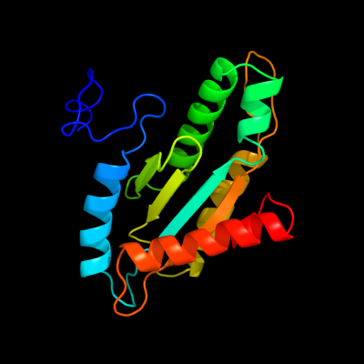
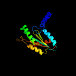
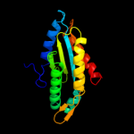
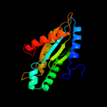
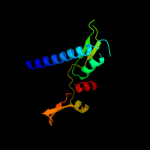
1 c1w25B_
100.0
31
PDB header: signaling proteinChain: B: PDB Molecule: stalked-cell differentiation controlling protein;PDBTitle: response regulator pled in complex with c-digmp
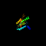
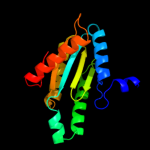
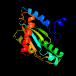
2 c3ezuA_
100.0
28
PDB header: signaling proteinChain: A: PDB Molecule: ggdef domain protein;PDBTitle: crystal structure of multidomain protein of unknown function with2 ggdef-domain (np_951600.1) from geobacter sulfurreducens at 1.95 a3 resolution
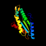
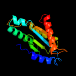
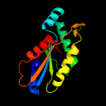
3 c3i5aA_
100.0
30
PDB header: signaling proteinChain: A: PDB Molecule: response regulator/ggdef domain protein;PDBTitle: crystal structure of full-length wpsr from pseudomonas syringae
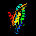
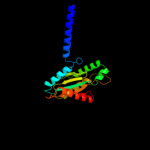
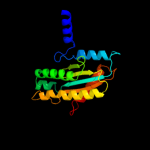
4 c3breA_
100.0
32
PDB header: signaling proteinChain: A: PDB Molecule: probable two-component response regulator;PDBTitle: crystal structure of p.aeruginosa pa3702
5 c3ignA_
100.0
26
PDB header: transferaseChain: A: PDB Molecule: diguanylate cyclase;PDBTitle: crystal structure of the ggdef domain from marinobacter2 aquaeolei diguanylate cyclase complexed with c-di-gmp -3 northeast structural genomics consortium target mqr89a
6 c3iclA_
100.0
30
PDB header: structural genomics, unknown functionChain: A: PDB Molecule: eal/ggdef domain protein;PDBTitle: x-ray structure of protein (eal/ggdef domain protein) from2 m.capsulatus, northeast structural genomics consortium3 target mcr174c
7 c3hvaA_
100.0
24
PDB header: transferaseChain: A: PDB Molecule: protein fimx;PDBTitle: crystal structure of fimx ggdef domain from pseudomonas2 aeruginosa
8 c3qyyB_
100.0
33
PDB header: signaling protein/inhibitorChain: B: PDB Molecule: response regulator;PDBTitle: a novel interaction mode between a microbial ggdef domain and the bis-2 (3, 5 )-cyclic di-gmp
9 d1w25a3
100.0
32
Fold: Ferredoxin-likeSuperfamily: Nucleotide cyclaseFamily: GGDEF domain10 c3i5cA_
100.0
27
PDB header: signaling proteinChain: A: PDB Molecule: fusion of general control protein gcn4 and wspr responsePDBTitle: crystal structure of a fusion protein containing the leucine zipper of2 gcn4 and the ggdef domain of wspr from pseudomonas aeruginosa
11 c3i5bA_
100.0
29
PDB header: signaling proteinChain: A: PDB Molecule: wspr response regulator;PDBTitle: crystal structure of the isolated ggdef domain of wpsr from2 pseudomonas aeruginosa
12 c3mtkA_
100.0
26
PDB header: transferaseChain: A: PDB Molecule: diguanylate cyclase/phosphodiesterase;PDBTitle: x-ray structure of diguanylate cyclase/phosphodiesterase from2 caldicellulosiruptor saccharolyticus, northeast structural genomics3 consortium target clr27c
13 c3hvwA_
100.0
16
PDB header: lyaseChain: A: PDB Molecule: diguanylate-cyclase (dgc);PDBTitle: crystal structure of the ggdef domain of the pa2567 protein2 from pseudomonas aeruginosa, northeast structural genomics3 consortium target par365c
14 c3pjwA_
100.0
22
PDB header: lyaseChain: A: PDB Molecule: cyclic dimeric gmp binding protein;PDBTitle: structure of pseudomonas fluorescence lapd ggdef-eal dual domain, i23
15 c3gfzB_
99.7
11
PDB header: hydrolase, signaling proteinChain: B: PDB Molecule: klebsiella pneumoniae blrp1;PDBTitle: klebsiella pneumoniae blrp1 ph 6 manganese/cy-digmp complex
16 c3p7nB_
98.4
16
PDB header: dna binding proteinChain: B: PDB Molecule: sensor histidine kinase;PDBTitle: crystal structure of light activated transcription factor el222 from2 erythrobacter litoralis
17 c2qv6D_
97.7
19
PDB header: hydrolaseChain: D: PDB Molecule: gtp cyclohydrolase iii;PDBTitle: gtp cyclohydrolase iii from m. jannaschii (mj0145)2 complexed with gtp and metal ions
18 c3lnrA_
95.9
13
PDB header: signaling proteinChain: A: PDB Molecule: aerotaxis transducer aer2;PDBTitle: crystal structure of poly-hamp domains from the p. aeruginosa soluble2 receptor aer2
19 c3uvjC_
95.7
17
PDB header: lyaseChain: C: PDB Molecule: guanylate cyclase soluble subunit alpha-3;PDBTitle: crystal structure of the catalytic domain of the heterodimeric human2 soluble guanylate cyclase 1.
20 c1cjkA_
95.6
13
PDB header: lyase/lyase/signaling proteinChain: A: PDB Molecule: adenylate cyclase, type v;PDBTitle: complex of gs-alpha with the catalytic domains of mammalian adenylyl2 cyclase: complex with adenosine 5'-(alpha thio)-triphosphate (rp),3 mg, and mn
21 d1azsa_
not modelled
95.3
13
Fold: Ferredoxin-likeSuperfamily: Nucleotide cyclaseFamily: Adenylyl and guanylyl cyclase catalytic domain22 d1fx2a_
not modelled
95.3
18
Fold: Ferredoxin-likeSuperfamily: Nucleotide cyclaseFamily: Adenylyl and guanylyl cyclase catalytic domain23 c3mr7B_
not modelled
95.1
10
PDB header: hydrolaseChain: B: PDB Molecule: adenylate/guanylate cyclase/hydrolase, alpha/beta foldPDBTitle: crystal structure of adenylate/guanylate cyclase/hydrolase from2 silicibacter pomeroyi
24 c3pjvD_
94.7
13
PDB header: lyaseChain: D: PDB Molecule: cyclic dimeric gmp binding protein;PDBTitle: structure of pseudomonas fluorescence lapd periplasmic domain
25 d1wc1a_
not modelled
94.7
13
Fold: Ferredoxin-likeSuperfamily: Nucleotide cyclaseFamily: Adenylyl and guanylyl cyclase catalytic domain26 d2asxa1
94.6
12
Fold: HAMP domain-likeSuperfamily: HAMP domain-likeFamily: HAMP domain27 c1wc6B_
not modelled
94.2
13
PDB header: lyaseChain: B: PDB Molecule: adenylate cyclase;PDBTitle: soluble adenylyl cyclase cyac from s. platensis in complex2 with rp-atpalphas in presence of bicarbonate
28 d1p0za_
93.9
13
Fold: Profilin-likeSuperfamily: Sensory domain-likeFamily: Sensory domain of two-component sensor kinase29 d3by8a1
93.3
13
Fold: Profilin-likeSuperfamily: Sensory domain-likeFamily: Sensory domain of two-component sensor kinase30 c1yk9A_
not modelled
91.8
16
PDB header: lyaseChain: A: PDB Molecule: adenylate cyclase;PDBTitle: crystal structure of a mutant form of the mycobacterial2 adenylyl cyclase rv1625c
31 c1ybuA_
not modelled
91.3
10
PDB header: hydrolaseChain: A: PDB Molecule: lipj;PDBTitle: mycobacterium tuberculosis adenylyl cyclase rv1900c chd, in complex2 with a substrate analog.
32 c1y10C_
not modelled
89.6
13
PDB header: lyaseChain: C: PDB Molecule: hypothetical protein rv1264/mt1302;PDBTitle: mycobacterial adenylyl cyclase rv1264, holoenzyme, inhibited state
33 c3et6A_
not modelled
89.4
9
PDB header: lyaseChain: A: PDB Molecule: soluble guanylyl cyclase beta;PDBTitle: the crystal structure of the catalytic domain of a eukaryotic2 guanylate cyclase
34 d1fx4a_
not modelled
88.7
14
Fold: Ferredoxin-likeSuperfamily: Nucleotide cyclaseFamily: Adenylyl and guanylyl cyclase catalytic domain35 c2aq4A_
not modelled
86.9
17
PDB header: transferaseChain: A: PDB Molecule: dna repair protein rev1;PDBTitle: ternary complex of the catalytic core of rev1 with dna and dctp.
36 c2rm8A_
not modelled
84.9
7
PDB header: signaling proteinChain: A: PDB Molecule: sensory rhodopsin ii transducer;PDBTitle: the solution structure of phototactic transducer protein2 htrii linker region from natronomonas pharaonis
37 c2w01C_
not modelled
83.9
15
PDB header: lyaseChain: C: PDB Molecule: adenylate cyclase;PDBTitle: crystal structure of the guanylyl cyclase cya2
38 c3b47A_
not modelled
79.6
12
PDB header: signaling proteinChain: A: PDB Molecule: methyl-accepting chemotaxis protein;PDBTitle: periplasmic sensor domain of chemotaxis protein gsu0582
39 c3zrwB_
not modelled
78.2
11
PDB header: signaling proteinChain: B: PDB Molecule: af1503 protein, osmolarity sensor protein envz;PDBTitle: the structure of the dimeric hamp-dhp fusion a291v mutant
40 c2wz1B_
not modelled
77.2
11
PDB header: lyaseChain: B: PDB Molecule: guanylate cyclase soluble subunit beta-1;PDBTitle: structure of the catalytic domain of human soluble2 guanylate cyclase 1 beta 3.
41 c3gqcB_
not modelled
74.9
16
PDB header: transferase/dnaChain: B: PDB Molecule: dna repair protein rev1;PDBTitle: structure of human rev1-dna-dntp ternary complex
42 c3b42B_
not modelled
65.5
12
PDB header: signaling proteinChain: B: PDB Molecule: methyl-accepting chemotaxis protein, putative;PDBTitle: periplasmic sensor domain of chemotaxis protein gsu0935
43 d1azsb_
not modelled
64.6
10
Fold: Ferredoxin-likeSuperfamily: Nucleotide cyclaseFamily: Adenylyl and guanylyl cyclase catalytic domain44 c1k1qA_
not modelled
61.7
16
PDB header: transcriptionChain: A: PDB Molecule: dbh protein;PDBTitle: crystal structure of a dinb family error prone dna2 polymerase from sulfolobus solfataricus
45 c1s97D_
not modelled
61.0
18
PDB header: transferase/dnaChain: D: PDB Molecule: dna polymerase iv;PDBTitle: dpo4 with gt mismatch
46 d1im4a_
not modelled
60.2
16
Fold: DNA/RNA polymerasesSuperfamily: DNA/RNA polymerasesFamily: Lesion bypass DNA polymerase (Y-family), catalytic domain47 d1k1sa2
not modelled
54.5
16
Fold: DNA/RNA polymerasesSuperfamily: DNA/RNA polymerasesFamily: Lesion bypass DNA polymerase (Y-family), catalytic domain48 d1yhta1
not modelled
45.7
22
Fold: TIM beta/alpha-barrelSuperfamily: (Trans)glycosidasesFamily: beta-N-acetylhexosaminidase catalytic domain49 d1jx4a2
not modelled
43.4
18
Fold: DNA/RNA polymerasesSuperfamily: DNA/RNA polymerasesFamily: Lesion bypass DNA polymerase (Y-family), catalytic domain50 d1vr6a1
not modelled
32.1
10
Fold: TIM beta/alpha-barrelSuperfamily: AldolaseFamily: Class I DAHP synthetase51 d1xxaa_
not modelled
26.9
18
Fold: DCoH-likeSuperfamily: C-terminal domain of arginine repressorFamily: C-terminal domain of arginine repressor52 c3l7xA_
not modelled
26.8
17
PDB header: cell cycleChain: A: PDB Molecule: putative hit-like protein involved in cell-cyclePDBTitle: the crystal structure of smu.412c from streptococcus mutans ua159
53 c3k30B_
not modelled
24.5
18
PDB header: oxidoreductaseChain: B: PDB Molecule: histamine dehydrogenase;PDBTitle: histamine dehydrogenase from nocardiodes simplex
54 c3hf3A_
not modelled
22.6
14
PDB header: oxidoreductaseChain: A: PDB Molecule: chromate reductase;PDBTitle: old yellow enzyme from thermus scotoductus sa-01
55 c3nuiA_
not modelled
21.6
12
PDB header: transferaseChain: A: PDB Molecule: pyruvate transaminase;PDBTitle: crystal structure of omega-transferase from vibrio fluvialis js17
56 c2kseA_
not modelled
20.2
17
PDB header: transferaseChain: A: PDB Molecule: sensor protein qsec;PDBTitle: backbone structure of the membrane domain of e. coli2 histidine kinase receptor qsec, center for structures of3 membrane proteins (csmp) target 4311c
57 c3i24B_
not modelled
19.3
14
PDB header: hydrolaseChain: B: PDB Molecule: hit family hydrolase;PDBTitle: crystal structure of a hit family hydrolase protein from2 vibrio fischeri. northeast structural genomics consortium3 target id vfr176
58 c3oheA_
not modelled
17.3
14
PDB header: hydrolaseChain: A: PDB Molecule: histidine triad (hit) protein;PDBTitle: crystal structure of a histidine triad protein (maqu_1709) from2 marinobacter aquaeolei vt8 at 1.20 a resolution
59 c2ylaA_
not modelled
16.2
14
PDB header: hydrolaseChain: A: PDB Molecule: beta-n-acetylhexosaminidase;PDBTitle: inhibition of the pneumococcal virulence factor strh and2 molecular insights into n-glycan recognition and3 hydrolysis
60 c3fosA_
not modelled
15.7
11
PDB header: transferaseChain: A: PDB Molecule: sensor protein;PDBTitle: crystal structure of two-component sensor histidine kinase domain from2 bacillus subtilis subsp. subtilis str. 168
61 d1szwa_
not modelled
15.3
25
Fold: Pseudouridine synthaseSuperfamily: Pseudouridine synthaseFamily: tRNA pseudouridine synthase TruD62 d1jiha2
not modelled
15.2
24
Fold: DNA/RNA polymerasesSuperfamily: DNA/RNA polymerasesFamily: Lesion bypass DNA polymerase (Y-family), catalytic domain63 c1qgeE_
not modelled
15.1
23
PDB header: hydrolaseChain: E: PDB Molecule: protein (triacylglycerol hydrolase);PDBTitle: new crystal form of pseudomonas glumae (formerly chromobacterium2 viscosum atcc 6918) lipase
64 c1sb7A_
not modelled
14.1
26
PDB header: lyaseChain: A: PDB Molecule: trna pseudouridine synthase d;PDBTitle: crystal structure of the e.coli pseudouridine synthase trud
65 d1wdia_
not modelled
13.3
19
Fold: QueA-likeSuperfamily: QueA-likeFamily: QueA-like66 c3iyuY_
not modelled
13.3
20
PDB header: virusChain: Y: PDB Molecule: outer capsid protein vp4;PDBTitle: atomic model of an infectious rotavirus particle
67 d1w6ta1
not modelled
12.7
14
Fold: TIM beta/alpha-barrelSuperfamily: Enolase C-terminal domain-likeFamily: Enolase68 d2c0ra1
not modelled
12.6
18
Fold: PLP-dependent transferase-likeSuperfamily: PLP-dependent transferasesFamily: GABA-aminotransferase-like69 d1ps9a1
not modelled
12.5
22
Fold: TIM beta/alpha-barrelSuperfamily: FMN-linked oxidoreductasesFamily: FMN-linked oxidoreductases70 c1t3nB_
not modelled
12.2
29
PDB header: replication/dnaChain: B: PDB Molecule: polymerase (dna directed) iota;PDBTitle: structure of the catalytic core of dna polymerase iota in2 complex with dna and dttp
71 c3nrdB_
not modelled
12.1
17
PDB header: nucleotide binding proteinChain: B: PDB Molecule: histidine triad (hit) protein;PDBTitle: crystal structure of a histidine triad (hit) protein (smc02904) from2 sinorhizobium meliloti 1021 at 2.06 a resolution
72 c1ps9A_
not modelled
11.8
24
PDB header: oxidoreductaseChain: A: PDB Molecule: 2,4-dienoyl-coa reductase;PDBTitle: the crystal structure and reaction mechanism of e. coli 2,4-2 dienoyl coa reductase
73 c3onqB_
not modelled
11.5
13
PDB header: structural genomics, unknown functionChain: B: PDB Molecule: regulator of polyketide synthase expression;PDBTitle: crystal structure of regulator of polyketide synthase expression2 bad_0249 from bifidobacterium adolescentis
74 c3nvtA_
not modelled
11.5
17
PDB header: transferase/isomeraseChain: A: PDB Molecule: 3-deoxy-d-arabino-heptulosonate 7-phosphate synthase;PDBTitle: 1.95 angstrom crystal structure of a bifunctional 3-deoxy-7-2 phosphoheptulonate synthase/chorismate mutase (aroa) from listeria3 monocytogenes egd-e
75 d2incc1
not modelled
10.9
21
Fold: beta-Grasp (ubiquitin-like)Superfamily: TmoB-likeFamily: TmoB-like76 d2p7ja2
not modelled
10.8
14
Fold: Profilin-likeSuperfamily: Sensory domain-likeFamily: YkuI C-terminal domain-like77 d2o5aa1
not modelled
10.7
19
Fold: NucleotidyltransferaseSuperfamily: NucleotidyltransferaseFamily: Iojap/YbeB-like78 c3rpmA_
not modelled
10.4
14
PDB header: hydrolaseChain: A: PDB Molecule: beta-n-acetyl-hexosaminidase;PDBTitle: crystal structure of the first gh20 domain of a novel beta-n-acetyl-2 hexosaminidase strh from streptococcus pneumoniae r6
79 c3v4gA_
not modelled
10.3
16
PDB header: dna binding proteinChain: A: PDB Molecule: arginine repressor;PDBTitle: 1.60 angstrom resolution crystal structure of an arginine repressor2 from vibrio vulnificus cmcp6
80 c2h90A_
not modelled
10.2
22
PDB header: oxidoreductaseChain: A: PDB Molecule: xenobiotic reductase a;PDBTitle: xenobiotic reductase a in complex with coumarin
81 d1j27a_
not modelled
10.2
14
Fold: Ferredoxin-likeSuperfamily: Hypothetical protein TT1725Family: Hypothetical protein TT172582 c3p0tB_
not modelled
10.2
11
PDB header: unknown functionChain: B: PDB Molecule: uncharacterized protein;PDBTitle: crystal structure of an hit-like protein from mycobacterium2 paratuberculosis
83 c3imiB_
not modelled
9.9
19
PDB header: structural genomics, unknown functionChain: B: PDB Molecule: hit family protein;PDBTitle: 2.01 angstrom resolution crystal structure of a hit family protein2 from bacillus anthracis str. 'ames ancestor'
84 c2zztA_
not modelled
9.7
9
PDB header: transport proteinChain: A: PDB Molecule: putative uncharacterized protein;PDBTitle: crystal structure of the cytosolic domain of the cation2 diffusion facilitator family protein
85 c3i5tB_
not modelled
9.3
12
PDB header: transferaseChain: B: PDB Molecule: aminotransferase;PDBTitle: crystal structure of aminotransferase prk07036 from rhodobacter2 sphaeroides kd131
86 d1b4ba_
not modelled
9.0
11
Fold: DCoH-likeSuperfamily: C-terminal domain of arginine repressorFamily: C-terminal domain of arginine repressor87 c3hmuA_
not modelled
8.3
14
PDB header: transferaseChain: A: PDB Molecule: aminotransferase, class iii;PDBTitle: crystal structure of a class iii aminotransferase from2 silicibacter pomeroyi
88 c1yy3A_
not modelled
8.3
19
PDB header: isomeraseChain: A: PDB Molecule: s-adenosylmethionine:trna ribosyltransferase-PDBTitle: structure of s-adenosylmethionine:trna ribosyltransferase-2 isomerase (quea)
89 d1zpda1
not modelled
8.2
15
Fold: DHS-like NAD/FAD-binding domainSuperfamily: DHS-like NAD/FAD-binding domainFamily: Pyruvate oxidase and decarboxylase, middle domain90 c1t94B_
not modelled
7.7
24
PDB header: replicationChain: B: PDB Molecule: polymerase (dna directed) kappa;PDBTitle: crystal structure of the catalytic core of human dna2 polymerase kappa
91 c3v4cB_
not modelled
7.4
15
PDB header: oxidoreductaseChain: B: PDB Molecule: aldehyde dehydrogenase (nadp+);PDBTitle: crystal structure of a semialdehyde dehydrogenase from sinorhizobium2 meliloti 1021
92 c2ordA_
not modelled
7.3
10
PDB header: transferaseChain: A: PDB Molecule: acetylornithine aminotransferase;PDBTitle: crystal structure of acetylornithine aminotransferase (ec 2.6.1.11)2 (acoat) (tm1785) from thermotoga maritima at 1.40 a resolution
93 c3i82A_
not modelled
7.2
13
PDB header: structural proteinChain: A: PDB Molecule: ethanolamine utilization protein eutl;PDBTitle: ethanolamine utilization microcompartment shell subunit, eutl closed2 form
94 c2zomC_
not modelled
6.9
8
PDB header: unknown functionChain: C: PDB Molecule: protein cuta, chloroplast, putative, expressed;PDBTitle: crystal structure of cuta1 from oryza sativa
95 c3dhhC_
not modelled
6.9
17
PDB header: oxidoreductaseChain: C: PDB Molecule: toluene 4-monooxygenase hydroxylase gammaPDBTitle: crystal structure of resting state toluene 4-monoxygenase2 hydroxylase complexed with effector protein
96 c2pfcA_
not modelled
6.7
17
PDB header: unknown functionChain: A: PDB Molecule: hypothetical protein rv0098/mt0107;PDBTitle: structure of mycobacterium tuberculosis rv0098
97 d1lxna_
not modelled
6.6
12
Fold: Ferredoxin-likeSuperfamily: MTH1187/YkoF-likeFamily: MTH1187-like98 c3nx3A_
not modelled
6.3
6
PDB header: transferaseChain: A: PDB Molecule: acetylornithine aminotransferase;PDBTitle: crystal structure of acetylornithine aminotransferase (argd) from2 campylobacter jejuni
99 d1s0aa_
not modelled
6.3
11
Fold: PLP-dependent transferase-likeSuperfamily: PLP-dependent transferasesFamily: GABA-aminotransferase-like