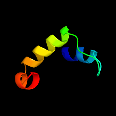
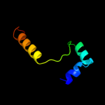
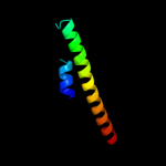
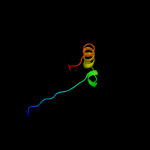
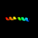
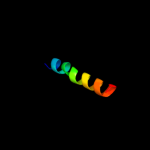
PDB header:structural genomics, unknown function Chain: A: PDB Molecule:uncharacterized protein; PDBTitle: solution nmr structure of the uncharacterized protein from2 rhodospirillum rubrum gene locus rru_a0810. northeast3 structural genomics target rrr43
Confidence and coverage
Confidence:
62.9%
Coverage:
23%
39 residues ( 23% of your sequence) have been modelled with 62.9% confidence by the single highest scoring template.
You may wish to submit your sequence to Phyrealarm. This will automatically scan your sequence every week for new potential templates as they appear in the Phyre2 library.
Please note: You must be registered and logged in to use Phyrealarm.
Warning: 55% of your sequence is predicted disordered. Disordered regions cannot be meaningfully predicted.
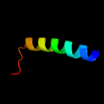
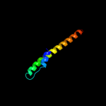
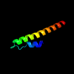
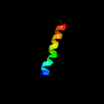
Region: 94 - 143 Aligned: 50 Modelled: 50 Confidence: 17.8% Identity: 20% PDB header:hydrolase Chain: D: PDB Molecule:exodeoxyribonuclease vii small subunit; PDBTitle: crystal structure of exodeoxyribonuclease vii small subunit2 (np_881400.1) from bordetella pertussis at 2.40 a resolution
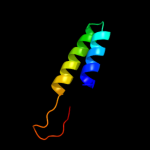
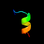
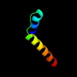
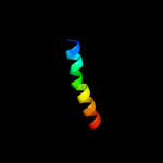
Region: 96 - 118 Aligned: 23 Modelled: 23 Confidence: 17.1% Identity: 39% PDB header:de novo protein Chain: C: PDB Molecule:coil ser l16l-pen; PDBTitle: switching the chirality of the metal environment alters the2 coordination mode in designed peptides.
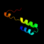
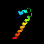
Region: 96 - 118 Aligned: 23 Modelled: 23 Confidence: 17.1% Identity: 39% PDB header:de novo protein Chain: A: PDB Molecule:coil ser l16d-pen; PDBTitle: switching the chirality of the metal environment alters the2 coordination mode in designed peptides.
Region: 96 - 118 Aligned: 23 Modelled: 23 Confidence: 17.1% Identity: 39% PDB header:de novo protein Chain: B: PDB Molecule:coil ser l16d-pen; PDBTitle: switching the chirality of the metal environment alters the2 coordination mode in designed peptides.
Region: 96 - 118 Aligned: 23 Modelled: 23 Confidence: 17.1% Identity: 39% PDB header:de novo protein Chain: C: PDB Molecule:coil ser l16d-pen; PDBTitle: switching the chirality of the metal environment alters the2 coordination mode in designed peptides.
Phyre2
21
22
23
24
25
26
27
28
29
30
31
32
33
34
35
36
37
38
39
40
41
42
43
44
45
46
47
48
49
50
51
52
53
54
55
56
57
58
59
60
61
62
63
64
65
66
Detailed template information
Binding site prediction
Due to computational demand, binding site predictions are not run for batch jobs
If you want to predict binding sites, please manually submit your model of choice to 3DLigandSite
Phyre is for academic use only
Please cite: Protein structure prediction on
the web: a case study using the Phyre server
Kelley LA and Sternberg MJE. Nature Protocols
4, 363 - 371 (2009) [pdf] [Import into BibTeX]
If you use the binding site
predictions from 3DLigandSite, please also cite:
3DLigandSite: predicting ligand-binding sites using similar structures.
Wass MN, Kelley LA and Sternberg
MJ Nucleic Acids Research 38, W469-73 (2010) [PubMed]