| 1 | c3stjC_
|
|
 |
100.0 |
15 |
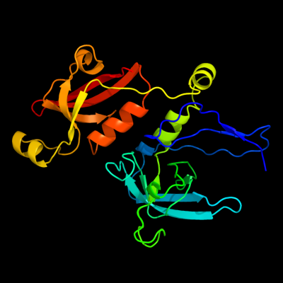
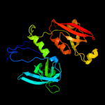
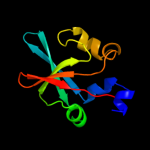
PDB header:hydrolase
Chain: C: PDB Molecule:protease degq;
PDBTitle: crystal structure of the protease + pdz1 domain of degq from2 escherichia coli
|
| 2 | c4a8aI_
|
|
 |
100.0 |
15 |
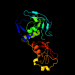
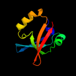
PDB header:hydrolase/hydrolase
Chain: I: PDB Molecule:periplasmic ph-dependent serine endoprotease degq;
PDBTitle: asymmetric cryo-em reconstruction of e. coli degq 12-mer in complex2 with lysozyme
|
| 3 | c3pv5B_
|
|
 |
100.0 |
15 |
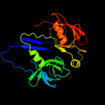
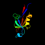
PDB header:hydrolase
Chain: B: PDB Molecule:degq;
PDBTitle: structure of legionella fallonii degq (n189g/p190g variant)
|
| 4 | c1ky9A_
|
|
 |
100.0 |
13 |
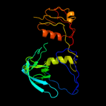
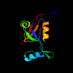
PDB header:hydrolase
Chain: A: PDB Molecule:protease do;
PDBTitle: crystal structure of degp (htra)
|
| 5 | c1lcyA_
|
|
 |
100.0 |
15 |
PDB header:hydrolase
Chain: A: PDB Molecule:htra2 serine protease;
PDBTitle: crystal structure of the mitochondrial serine protease htra2
|
| 6 | c3gdsA_
|
|
 |
100.0 |
11 |
PDB header:hydrolase/hydrolase activator
Chain: A: PDB Molecule:protease degs;
PDBTitle: crystal structure of degs h198p/d320a mutant modified by dfp in2 complex with dnrdgnvyyf peptide
|
| 7 | c3pv4A_
|
|
 |
100.0 |
15 |
PDB header:hydrolase
Chain: A: PDB Molecule:degq;
PDBTitle: structure of legionella fallonii degq (delta-pdz2 variant)
|
| 8 | c3qo6B_
|
|
 |
100.0 |
12 |
PDB header:photosynthesis
Chain: B: PDB Molecule:protease do-like 1, chloroplastic;
PDBTitle: crystal structure analysis of the plant protease deg1
|
| 9 | c2z9iB_
|
|
 |
100.0 |
12 |
PDB header:hydrolase
Chain: B: PDB Molecule:probable serine protease pepd;
PDBTitle: crystal structure of rv0983 from mycobacterium tuberculosis-2 proteolytically active form
|
| 10 | c2r3yC_
|
|
 |
99.9 |
13 |
PDB header:hydrolase/hydrolase activator
Chain: C: PDB Molecule:protease degs;
PDBTitle: crystal structure of the degs protease in complex with the2 ywf activating peptide
|
| 11 | d2i4sa1
|
|
 |
99.8 |
20 |
Fold:PDZ domain-like
Superfamily:PDZ domain-like
Family:EpsC C-terminal domain-like |
| 12 | d2i6va1
|
|
 |
99.7 |
22 |
Fold:PDZ domain-like
Superfamily:PDZ domain-like
Family:EpsC C-terminal domain-like |
| 13 | c2p3wB_
|
|
 |
99.6 |
19 |
PDB header:protein binding
Chain: B: PDB Molecule:probable serine protease htra3;
PDBTitle: crystal structure of the htra3 pdz domain bound to a phage-derived2 ligand (fgrwv)
|
| 14 | c2joaA_
|
|
 |
99.6 |
18 |
PDB header:protein binding
Chain: A: PDB Molecule:serine protease htra1;
PDBTitle: htra1 bound to an optimized peptide: nmr assignment of pdz2 domain and ligand resonances
|
| 15 | d1lcya1
|
|
 |
99.6 |
17 |
Fold:PDZ domain-like
Superfamily:PDZ domain-like
Family:HtrA-like serine proteases |
| 16 | d2z9ia1
|
|
 |
99.5 |
13 |
Fold:PDZ domain-like
Superfamily:PDZ domain-like
Family:HtrA-like serine proteases |
| 17 | d1ky9a1
|
|
 |
99.5 |
11 |
Fold:PDZ domain-like
Superfamily:PDZ domain-like
Family:HtrA-like serine proteases |
| 18 | c2kl1A_
|
|
 |
99.5 |
9 |
PDB header:protein binding
Chain: A: PDB Molecule:ylbl protein;
PDBTitle: solution structure of gtr34c from geobacillus thermodenitrificans.2 northeast structural genomics consortium target gtr34c
|
| 19 | c3i18A_
|
|
 |
99.5 |
10 |
PDB header:structural genomics, unknown function
Chain: A: PDB Molecule:lmo2051 protein;
PDBTitle: crystal structure of the pdz domain of the sdrc-like protein2 (lmo2051) from listeria monocytogenes, northeast structural3 genomics consortium target lmr166b
|
| 20 | c2kjpA_
|
|
 |
99.4 |
8 |
PDB header:structural genomics, unknown function
Chain: A: PDB Molecule:uncharacterized protein ylbl;
PDBTitle: solution structure of protein ylbl (bsu15050) from bacillus2 subtilis, northeast structural genomics consortium target3 sr713a
|
| 21 | d1ky9a2 |
|
not modelled |
99.4 |
14 |
Fold:Trypsin-like serine proteases
Superfamily:Trypsin-like serine proteases
Family:Prokaryotic proteases |
| 22 | c2rceI_ |
|
not modelled |
99.3 |
13 |
PDB header:hydrolase
Chain: I: PDB Molecule:protease degs;
PDBTitle: dfp modified degs delta pdz
|
| 23 | c3ossC_ |
|
not modelled |
99.3 |
30 |
PDB header:protein transport
Chain: C: PDB Molecule:type 2 secretion system, gspc;
PDBTitle: the crystal structure of enterotoxigenic escherichia coli gspc-gspd2 complex from the type ii secretion system
|
| 24 | d1sota1 |
|
not modelled |
99.3 |
12 |
Fold:PDZ domain-like
Superfamily:PDZ domain-like
Family:HtrA-like serine proteases |
| 25 | d2hgaa1 |
|
not modelled |
99.3 |
16 |
Fold:PDZ domain-like
Superfamily:PDZ domain-like
Family:MTH1368 C-terminal domain-like |
| 26 | d1ky9b2 |
|
not modelled |
99.2 |
16 |
Fold:PDZ domain-like
Superfamily:PDZ domain-like
Family:HtrA-like serine proteases |
| 27 | c3mmgB_ |
|
not modelled |
99.2 |
9 |
PDB header:viral protein, hydrolase
Chain: B: PDB Molecule:nuclear inclusion protein a;
PDBTitle: crystal structure of tobacco vein mottling virus protease
|
| 28 | d1fc6a3 |
|
not modelled |
99.2 |
11 |
Fold:PDZ domain-like
Superfamily:PDZ domain-like
Family:Tail specific protease PDZ domain |
| 29 | c3rleA_ |
|
not modelled |
99.1 |
17 |
PDB header:membrane protein
Chain: A: PDB Molecule:golgi reassembly-stacking protein 2;
PDBTitle: crystal structure of grasp55 grasp domain (residues 7-208)
|
| 30 | c2zplA_ |
|
not modelled |
99.1 |
12 |
PDB header:hydrolase
Chain: A: PDB Molecule:regulator of sigma e protease;
PDBTitle: crystal structure analysis of pdz domain a
|
| 31 | d1l1ja_ |
|
not modelled |
99.1 |
15 |
Fold:Trypsin-like serine proteases
Superfamily:Trypsin-like serine proteases
Family:Prokaryotic proteases |
| 32 | c2zpmA_ |
|
not modelled |
99.1 |
19 |
PDB header:hydrolase
Chain: A: PDB Molecule:regulator of sigma e protease;
PDBTitle: crystal structure analysis of pdz domain b
|
| 33 | d1q31a_ |
|
not modelled |
99.1 |
11 |
Fold:Trypsin-like serine proteases
Superfamily:Trypsin-like serine proteases
Family:Viral proteases |
| 34 | d2qf3a1 |
|
not modelled |
99.0 |
14 |
Fold:Trypsin-like serine proteases
Superfamily:Trypsin-like serine proteases
Family:Prokaryotic proteases |
| 35 | d2z9ia2 |
|
not modelled |
98.9 |
12 |
Fold:Trypsin-like serine proteases
Superfamily:Trypsin-like serine proteases
Family:Prokaryotic proteases |
| 36 | d1lvmb_ |
|
not modelled |
98.9 |
11 |
Fold:Trypsin-like serine proteases
Superfamily:Trypsin-like serine proteases
Family:Viral proteases |
| 37 | c3nwuB_ |
|
not modelled |
98.8 |
15 |
PDB header:hydrolase
Chain: B: PDB Molecule:serine protease htra1;
PDBTitle: substrate induced remodeling of the active site regulates htra12 activity
|
| 38 | c3nziA_ |
|
not modelled |
98.8 |
13 |
PDB header:hydrolase/hydrolase substrate
Chain: A: PDB Molecule:serine protease htra1;
PDBTitle: substrate induced remodeling of the active site regulates htra12 activity
|
| 39 | c2yuyA_ |
|
not modelled |
98.8 |
16 |
PDB header:signaling protein
Chain: A: PDB Molecule:rho gtpase activating protein 21;
PDBTitle: solution structure of pdz domain of rho gtpase activating2 protein 21
|
| 40 | c3stiC_ |
|
not modelled |
98.8 |
15 |
PDB header:hydrolase
Chain: C: PDB Molecule:protease degq;
PDBTitle: crystal structure of the protease domain of degq from escherichia coli
|
| 41 | c2eaqA_ |
|
not modelled |
98.8 |
22 |
PDB header:metal binding protein
Chain: A: PDB Molecule:lim domain only protein 7;
PDBTitle: crystal structure of pdz domain of kiaa0858 (lim), ms07932 from homo sapiens
|
| 42 | d1qtfa_ |
|
not modelled |
98.7 |
10 |
Fold:Trypsin-like serine proteases
Superfamily:Trypsin-like serine proteases
Family:Prokaryotic proteases |
| 43 | d1wifa_ |
|
not modelled |
98.7 |
20 |
Fold:PDZ domain-like
Superfamily:PDZ domain-like
Family:PDZ domain |
| 44 | d1x5qa1 |
|
not modelled |
98.7 |
18 |
Fold:PDZ domain-like
Superfamily:PDZ domain-like
Family:PDZ domain |
| 45 | c3shuB_ |
|
not modelled |
98.6 |
20 |
PDB header:cell adhesion
Chain: B: PDB Molecule:tight junction protein zo-1;
PDBTitle: crystal structure of zo-1 pdz3
|
| 46 | d1wfga_ |
|
not modelled |
98.6 |
22 |
Fold:PDZ domain-like
Superfamily:PDZ domain-like
Family:PDZ domain |
| 47 | c2b0fA_ |
|
not modelled |
98.6 |
3 |
PDB header:hydrolase/hydrolase inhibitor
Chain: A: PDB Molecule:picornain 3c (protease 3c) (p3c);
PDBTitle: nmr structure of the human rhinovirus 3c protease (serotype 14) with2 covalently bound ace-lealfq-ethylpropionate inhibitor
|
| 48 | c3diwB_ |
|
not modelled |
98.6 |
16 |
PDB header:signaling protein/cell adhesion
Chain: B: PDB Molecule:tax1-binding protein 3;
PDBTitle: c-terminal beta-catenin bound tip-1 structure
|
| 49 | c3l4fD_ |
|
not modelled |
98.6 |
10 |
PDB header:signaling protein/protein binding
Chain: D: PDB Molecule:sh3 and multiple ankyrin repeat domains protein
PDBTitle: crystal structure of betapix coiled-coil domain and shank2 pdz complex
|
| 50 | c2krgA_ |
|
not modelled |
98.6 |
13 |
PDB header:signaling protein
Chain: A: PDB Molecule:na(+)/h(+) exchange regulatory cofactor nhe-rf1;
PDBTitle: solution structure of human sodium/ hydrogen exchange2 regulatory factor 1(150-358)
|
| 51 | c2komA_ |
|
not modelled |
98.6 |
15 |
PDB header:signaling protein
Chain: A: PDB Molecule:partitioning defective 3 homolog;
PDBTitle: solution structure of humar par-3b pdz2 (residues 451-549)
|
| 52 | d1ueqa_ |
|
not modelled |
98.6 |
16 |
Fold:PDZ domain-like
Superfamily:PDZ domain-like
Family:PDZ domain |
| 53 | c2q3gA_ |
|
not modelled |
98.6 |
10 |
PDB header:structural genomics
Chain: A: PDB Molecule:pdz and lim domain protein 7;
PDBTitle: structure of the pdz domain of human pdlim7 bound to a c-2 terminal extension from human beta-tropomyosin
|
| 54 | d1q3oa_ |
|
not modelled |
98.6 |
11 |
Fold:PDZ domain-like
Superfamily:PDZ domain-like
Family:PDZ domain |
| 55 | d1rgwa_ |
|
not modelled |
98.6 |
12 |
Fold:PDZ domain-like
Superfamily:PDZ domain-like
Family:PDZ domain |
| 56 | d1m5za_ |
|
not modelled |
98.6 |
17 |
Fold:PDZ domain-like
Superfamily:PDZ domain-like
Family:PDZ domain |
| 57 | c2eehA_ |
|
not modelled |
98.6 |
22 |
PDB header:metal binding protein
Chain: A: PDB Molecule:pdz domain-containing protein 7;
PDBTitle: solution structure of first pdz domain of pdz domain2 containing protein 7
|
| 58 | d2f5ya1 |
|
not modelled |
98.5 |
17 |
Fold:PDZ domain-like
Superfamily:PDZ domain-like
Family:PDZ domain |
| 59 | d1ozia_ |
|
not modelled |
98.5 |
18 |
Fold:PDZ domain-like
Superfamily:PDZ domain-like
Family:PDZ domain |
| 60 | c2kjdA_ |
|
not modelled |
98.5 |
12 |
PDB header:signaling protein
Chain: A: PDB Molecule:sodium/hydrogen exchange regulatory cofactor nhe-
PDBTitle: solution structure of extended pdz2 domain from nherf1 (150-2 270)
|
| 61 | d1uf1a_ |
|
not modelled |
98.5 |
22 |
Fold:PDZ domain-like
Superfamily:PDZ domain-like
Family:PDZ domain |
| 62 | d1lcya2 |
|
not modelled |
98.5 |
14 |
Fold:Trypsin-like serine proteases
Superfamily:Trypsin-like serine proteases
Family:Prokaryotic proteases |
| 63 | c3khfA_ |
|
not modelled |
98.5 |
13 |
PDB header:transferase
Chain: A: PDB Molecule:microtubule-associated serine/threonine-protein
PDBTitle: the crystal structure of the pdz domain of human microtubule2 associated serine/threonine kinase 3 (mast3)
|
| 64 | c3shwA_ |
|
not modelled |
98.5 |
21 |
PDB header:cell adhesion
Chain: A: PDB Molecule:tight junction protein zo-1;
PDBTitle: crystal structure of zo-1 pdz3-sh3-guk supramodule complex with2 connexin-45 peptide
|
| 65 | d1p1da2 |
|
not modelled |
98.5 |
23 |
Fold:PDZ domain-like
Superfamily:PDZ domain-like
Family:PDZ domain |
| 66 | c2vwrA_ |
|
not modelled |
98.5 |
24 |
PDB header:protein-binding
Chain: A: PDB Molecule:ligand of numb protein x 2;
PDBTitle: crystal structure of the second pdz domain of numb-binding2 protein 2
|
| 67 | c2v90E_ |
|
not modelled |
98.5 |
13 |
PDB header:protein-binding
Chain: E: PDB Molecule:pdz domain-containing protein 3;
PDBTitle: crystal structure of the 3rd pdz domain of intestine- and2 kidney-enriched pdz domain ikepp (pdzd3)
|
| 68 | c2w5eB_ |
|
not modelled |
98.5 |
9 |
PDB header:hydrolase
Chain: B: PDB Molecule:putative serine protease;
PDBTitle: structural and biochemical analysis of human pathogenic2 astrovirus serine protease at 2.0 angstrom resolution
|
| 69 | c2qt5A_ |
|
not modelled |
98.5 |
18 |
PDB header:peptide binding protein
Chain: A: PDB Molecule:glutamate receptor-interacting protein 1;
PDBTitle: crystal structure of grip1 pdz12 in complex with the fras12 peptide
|
| 70 | d1t2ma1 |
|
not modelled |
98.5 |
18 |
Fold:PDZ domain-like
Superfamily:PDZ domain-like
Family:PDZ domain |
| 71 | d2fe5a1 |
|
not modelled |
98.5 |
22 |
Fold:PDZ domain-like
Superfamily:PDZ domain-like
Family:PDZ domain |
| 72 | c3k6zA_ |
|
not modelled |
98.5 |
14 |
PDB header:hydrolase
Chain: A: PDB Molecule:possible membrane-associated serine protease;
PDBTitle: crystal structure of rv3671c protease, inactive form
|
| 73 | d1ihja_ |
|
not modelled |
98.5 |
13 |
Fold:PDZ domain-like
Superfamily:PDZ domain-like
Family:PDZ domain |
| 74 | c2egkC_ |
|
not modelled |
98.5 |
12 |
PDB header:protein binding
Chain: C: PDB Molecule:general receptor for phosphoinositides 1-
PDBTitle: crystal structure of tamalin pdz-intrinsic ligand fusion2 protein
|
| 75 | c1p1dA_ |
|
not modelled |
98.5 |
22 |
PDB header:protein binding
Chain: A: PDB Molecule:glutamate receptor interacting protein;
PDBTitle: structural insights into the inter-domain chaperoning of2 tandem pdz domains in glutamate receptor interacting3 proteins
|
| 76 | c2v1wB_ |
|
not modelled |
98.5 |
14 |
PDB header:structural protein
Chain: B: PDB Molecule:pdz and lim domain protein 4;
PDBTitle: crystal structure of human lim protein ril (pdlim4) pdz2 domain bound to the c-terminal peptide of human alpha-3 actinin-1
|
| 77 | d1wf7a_ |
|
not modelled |
98.5 |
16 |
Fold:PDZ domain-like
Superfamily:PDZ domain-like
Family:PDZ domain |
| 78 | d2cssa1 |
|
not modelled |
98.5 |
18 |
Fold:PDZ domain-like
Superfamily:PDZ domain-like
Family:PDZ domain |
| 79 | d1w9ea1 |
|
not modelled |
98.4 |
21 |
Fold:PDZ domain-like
Superfamily:PDZ domain-like
Family:PDZ domain |
| 80 | c1u38A_ |
|
not modelled |
98.4 |
21 |
PDB header:protein transport
Chain: A: PDB Molecule:amyloid beta a4 precursor protein-binding,
PDBTitle: auto-inhibition mechanism of x11s/mints family scaffold2 proteins revealed by the closed conformation of the tandem3 pdz domains
|
| 81 | c3qglD_ |
|
not modelled |
98.4 |
13 |
PDB header:protein binding
Chain: D: PDB Molecule:sorting nexin-27;
PDBTitle: crystal structure of pdz domain of sorting nexin 27 (snx27) in complex2 with the eseskv peptide corresponding to the c-terminal tail of girk3
|
| 82 | d1q7xa_ |
|
not modelled |
98.4 |
20 |
Fold:PDZ domain-like
Superfamily:PDZ domain-like
Family:PDZ domain |
| 83 | c2dm8A_ |
|
not modelled |
98.4 |
21 |
PDB header:protein binding
Chain: A: PDB Molecule:inad-like protein;
PDBTitle: solution structure of the eighth pdz domain of human inad-2 like protein
|
| 84 | c2o2tB_ |
|
not modelled |
98.4 |
22 |
PDB header:structural protein
Chain: B: PDB Molecule:multiple pdz domain protein;
PDBTitle: the crystal structure of the 1st pdz domain of mpdz
|
| 85 | d1p1da1 |
|
not modelled |
98.4 |
16 |
Fold:PDZ domain-like
Superfamily:PDZ domain-like
Family:PDZ domain |
| 86 | c1u37A_ |
|
not modelled |
98.4 |
21 |
PDB header:protein transport
Chain: A: PDB Molecule:amyloid beta a4 precursor protein-binding,
PDBTitle: auto-inhibition mechanism of x11s/mints family scaffold2 proteins revealed by the closed conformation of the tandem3 pdz domains
|
| 87 | d1wf8a1 |
|
not modelled |
98.4 |
20 |
Fold:PDZ domain-like
Superfamily:PDZ domain-like
Family:PDZ domain |
| 88 | d2byga1 |
|
not modelled |
98.4 |
22 |
Fold:PDZ domain-like
Superfamily:PDZ domain-like
Family:PDZ domain |
| 89 | c3eggC_ |
|
not modelled |
98.4 |
20 |
PDB header:hydrolase
Chain: C: PDB Molecule:spinophilin;
PDBTitle: crystal structure of a complex between protein phosphatase 1 alpha2 (pp1) and the pp1 binding and pdz domains of spinophilin
|
| 90 | c2ogpA_ |
|
not modelled |
98.4 |
17 |
PDB header:signaling protein
Chain: A: PDB Molecule:partitioning-defective 3 homolog;
PDBTitle: solution structure of the second pdz domain of par-3
|
| 91 | c2iwnA_ |
|
not modelled |
98.4 |
18 |
PDB header:signaling protein
Chain: A: PDB Molecule:multiple pdz domain protein;
PDBTitle: 3rd pdz domain of multiple pdz domain protein mpdz (casp2 target)
|
| 92 | c2he4A_ |
|
not modelled |
98.4 |
12 |
PDB header:structural genomics, unknown function
Chain: A: PDB Molecule:na(+)/h(+) exchange regulatory cofactor nhe-rf2;
PDBTitle: the crystal structure of the second pdz domain of human2 nherf-2 (slc9a3r2) interacting with a mode 1 pdz binding3 motif
|
| 93 | c2gzvA_ |
|
not modelled |
98.4 |
11 |
PDB header:signaling protein
Chain: A: PDB Molecule:prkca-binding protein;
PDBTitle: the cystal structure of the pdz domain of human pick1 (casp target)
|
| 94 | c2omjA_ |
|
not modelled |
98.4 |
18 |
PDB header:cell adhesion
Chain: A: PDB Molecule:rho guanine nucleotide exchange factor 12;
PDBTitle: solution structure of larg pdz domain
|
| 95 | c3cyyA_ |
|
not modelled |
98.4 |
13 |
PDB header:peptide binding protein
Chain: A: PDB Molecule:tight junction protein zo-1;
PDBTitle: the crystal structure of zo-1 pdz2 in complex with the cx43 peptide
|
| 96 | d1uhpa_ |
|
not modelled |
98.4 |
17 |
Fold:PDZ domain-like
Superfamily:PDZ domain-like
Family:PDZ domain |
| 97 | c2i04B_ |
|
not modelled |
98.4 |
13 |
PDB header:peptide binding protein
Chain: B: PDB Molecule:membrane-associated guanylate kinase, ww and pdz
PDBTitle: x-ray crystal structure of magi-1 pdz1 bound to the c-2 terminal peptide of hpv18 e6
|
| 98 | c2osgB_ |
|
not modelled |
98.4 |
18 |
PDB header:cell adhesion
Chain: B: PDB Molecule:tight junction protein zo-2;
PDBTitle: solution structure and binding property of the domain-2 swapped dimer of zo2pdz2
|
| 99 | c2jilA_ |
|
not modelled |
98.4 |
15 |
PDB header:membrane protein
Chain: A: PDB Molecule:glutamate receptor interacting protein-1;
PDBTitle: crystal structure of 2nd pdz domain of glutamate receptor2 interacting protein-1 (grip1)
|
| 100 | d1ry4a_ |
|
not modelled |
98.4 |
14 |
Fold:PDZ domain-like
Superfamily:PDZ domain-like
Family:PDZ domain |
| 101 | d1d5ga_ |
|
not modelled |
98.4 |
20 |
Fold:PDZ domain-like
Superfamily:PDZ domain-like
Family:PDZ domain |
| 102 | c2ka9A_ |
|
not modelled |
98.4 |
19 |
PDB header:cell adhesion
Chain: A: PDB Molecule:disks large homolog 4;
PDBTitle: solution structure of psd-95 pdz12 complexed with cypin2 peptide
|
| 103 | c2z17A_ |
|
not modelled |
98.4 |
13 |
PDB header:protein binding
Chain: A: PDB Molecule:pleckstrin homology sec7 and coiled-coil domains-
PDBTitle: crystal sturcture of pdz domain from human pleckstrin2 homology, sec7
|
| 104 | d1g9oa_ |
|
not modelled |
98.4 |
9 |
Fold:PDZ domain-like
Superfamily:PDZ domain-like
Family:PDZ domain |
| 105 | c2jreA_ |
|
not modelled |
98.4 |
15 |
PDB header:de novo protein
Chain: A: PDB Molecule:c60-1 pdz domain peptide;
PDBTitle: c60-1, a pdz domain designed using statistical coupling2 analysis
|
| 106 | c2iwoA_ |
|
not modelled |
98.4 |
14 |
PDB header:signaling protein
Chain: A: PDB Molecule:multiple pdz domain protein;
PDBTitle: 12th pdz domain of multiple pdz domain protein mpdz (casp2 target)
|
| 107 | c2vsvB_ |
|
not modelled |
98.3 |
11 |
PDB header:protein-binding
Chain: B: PDB Molecule:rhophilin-2;
PDBTitle: crystal structure of the pdz domain of human rhophilin-2
|
| 108 | c3r0hA_ |
|
not modelled |
98.3 |
16 |
PDB header:peptide binding protein
Chain: A: PDB Molecule:inactivation-no-after-potential d protein;
PDBTitle: structure of inad pdz45 in complex with ng2 peptide
|
| 109 | c2k1zA_ |
|
not modelled |
98.3 |
19 |
PDB header:signaling protein
Chain: A: PDB Molecule:partitioning-defective 3 homolog;
PDBTitle: solution structure of par-3 pdz3
|
| 110 | d1qaua_ |
|
not modelled |
98.3 |
18 |
Fold:PDZ domain-like
Superfamily:PDZ domain-like
Family:PDZ domain |
| 111 | d1b8qa_ |
|
not modelled |
98.3 |
21 |
Fold:PDZ domain-like
Superfamily:PDZ domain-like
Family:PDZ domain |
| 112 | c2jxoA_ |
|
not modelled |
98.3 |
9 |
PDB header:protein binding
Chain: A: PDB Molecule:ezrin-radixin-moesin-binding phosphoprotein 50;
PDBTitle: structure of the second pdz domain of nherf-1
|
| 113 | d1um1a_ |
|
not modelled |
98.3 |
18 |
Fold:PDZ domain-like
Superfamily:PDZ domain-like
Family:PDZ domain |
| 114 | d1uepa_ |
|
not modelled |
98.3 |
15 |
Fold:PDZ domain-like
Superfamily:PDZ domain-like
Family:PDZ domain |
| 115 | c3k1rA_ |
|
not modelled |
98.3 |
23 |
PDB header:structural protein
Chain: A: PDB Molecule:harmonin;
PDBTitle: structure of harmonin npdz1 in complex with the sam-pbm of2 sans
|
| 116 | c2jikB_ |
|
not modelled |
98.3 |
17 |
PDB header:membrane protein
Chain: B: PDB Molecule:synaptojanin-2 binding protein;
PDBTitle: crystal structure of pdz domain of synaptojanin-2 binding2 protein
|
| 117 | d1whda_ |
|
not modelled |
98.3 |
16 |
Fold:PDZ domain-like
Superfamily:PDZ domain-like
Family:PDZ domain |
| 118 | d1wh1a_ |
|
not modelled |
98.3 |
16 |
Fold:PDZ domain-like
Superfamily:PDZ domain-like
Family:PDZ domain |
| 119 | c3ggeA_ |
|
not modelled |
98.3 |
13 |
PDB header:protein binding
Chain: A: PDB Molecule:pdz domain-containing protein gipc2;
PDBTitle: crystal structure of the pdz domain of pdz domain-containing protein2 gipc2
|
| 120 | d1x5ra1 |
|
not modelled |
98.3 |
17 |
Fold:PDZ domain-like
Superfamily:PDZ domain-like
Family:PDZ domain |