Phyre Home Retrieve Phyre Job Id
Emaill.a.kelley@imperial.ac.uk
DescriptionP77657
DateThu Jan 5 12:31:25 GMT 2012
Unique Job IDa9d9b66aa25b0774

Summary 

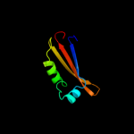
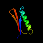
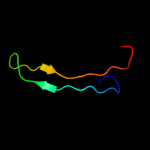
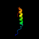
Top model
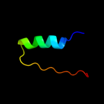
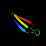
Image coloured by rainbow N → C terminus
Model (left) based on template d1l5ia_
Top template information
Fold:Origin of replication-binding domain, RBD-like
Superfamily:Origin of replication-binding domain, RBD-like
Family:DNA-binding domain of REP protein
Confidence and coverage
Confidence: 81.6% Coverage: 29%
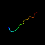
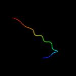
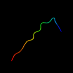
61 residues ( 29% of your sequence) have been modelled with 81.6% confidence by the single highest scoring template.
You may wish to submit your sequence to Phyrealarm. This will automatically scan your sequence every week for new potential templates as they appear in the Phyre2 library.
Please note: You must be registered and logged in to use Phyrealarm.
3D viewing
Interactive 3D view in Jmol

Sequence analysis 

Secondary structure and disorder prediction 

   1........10.........20.........30.........40.........50.........60
Sequence  MKTYQGTHGVHILEYQSKINKLLCYLTNRYPRLIAVRVDLHYPKIVDNGDNICCFPNLEP
Secondary structure 


SS confidence 


Disorder  ???????


?

Disorder confidence 


   .........70.........80.........90.........100.........110.........120
Sequence  GVISRMRESLRAKLEADRTRKVREDKRIYRCPLFIIWAKEYSESGKCHYHICLLFNKDAY
Secondary structure 


SS confidence 


Disorder 


?????????


Disorder confidence 


   .........130.........140.........150.........160.........170.........180
Sequence  YHLGDYDQDDNLRGMITGAWYSALRLERDDHPGLVHFPENCKYVLDTNSTDFQQNYQALL
Secondary structure 


SS confidence 


Disorder 


?


?


?


Disorder confidence 


   .........190.........200........
Sequence  TRLDYLTKVESKVFGEGDRNFGCSQIEL
Secondary structure 


SS confidence 


Disorder 


????
????
???????
Disorder confidence 


Confidence Key
High(9)                    Low (0)
?Disordered
Alpha helix
Beta strand

Domain analysis 

Hover over an aligned region to see model and summary info

Please note, only up to the top 20 hits are modelled to reduce computer load

RankAligned region

PDB 1l5i chain A

3D model

Region: 32 - 119
Aligned: 61
Modelled: 72
Confidence: 81.6%
Identity: 30%
Fold: Origin of replication-binding domain, RBD-like
Superfamily: Origin of replication-binding domain, RBD-like
Family: DNA-binding domain of REP protein

Phyre2

PDB 2ns6 chain A

3D model

Region: 34 - 113
Aligned: 55
Modelled: 55
Confidence: 73.8%
Identity: 15%
PDB header:hydrolase
Chain: A: PDB Molecule:mobilization protein a;
PDBTitle: crystal structure of the minimal relaxase domain of moba2 from plasmid r1162

Phyre2

PDB 3dkx chain A

3D model

Region: 102 - 117
Aligned: 16
Modelled: 16
Confidence: 71.5%
Identity: 25%
PDB header:replication
Chain: A: PDB Molecule:replication protein repb;
PDBTitle: crystal structure of the replication initiator protein2 encoded on plasmid pmv158 (repb), trigonal form, to 2.7 ang3 resolution

Phyre2

PDB 2x3g chain A

3D model

Region: 101 - 117
Aligned: 17
Modelled: 17
Confidence: 63.0%
Identity: 41%
PDB header:viral protein
Chain: A: PDB Molecule:sirv1 hypothetical protein orf119;
PDBTitle: crystal structure of the hypothetical protein orf119 from2 sulfolobus islandicus rod-shaped virus 1

Phyre2

PDB 2hwt chain A

3D model

Region: 89 - 119
Aligned: 31
Modelled: 31
Confidence: 39.9%
Identity: 16%
PDB header:replication, hydrolase
Chain: A: PDB Molecule:putative replicase-associated protein;
PDBTitle: nmr solution structure of the master-rep protein nuclease2 domain (2-95) from the faba bean necrotic yellows virus

Phyre2

PDB 2nzc chain A domain 1

3D model

Region: 21 - 76
Aligned: 50
Modelled: 56
Confidence: 16.9%
Identity: 14%
Fold: Ferredoxin-like
Superfamily: ACT-like
Family: TM1266-like

Phyre2

PDB 1f86 chain A

3D model

Region: 100 - 116
Aligned: 17
Modelled: 17
Confidence: 16.5%
Identity: 29%
Fold: Prealbumin-like
Superfamily: Transthyretin (synonym: prealbumin)
Family: Transthyretin (synonym: prealbumin)

Phyre2

PDB 1tta chain A

3D model

Region: 100 - 116
Aligned: 17
Modelled: 17
Confidence: 15.4%
Identity: 29%
Fold: Prealbumin-like
Superfamily: Transthyretin (synonym: prealbumin)
Family: Transthyretin (synonym: prealbumin)

Phyre2

PDB 1tfp chain A

3D model

Region: 100 - 116
Aligned: 17
Modelled: 17
Confidence: 12.5%
Identity: 35%
Fold: Prealbumin-like
Superfamily: Transthyretin (synonym: prealbumin)
Family: Transthyretin (synonym: prealbumin)

Phyre2

PDB 2h1x chain B

3D model

Region: 103 - 116
Aligned: 14
Modelled: 14
Confidence: 10.7%
Identity: 36%
PDB header:hydrolase
Chain: B: PDB Molecule:5-hydroxyisourate hydrolase (formerly known as
PDBTitle: crystal structure of 5-hydroxyisourate hydrolase (formerly2 known as trp, transthyretin related protein)

Phyre2

PDB 2h0e chain A

3D model

Region: 103 - 116
Aligned: 14
Modelled: 14
Confidence: 10.4%
Identity: 43%
PDB header:hydrolase
Chain: A: PDB Molecule:transthyretin-like protein pucm;
PDBTitle: crystal structure of pucm in the absence of substrate

Phyre2

PDB 2gpz chain C

3D model

Region: 103 - 116
Aligned: 14
Modelled: 14
Confidence: 9.6%
Identity: 36%
PDB header:hydrolase
Chain: C: PDB Molecule:transthyretin-like protein;
PDBTitle: transthyretin-like protein from salmonella dublin

Phyre2

PDB 4a69 chain A

3D model

Region: 15 - 41
Aligned: 27
Modelled: 27
Confidence: 9.3%
Identity: 26%
PDB header:transcription
Chain: A: PDB Molecule:histone deacetylase 3,;
PDBTitle: structure of hdac3 bound to corepressor and inositol tetraphosphate

Phyre2

PDB 2ljh chain A

3D model

Region: 93 - 125
Aligned: 33
Modelled: 33
Confidence: 9.2%
Identity: 12%
PDB header:hydrolase
Chain: A: PDB Molecule:double-stranded rna-specific editase adar;
PDBTitle: nmr structure of double-stranded rna-specific editase adar

Phyre2

PDB 2hw0 chain A

3D model

Region: 88 - 119
Aligned: 32
Modelled: 32
Confidence: 8.8%
Identity: 22%
PDB header:hydrolase, replication
Chain: A: PDB Molecule:replicase;
PDBTitle: nmr solution structure of the nuclease domain from the2 replicator initiator protein from porcine circovirus pcv2

Phyre2

PDB 1oo2 chain A

3D model

Region: 100 - 116
Aligned: 17
Modelled: 17
Confidence: 8.7%
Identity: 29%
Fold: Prealbumin-like
Superfamily: Transthyretin (synonym: prealbumin)
Family: Transthyretin (synonym: prealbumin)

Phyre2

PDB 1kgi chain A

3D model

Region: 100 - 116
Aligned: 17
Modelled: 17
Confidence: 8.6%
Identity: 35%
Fold: Prealbumin-like
Superfamily: Transthyretin (synonym: prealbumin)
Family: Transthyretin (synonym: prealbumin)

Phyre2

PDB 1ohe chain A domain 1

3D model

Region: 4 - 36
Aligned: 33
Modelled: 33
Confidence: 8.3%
Identity: 9%
Fold: (Phosphotyrosine protein) phosphatases II
Superfamily: (Phosphotyrosine protein) phosphatases II
Family: Dual specificity phosphatase-like

Phyre2

PDB 1luz chain A

3D model

Region: 4 - 39
Aligned: 35
Modelled: 36
Confidence: 8.1%
Identity: 17%
Fold: OB-fold
Superfamily: Nucleic acid-binding proteins
Family: Cold shock DNA-binding domain-like

Phyre2

PDB 3max chain B

3D model

Region: 15 - 41
Aligned: 27
Modelled: 27
Confidence: 7.7%
Identity: 22%
PDB header:hydrolase
Chain: B: PDB Molecule:histone deacetylase 2;
PDBTitle: crystal structure of human hdac2 complexed with an n-(2-aminophenyl)2 benzamide

Phyre2
1

d1l5ia_
2

c2ns6A_
3

c3dkxA_
4

c2x3gA_
5

c2hwtA_
6

d2nzca1
7

d1f86a_
8

d1ttaa_
9

d1tfpa_
10

c2h1xB_
11

c2h0eA_
12

c2gpzC_
13

c4a69A_
14

c2ljhA_
15

c2hw0A_
16

d1oo2a_
17

d1kgia_
18

d1ohea1
19

d1luza_
20

c3maxB_
21


22


23


24


25


26


27


28


Detailed template information 

#
Template Alignment Coverage3D Model Confidence
% i.d. Template Information
1d1l5ia_


81.6 30 Fold:Origin of replication-binding domain, RBD-like
Superfamily:Origin of replication-binding domain, RBD-like
Family:DNA-binding domain of REP protein
2c2ns6A_


73.8 15 PDB header:hydrolase
Chain: A: PDB Molecule:mobilization protein a;
PDBTitle: crystal structure of the minimal relaxase domain of moba2 from plasmid r1162
3c3dkxA_


71.5 25 PDB header:replication
Chain: A: PDB Molecule:replication protein repb;
PDBTitle: crystal structure of the replication initiator protein2 encoded on plasmid pmv158 (repb), trigonal form, to 2.7 ang3 resolution
4c2x3gA_


63.0 41 PDB header:viral protein
Chain: A: PDB Molecule:sirv1 hypothetical protein orf119;
PDBTitle: crystal structure of the hypothetical protein orf119 from2 sulfolobus islandicus rod-shaped virus 1
5c2hwtA_


39.9 16 PDB header:replication, hydrolase
Chain: A: PDB Molecule:putative replicase-associated protein;
PDBTitle: nmr solution structure of the master-rep protein nuclease2 domain (2-95) from the faba bean necrotic yellows virus
6d2nzca1


16.9 14 Fold:Ferredoxin-like
Superfamily:ACT-like
Family:TM1266-like
7d1f86a_


16.5 29 Fold:Prealbumin-like
Superfamily:Transthyretin (synonym: prealbumin)
Family:Transthyretin (synonym: prealbumin)
8d1ttaa_


15.4 29 Fold:Prealbumin-like
Superfamily:Transthyretin (synonym: prealbumin)
Family:Transthyretin (synonym: prealbumin)
9d1tfpa_


12.5 35 Fold:Prealbumin-like
Superfamily:Transthyretin (synonym: prealbumin)
Family:Transthyretin (synonym: prealbumin)
10c2h1xB_


10.7 36 PDB header:hydrolase
Chain: B: PDB Molecule:5-hydroxyisourate hydrolase (formerly known as
PDBTitle: crystal structure of 5-hydroxyisourate hydrolase (formerly2 known as trp, transthyretin related protein)
11c2h0eA_


10.4 43 PDB header:hydrolase
Chain: A: PDB Molecule:transthyretin-like protein pucm;
PDBTitle: crystal structure of pucm in the absence of substrate
12c2gpzC_


9.6 36 PDB header:hydrolase
Chain: C: PDB Molecule:transthyretin-like protein;
PDBTitle: transthyretin-like protein from salmonella dublin
13c4a69A_


9.3 26 PDB header:transcription
Chain: A: PDB Molecule:histone deacetylase 3,;
PDBTitle: structure of hdac3 bound to corepressor and inositol tetraphosphate
14c2ljhA_


9.2 12 PDB header:hydrolase
Chain: A: PDB Molecule:double-stranded rna-specific editase adar;
PDBTitle: nmr structure of double-stranded rna-specific editase adar
15c2hw0A_


8.8 22 PDB header:hydrolase, replication
Chain: A: PDB Molecule:replicase;
PDBTitle: nmr solution structure of the nuclease domain from the2 replicator initiator protein from porcine circovirus pcv2
16d1oo2a_


8.7 29 Fold:Prealbumin-like
Superfamily:Transthyretin (synonym: prealbumin)
Family:Transthyretin (synonym: prealbumin)
17d1kgia_


8.6 35 Fold:Prealbumin-like
Superfamily:Transthyretin (synonym: prealbumin)
Family:Transthyretin (synonym: prealbumin)
18d1ohea1


8.3 9 Fold:(Phosphotyrosine protein) phosphatases II
Superfamily:(Phosphotyrosine protein) phosphatases II
Family:Dual specificity phosphatase-like
19d1luza_


8.1 17 Fold:OB-fold
Superfamily:Nucleic acid-binding proteins
Family:Cold shock DNA-binding domain-like
20c3maxB_


7.7 22 PDB header:hydrolase
Chain: B: PDB Molecule:histone deacetylase 2;
PDBTitle: crystal structure of human hdac2 complexed with an n-(2-aminophenyl)2 benzamide
21c3ew8A_


not modelled 7.7 26 PDB header:hydrolase
Chain: A: PDB Molecule:histone deacetylase 8;
PDBTitle: crystal structure analysis of human hdac8 d101l variant
22d1m55a_


not modelled 7.6 13 Fold:Origin of replication-binding domain, RBD-like
Superfamily:Origin of replication-binding domain, RBD-like
Family:Replication protein Rep, nuclease domain
23d1t64a_


not modelled 7.1 26 Fold:Arginase/deacetylase
Superfamily:Arginase/deacetylase
Family:Histone deacetylase, HDAC
24c3qvaB_


not modelled 6.0 17 PDB header:hydrolase
Chain: B: PDB Molecule:transthyretin-like protein;
PDBTitle: structure of klebsiella pneumoniae 5-hydroxyisourate hydrolase
25c1oheA_


not modelled 5.9 9 PDB header:hydrolase
Chain: A: PDB Molecule:cdc14b2 phosphatase;
PDBTitle: structure of cdc14b phosphatase with a peptide ligand
26c3swfA_


not modelled 5.7 21 PDB header:transport protein
Chain: A: PDB Molecule:cgmp-gated cation channel alpha-1;
PDBTitle: cnga1 621-690 containing clz domain
27c2jepB_


not modelled 5.4 8 PDB header:hydrolase
Chain: B: PDB Molecule:xyloglucanase;
PDBTitle: native family 5 xyloglucanase from paenibacillus pabuli
28c3ugsB_


not modelled 5.3 3 PDB header:transferase
Chain: B: PDB Molecule:undecaprenyl pyrophosphate synthase;
PDBTitle: crystal structure of a probable undecaprenyl diphosphate synthase2 (upps) from campylobacter jejuni

Binding site prediction 

Due to computational demand, binding site predictions are not run for batch jobs

If you want to predict binding sites, please manually submit your model of choice to 3DLigandSite


Phyre is for academic use only

Please cite: Protein structure prediction on the web: a case study using the Phyre server
Kelley LA and Sternberg MJE. Nature Protocols 4, 363 - 371 (2009) [pdf] [Import into BibTeX]
 
If you use the binding site predictions from 3DLigandSite, please also cite:
3DLigandSite: predicting ligand-binding sites using similar structures.
Wass MN, Kelley LA and Sternberg MJ Nucleic Acids Research 38, W469-73 (2010) [PubMed]
 
© Structural Bioinformatics Group
Imperial College London
Lawrence Kelley, Benjamin Jefferys
Disclaimer
Terms and Conditions
Component software
Template detection: HHpred 1.51
Secondary structure prediction: Psi-pred 2.5
Disorder prediction: Disopred 2.4
Transmembrane prediction: Memsat_SVM
Multi-template modelling and ab initio: Poing 1.0