Phyre Home Retrieve Phyre Job Id
Emaill.a.kelley@imperial.ac.uk
DescriptionP25736
DateThu Jan 5 11:42:21 GMT 2012
Unique Job ID936fdeb6dff33c7b

Summary 

Top model
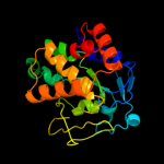
Image coloured by rainbow N → C terminus
Model (left) based on template d1ouoa_
Top template information
Fold:His-Me finger endonucleases
Superfamily:His-Me finger endonucleases
Family:Endonuclease I
Confidence and coverage
Confidence:100.0% Coverage: 89%
208 residues ( 89% of your sequence) have been modelled with 100.0% confidence by the single highest scoring template.
3D viewing
Interactive 3D view in Jmol

Sequence analysis 

Secondary structure and disorder prediction 

   1........10.........20.........30.........40.........50.........60
Sequence  MYRYLSIAAVVLSAAFSGPALAEGINSFSQAKAAAVKVHADAPGTFYCGCKINWQGKKGV
Secondary structure 


SS confidence 


Disorder  ???


??????????


Disorder confidence 


   .........70.........80.........90.........100.........110.........120
Sequence  VDLQSCGYQVRKNENRASRVEWEHVVPAWQFGHQRQCWQDGGRKNCAKDPVYRKMESDMH
Secondary structure 


SS confidence 


Disorder 


??

???????


????????????????????????


Disorder confidence 


   .........130.........140.........150.........160.........170.........180
Sequence  NLQPSVGEVNGDRGNFMYSQWNGGEGQYGQCAMKVDFKEKAAEPPARARGAIARTYFYMR
Secondary structure 


SS confidence 


Disorder 


????


Disorder confidence 


   .........190.........200.........210.........220.........230.....
Sequence  DQYNLTLSRQQTQLFNAWNKMYPVTDWECERDERIAKVQGNHNPYVQRACQARKS
Secondary structure 


SS confidence 


Disorder 


?


????
Disorder confidence 


Confidence Key
High(9)                    Low (0)
?Disordered
Alpha helix
Beta strand

Domain analysis 

Hover over an aligned region to see model and summary info

Please note, only up to the top 20 hits are modelled to reduce computer load

RankAligned region

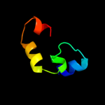
PDB 1ouo chain A

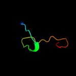
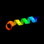
3D model

Region: 23 - 230
Aligned: 208
Modelled: 207
Confidence: 100.0%
Identity: 63%
Fold: His-Me finger endonucleases
Superfamily: His-Me finger endonucleases
Family: Endonuclease I

Phyre2

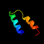
PDB 2qgp chain A

3D model

Region: 46 - 141
Aligned: 50
Modelled: 50
Confidence: 93.5%
Identity: 32%
PDB header:hydrolase
Chain: A: PDB Molecule:hnh endonuclease;
PDBTitle: x-ray structure of the nhn endonuclease from geobacter2 metallireducens. northeast structural genomics consortium3 target gmr87.

Phyre2

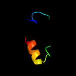
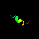
PDB 2ebf chain X domain 3

3D model

Region: 203 - 227
Aligned: 19
Modelled: 25
Confidence: 29.5%
Identity: 47%
Fold: Cysteine proteinases
Superfamily: Cysteine proteinases
Family: PMT C-terminal domain like

Phyre2

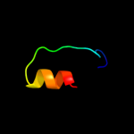
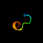
PDB 2e72 chain A

3D model

Region: 92 - 117
Aligned: 26
Modelled: 26
Confidence: 29.2%
Identity: 38%
PDB header:structural genomics, unknown function
Chain: A: PDB Molecule:pogo transposable element with znf domain;
PDBTitle: solution structure of the zinc finger domain of human2 kiaa0461

Phyre2

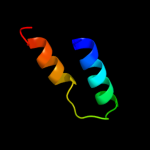
PDB 3c4r chain C

3D model

Region: 171 - 209
Aligned: 38
Modelled: 39
Confidence: 23.4%
Identity: 24%
PDB header:structural genomics, unknown function
Chain: C: PDB Molecule:uncharacterized protein;
PDBTitle: crystal structure of an uncharacterized protein encoded by2 cryptic prophage

Phyre2

PDB 2ebf chain X domain 1

3D model

Region: 171 - 204
Aligned: 34
Modelled: 34
Confidence: 20.9%
Identity: 12%
Fold: PMT central region-like
Superfamily: PMT central region-like
Family: PMT central region-like

Phyre2

PDB 2lmd chain A

3D model

Region: 176 - 198
Aligned: 23
Modelled: 23
Confidence: 18.9%
Identity: 22%
PDB header:transcription
Chain: A: PDB Molecule:prospero homeobox protein 1;
PDBTitle: minimal constraints solution nmr structure of prospero homeobox2 protein 1 from homo sapiens, northeast structural genomics consortium3 target hr4660b

Phyre2

PDB 1mij chain A

3D model

Region: 176 - 198
Aligned: 23
Modelled: 21
Confidence: 15.7%
Identity: 17%
Fold: DNA/RNA-binding 3-helical bundle
Superfamily: Homeodomain-like
Family: Homeodomain

Phyre2

PDB 3gqe chain A

3D model

Region: 108 - 139
Aligned: 31
Modelled: 32
Confidence: 15.4%
Identity: 16%
PDB header:viral protein
Chain: A: PDB Molecule:non-structural protein 3;
PDBTitle: crystal structure of macro domain of venezuelan equine encephalitis2 virus

Phyre2

PDB 2evr chain A domain 1

3D model

Region: 96 - 126
Aligned: 16
Modelled: 16
Confidence: 14.7%
Identity: 38%
Fold: SH3-like barrel
Superfamily: Prokaryotic SH3-related domain
Family: Spr N-terminal domain-like

Phyre2

PDB 1tr8 chain A

3D model

Region: 166 - 177
Aligned: 12
Modelled: 11
Confidence: 13.7%
Identity: 17%
PDB header:chaperone
Chain: A: PDB Molecule:conserved protein (mth177);
PDBTitle: crystal structure of archaeal nascent polypeptide-associated complex2 (aenac)

Phyre2

PDB 2ec5 chain B

3D model

Region: 171 - 204
Aligned: 34
Modelled: 34
Confidence: 11.2%
Identity: 12%
PDB header:toxin
Chain: B: PDB Molecule:dermonecrotic toxin;
PDBTitle: crystal structures reveal a thiol-protease like catalytic triad in the2 c-terminal region of pasteurella multocida toxin

Phyre2

PDB 1em8 chain B

3D model

Region: 29 - 47
Aligned: 19
Modelled: 19
Confidence: 11.1%
Identity: 21%
Fold: DNA polymerase III psi subunit
Superfamily: DNA polymerase III psi subunit
Family: DNA polymerase III psi subunit

Phyre2

PDB 1m8o chain B

3D model

Region: 194 - 229
Aligned: 34
Modelled: 36
Confidence: 10.6%
Identity: 26%
PDB header:membrane protein
Chain: B: PDB Molecule:platele integrin beta3 subunit: cytoplasmic
PDBTitle: platelet integrin alfaiib-beta3 cytoplasmic domain

Phyre2

PDB 3gpq chain A

3D model

Region: 110 - 139
Aligned: 29
Modelled: 30
Confidence: 10.6%
Identity: 21%
PDB header:viral protein/rna
Chain: A: PDB Molecule:non-structural protein 3;
PDBTitle: crystal structure of macro domain of chikungunya virus in complex with2 rna

Phyre2

PDB 1yc6 chain 1 domain 1

3D model

Region: 61 - 87
Aligned: 25
Modelled: 27
Confidence: 9.7%
Identity: 28%
Fold: Nucleoplasmin-like/VP (viral coat and capsid proteins)
Superfamily: Positive stranded ssRNA viruses
Family: Bromoviridae-like VP

Phyre2

PDB 2bpb chain B

3D model

Region: 165 - 198
Aligned: 30
Modelled: 34
Confidence: 9.6%
Identity: 10%
PDB header:oxidoreductase
Chain: B: PDB Molecule:sulfite\:cytochrome c oxidoreductase subunit b;
PDBTitle: sulfite dehydrogenase from starkeya novella

Phyre2

PDB 3g9w chain C

3D model

Region: 193 - 229
Aligned: 35
Modelled: 37
Confidence: 9.1%
Identity: 20%
PDB header:cell adhesion
Chain: C: PDB Molecule:integrin beta-1d;
PDBTitle: crystal structure of talin2 f2-f3 in complex with the integrin beta1d2 cytoplasmic tail

Phyre2

PDB 2bcg chain G domain 3

3D model

Region: 85 - 124
Aligned: 26
Modelled: 26
Confidence: 8.4%
Identity: 42%
Fold: FAD-linked reductases, C-terminal domain
Superfamily: FAD-linked reductases, C-terminal domain
Family: GDI-like

Phyre2

PDB 3t4a chain G

3D model

Region: 27 - 41
Aligned: 15
Modelled: 15
Confidence: 8.0%
Identity: 33%
PDB header:immune system
Chain: G: PDB Molecule:fibrinogen-binding protein;
PDBTitle: structure of a truncated form of staphylococcal complement inhibitor b2 bound to human c3c at 3.4 angstrom resolution

Phyre2
1

d1ouoa_
2

c2qgpA_
3

d2ebfx3
4

c2e72A_
5

c3c4rC_
6

d2ebfx1
7

c2lmdA_
8

d1mija_
9

c3gqeA_
10

d2evra1
11

c1tr8A_
12

c2ec5B_
13

d1em8b_
14

c1m8oB_
15

c3gpqA_
16

d1yc611
17

c2bpbB_
18

c3g9wC_
19

d2bcgg3
20

c3t4aG_
21


22


23


24


25


26


27


28


29


30


31


32


33


34


35


36


37


38


39


40


41


42


43


44


45


Detailed template information 

#
Template Alignment Coverage3D Model Confidence
% i.d. Template Information
1d1ouoa_


100.0 63 Fold:His-Me finger endonucleases
Superfamily:His-Me finger endonucleases
Family:Endonuclease I
2c2qgpA_


93.5 32 PDB header:hydrolase
Chain: A: PDB Molecule:hnh endonuclease;
PDBTitle: x-ray structure of the nhn endonuclease from geobacter2 metallireducens. northeast structural genomics consortium3 target gmr87.
3d2ebfx3


29.5 47 Fold:Cysteine proteinases
Superfamily:Cysteine proteinases
Family:PMT C-terminal domain like
4c2e72A_


29.2 38 PDB header:structural genomics, unknown function
Chain: A: PDB Molecule:pogo transposable element with znf domain;
PDBTitle: solution structure of the zinc finger domain of human2 kiaa0461
5c3c4rC_


23.4 24 PDB header:structural genomics, unknown function
Chain: C: PDB Molecule:uncharacterized protein;
PDBTitle: crystal structure of an uncharacterized protein encoded by2 cryptic prophage
6d2ebfx1


20.9 12 Fold:PMT central region-like
Superfamily:PMT central region-like
Family:PMT central region-like
7c2lmdA_


18.9 22 PDB header:transcription
Chain: A: PDB Molecule:prospero homeobox protein 1;
PDBTitle: minimal constraints solution nmr structure of prospero homeobox2 protein 1 from homo sapiens, northeast structural genomics consortium3 target hr4660b
8d1mija_


15.7 17 Fold:DNA/RNA-binding 3-helical bundle
Superfamily:Homeodomain-like
Family:Homeodomain
9c3gqeA_


15.4 16 PDB header:viral protein
Chain: A: PDB Molecule:non-structural protein 3;
PDBTitle: crystal structure of macro domain of venezuelan equine encephalitis2 virus
10d2evra1


14.7 38 Fold:SH3-like barrel
Superfamily:Prokaryotic SH3-related domain
Family:Spr N-terminal domain-like
11c1tr8A_


13.7 17 PDB header:chaperone
Chain: A: PDB Molecule:conserved protein (mth177);
PDBTitle: crystal structure of archaeal nascent polypeptide-associated complex2 (aenac)
12c2ec5B_


11.2 12 PDB header:toxin
Chain: B: PDB Molecule:dermonecrotic toxin;
PDBTitle: crystal structures reveal a thiol-protease like catalytic triad in the2 c-terminal region of pasteurella multocida toxin
13d1em8b_


11.1 21 Fold:DNA polymerase III psi subunit
Superfamily:DNA polymerase III psi subunit
Family:DNA polymerase III psi subunit
14c1m8oB_


10.6 26 PDB header:membrane protein
Chain: B: PDB Molecule:platele integrin beta3 subunit: cytoplasmic
PDBTitle: platelet integrin alfaiib-beta3 cytoplasmic domain
15c3gpqA_


10.6 21 PDB header:viral protein/rna
Chain: A: PDB Molecule:non-structural protein 3;
PDBTitle: crystal structure of macro domain of chikungunya virus in complex with2 rna
16d1yc611


9.7 28 Fold:Nucleoplasmin-like/VP (viral coat and capsid proteins)
Superfamily:Positive stranded ssRNA viruses
Family:Bromoviridae-like VP
17c2bpbB_


9.6 10 PDB header:oxidoreductase
Chain: B: PDB Molecule:sulfite\:cytochrome c oxidoreductase subunit b;
PDBTitle: sulfite dehydrogenase from starkeya novella
18c3g9wC_


9.1 20 PDB header:cell adhesion
Chain: C: PDB Molecule:integrin beta-1d;
PDBTitle: crystal structure of talin2 f2-f3 in complex with the integrin beta1d2 cytoplasmic tail
19d2bcgg3


8.4 42 Fold:FAD-linked reductases, C-terminal domain
Superfamily:FAD-linked reductases, C-terminal domain
Family:GDI-like
20c3t4aG_


8.0 33 PDB header:immune system
Chain: G: PDB Molecule:fibrinogen-binding protein;
PDBTitle: structure of a truncated form of staphylococcal complement inhibitor b2 bound to human c3c at 3.4 angstrom resolution
21d1js9c_


not modelled 7.8 28 Fold:Nucleoplasmin-like/VP (viral coat and capsid proteins)
Superfamily:Positive stranded ssRNA viruses
Family:Bromoviridae-like VP
22d1i5za1


not modelled 7.8 20 Fold:DNA/RNA-binding 3-helical bundle
Superfamily:"Winged helix" DNA-binding domain
Family:CAP C-terminal domain-like
23d1qbaa2


not modelled 7.7 18 Fold:Common fold of diphtheria toxin/transcription factors/cytochrome f
Superfamily:Carbohydrate-binding domain
Family:Bacterial chitobiase, n-terminal domain
24d2dk1a1


not modelled 7.6 18 Fold:WW domain-like
Superfamily:WW domain
Family:WW domain
25c2a45L_


not modelled 7.5 25 PDB header:hydrolase/hydrolase inhibitor
Chain: L: PDB Molecule:fibrinogen gamma chain;
PDBTitle: crystal structure of the complex between thrombin and the central "e"2 region of fibrin
26c3gnzP_


not modelled 7.5 50 PDB header:toxin
Chain: P: PDB Molecule:25 kda protein elicitor;
PDBTitle: toxin fold for microbial attack and plant defense
27c3bunB_


not modelled 7.3 24 PDB header:ligase/signaling protein
Chain: B: PDB Molecule:e3 ubiquitin-protein ligase cbl;
PDBTitle: crystal structure of c-cbl-tkb domain complexed with its2 binding motif in sprouty4
28c2cblA_


not modelled 7.0 24 PDB header:complex (proto-oncogene/peptide)
Chain: A: PDB Molecule:proto-oncogene cbl;
PDBTitle: n-terminal domain of cbl in complex with its binding site2 on zap-70
29d1yy9a4


not modelled 6.9 30 Fold:Knottins (small inhibitors, toxins, lectins)
Superfamily:Growth factor receptor domain
Family:Growth factor receptor domain
30c2qffA_


not modelled 6.8 27 PDB header:hydrolase inhibitor
Chain: A: PDB Molecule:hypothetical protein;
PDBTitle: crystal structure of staphylococcal complement inhibitor
31d1cwpa_


not modelled 6.7 56 Fold:Nucleoplasmin-like/VP (viral coat and capsid proteins)
Superfamily:Positive stranded ssRNA viruses
Family:Bromoviridae-like VP
32c3st1A_


not modelled 6.3 40 PDB header:toxin
Chain: A: PDB Molecule:necrosis-and ethylene-inducing protein;
PDBTitle: crystal structure of necrosis and ethylene inducing protein 2 from the2 causal agent of cocoa's witches broom disease
33c3nzqB_


not modelled 6.2 32 PDB header:lyase
Chain: B: PDB Molecule:biosynthetic arginine decarboxylase;
PDBTitle: crystal structure of biosynthetic arginine decarboxylase adc (spea)2 from escherichia coli, northeast structural genomics consortium3 target er600
34c2kncB_


not modelled 6.1 27 PDB header:cell adhesion
Chain: B: PDB Molecule:integrin beta-3;
PDBTitle: platelet integrin alfaiib-beta3 transmembrane-cytoplasmic2 heterocomplex
35c2y69S_


not modelled 6.0 43 PDB header:electron transport
Chain: S: PDB Molecule:cytochrome c oxidase subunit 5b;
PDBTitle: bovine heart cytochrome c oxidase re-refined with molecular2 oxygen
36d1d5ta2


not modelled 6.0 35 Fold:FAD-linked reductases, C-terminal domain
Superfamily:FAD-linked reductases, C-terminal domain
Family:GDI-like
37d3buxb2


not modelled 6.0 17 Fold:N-cbl like
Superfamily:N-terminal domain of cbl (N-cbl)
Family:N-terminal domain of cbl (N-cbl)
38d2gaua1


not modelled 5.9 20 Fold:DNA/RNA-binding 3-helical bundle
Superfamily:"Winged helix" DNA-binding domain
Family:CAP C-terminal domain-like
39c2yqfA_


not modelled 5.9 23 PDB header:protein binding
Chain: A: PDB Molecule:ankyrin-1;
PDBTitle: solution structure of the death domain of ankyrin-1
40d1cwva5


not modelled 5.8 22 Fold:C-type lectin-like
Superfamily:C-type lectin-like
Family:Invasin/intimin cell-adhesion fragment, C-terminal domain
41c1cwpB_


not modelled 5.7 56 PDB header:virus/rna
Chain: B: PDB Molecule:coat protein;
PDBTitle: structures of the native and swollen forms of cowpea2 chlorotic mottle virus determined by x-ray crystallography3 and cryo-electron microscopy
42c1hp9A_


not modelled 5.7 60 PDB header:toxin
Chain: A: PDB Molecule:kappa-hefutoxin 1;
PDBTitle: kappa-hefutoxins: a novel class of potassium channel toxins2 from scorpion venom
43d1pbya1


not modelled 5.3 13 Fold:Cytochrome c
Superfamily:Cytochrome c
Family:Quinohemoprotein amine dehydrogenase A chain, domains 1 and 2
44c3rnvA_


not modelled 5.2 13 PDB header:hydrolase
Chain: A: PDB Molecule:helper component proteinase;
PDBTitle: structure of the autocatalytic cysteine protease domain of potyvirus2 helper-component proteinase
45c2q4pA_


not modelled 5.0 11 PDB header:structural genomics, unknown function
Chain: A: PDB Molecule:protein rs21-c6;
PDBTitle: ensemble refinement of the crystal structure of protein from mus2 musculus mm.29898

Binding site prediction 

Due to computational demand, binding site predictions are not run for batch jobs

If you want to predict binding sites, please manually submit your model of choice to 3DLigandSite


Phyre is for academic use only

Please cite: Protein structure prediction on the web: a case study using the Phyre server
Kelley LA and Sternberg MJE. Nature Protocols 4, 363 - 371 (2009) [pdf] [Import into BibTeX]
 
If you use the binding site predictions from 3DLigandSite, please also cite:
3DLigandSite: predicting ligand-binding sites using similar structures.
Wass MN, Kelley LA and Sternberg MJ Nucleic Acids Research 38, W469-73 (2010) [PubMed]
 
© Structural Bioinformatics Group
Imperial College London
Lawrence Kelley, Benjamin Jefferys
Disclaimer
Terms and Conditions
Component software
Template detection: HHpred 1.51
Secondary structure prediction: Psi-pred 2.5
Disorder prediction: Disopred 2.4
Transmembrane prediction: Memsat_SVM
Multi-template modelling and ab initio: Poing 1.0