Phyre Home Retrieve Phyre Job Id
Emaill.a.kelley@imperial.ac.uk
DescriptionP62768
DateThu Jan 5 12:07:50 GMT 2012
Unique Job ID8b0e5b8225681ef0

Summary 

Top model
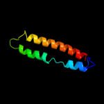
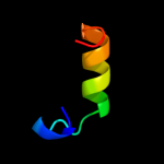
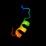
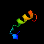
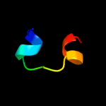
Image coloured by rainbow N → C terminus
Model (left) based on template c2dnxA_
Top template information
PDB header:transport protein
Chain: A: PDB Molecule:syntaxin-12;
PDBTitle: solution structure of rsgi ruh-063, an n-terminal domain of2 syntaxin 12 from human cdna
Confidence and coverage
Confidence: 32.4% Coverage: 52%
67 residues ( 52% of your sequence) have been modelled with 32.4% confidence by the single highest scoring template.
You may wish to submit your sequence to Phyrealarm. This will automatically scan your sequence every week for new potential templates as they appear in the Phyre2 library.
Please note: You must be registered and logged in to use Phyrealarm.
3D viewing
Interactive 3D view in Jmol

Sequence analysis 

Secondary structure and disorder prediction 

   1........10.........20.........30.........40.........50.........60
Sequence  MYDNLKSLGITNPEEIDRYSLRQEANNDILKIYFQKDKGEFFAKSVKFKYPRQRKTVVAD
Secondary structure 


SS confidence 


Disorder  ????


?


?


???
?


??

????
Disorder confidence 


   .........70.........80.........90.........100.........110.........120
Sequence  GVGQGYKEVQEISPNLRYIIDELDQICQRDRSEVDLKRKILDDLRHLESVVTNKISEIEA
Secondary structure 


SS confidence 


Disorder  ????


??
??


???


Disorder confidence 


   ........
Sequence  DLEKLTRK
Secondary structure 

SS confidence 


Disorder 


???
Disorder confidence 


Confidence Key
High(9)                    Low (0)
?Disordered
Alpha helix
Beta strand

Domain analysis 

Hover over an aligned region to see model and summary info

Please note, only up to the top 20 hits are modelled to reduce computer load

RankAligned region

PDB 2dnx chain A

3D model

Region: 58 - 125
Aligned: 67
Modelled: 68
Confidence: 32.4%
Identity: 16%
PDB header:transport protein
Chain: A: PDB Molecule:syntaxin-12;
PDBTitle: solution structure of rsgi ruh-063, an n-terminal domain of2 syntaxin 12 from human cdna

Phyre2

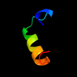
PDB 3t97 chain B

3D model

Region: 87 - 126
Aligned: 40
Modelled: 40
Confidence: 27.8%
Identity: 15%
PDB header:protein transport
Chain: B: PDB Molecule:nuclear pore complex protein nup54;
PDBTitle: molecular architecture of the transport channel of the nuclear pore2 complex: nup62/nup54

Phyre2

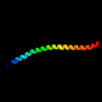
PDB 1s94 chain A

3D model

Region: 66 - 125
Aligned: 56
Modelled: 60
Confidence: 24.8%
Identity: 18%
Fold: STAT-like
Superfamily: t-snare proteins
Family: t-snare proteins

Phyre2

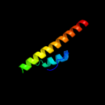
PDB 1s94 chain A

3D model

Region: 66 - 125
Aligned: 56
Modelled: 60
Confidence: 24.8%
Identity: 18%
PDB header:endocytosis/exocytosis
Chain: A: PDB Molecule:s-syntaxin;
PDBTitle: crystal structure of the habc domain of neuronal syntaxin from the2 squid loligo pealei

Phyre2

PDB 2dah chain A domain 1

3D model

Region: 5 - 26
Aligned: 22
Modelled: 22
Confidence: 24.2%
Identity: 23%
Fold: RuvA C-terminal domain-like
Superfamily: UBA-like
Family: UBA domain

Phyre2

PDB 2dna chain A domain 1

3D model

Region: 5 - 26
Aligned: 22
Modelled: 22
Confidence: 23.6%
Identity: 14%
Fold: RuvA C-terminal domain-like
Superfamily: UBA-like
Family: UBA domain

Phyre2

PDB 2bwb chain A domain 1

3D model

Region: 5 - 26
Aligned: 22
Modelled: 22
Confidence: 21.9%
Identity: 14%
Fold: RuvA C-terminal domain-like
Superfamily: UBA-like
Family: UBA domain

Phyre2

PDB 2cwb chain A

3D model

Region: 5 - 26
Aligned: 22
Modelled: 22
Confidence: 21.8%
Identity: 23%
PDB header:protein binding
Chain: A: PDB Molecule:chimera of immunoglobulin g binding protein g
PDBTitle: solution structure of the ubiquitin-associated domain of2 human bmsc-ubp and its complex with ubiquitin

Phyre2

PDB 1vej chain A domain 1

3D model

Region: 5 - 26
Aligned: 22
Modelled: 22
Confidence: 21.3%
Identity: 23%
Fold: RuvA C-terminal domain-like
Superfamily: UBA-like
Family: UBA domain

Phyre2

PDB 1wr1 chain B

3D model

Region: 5 - 26
Aligned: 22
Modelled: 22
Confidence: 20.4%
Identity: 14%
PDB header:signaling protein
Chain: B: PDB Molecule:ubiquitin-like protein dsk2;
PDBTitle: the complex sturcture of dsk2p uba with ubiquitin

Phyre2

PDB 1m1j chain A

3D model

Region: 58 - 123
Aligned: 66
Modelled: 66
Confidence: 18.5%
Identity: 14%
PDB header:blood clotting
Chain: A: PDB Molecule:fibrinogen alpha subunit;
PDBTitle: crystal structure of native chicken fibrinogen with two different2 bound ligands

Phyre2

PDB 1vp7 chain D

3D model

Region: 69 - 126
Aligned: 58
Modelled: 58
Confidence: 18.5%
Identity: 10%
PDB header:hydrolase
Chain: D: PDB Molecule:exodeoxyribonuclease vii small subunit;
PDBTitle: crystal structure of exodeoxyribonuclease vii small subunit2 (np_881400.1) from bordetella pertussis at 2.40 a resolution

Phyre2

PDB 2dna chain A

3D model

Region: 5 - 26
Aligned: 22
Modelled: 22
Confidence: 17.6%
Identity: 14%
PDB header:structural genomics, unknown function
Chain: A: PDB Molecule:unnamed protein product;
PDBTitle: solution structure of rsgi ruh-056, a uba domain from mouse2 cdna

Phyre2

PDB 2dah chain A

3D model

Region: 5 - 26
Aligned: 22
Modelled: 22
Confidence: 16.8%
Identity: 23%
PDB header:structural genomics, unknown function
Chain: A: PDB Molecule:ubiquilin-3;
PDBTitle: solution structure of the c-terminal uba domain in the2 human ubiquilin 3

Phyre2

PDB 2jy5 chain A

3D model

Region: 5 - 26
Aligned: 22
Modelled: 22
Confidence: 16.0%
Identity: 18%
PDB header:signaling protein
Chain: A: PDB Molecule:ubiquilin-1;
PDBTitle: nmr structure of ubiquilin 1 uba domain

Phyre2

PDB 3a2a chain C

3D model

Region: 94 - 128
Aligned: 34
Modelled: 35
Confidence: 15.8%
Identity: 32%
PDB header:transport protein
Chain: C: PDB Molecule:voltage-gated hydrogen channel 1;
PDBTitle: the structure of the carboxyl-terminal domain of the human voltage-2 gated proton channel hv1

Phyre2

PDB 1deq chain O

3D model

Region: 67 - 125
Aligned: 59
Modelled: 59
Confidence: 14.3%
Identity: 10%
PDB header:
PDB COMPND:

Phyre2

PDB 2eam chain A

3D model

Region: 3 - 18
Aligned: 16
Modelled: 16
Confidence: 11.7%
Identity: 31%
PDB header:signaling protein
Chain: A: PDB Molecule:putative 47 kda protein;
PDBTitle: solution structure of the n-terminal sam-domain of a human2 putative 47 kda protein

Phyre2

PDB 1ydi chain B

3D model

Region: 105 - 122
Aligned: 18
Modelled: 18
Confidence: 11.6%
Identity: 22%
PDB header:cell adhesion, structural protein
Chain: B: PDB Molecule:alpha-actinin 4;
PDBTitle: human vinculin head domain (vh1, 1-258) in complex with2 human alpha-actinin's vinculin-binding site (residues 731-3 760)

Phyre2

PDB 1o5h chain A

3D model

Region: 54 - 124
Aligned: 66
Modelled: 71
Confidence: 10.4%
Identity: 15%
Fold: Methenyltetrahydrofolate cyclohydrolase-like
Superfamily: Methenyltetrahydrofolate cyclohydrolase-like
Family: Methenyltetrahydrofolate cyclohydrolase-like

Phyre2
1

c2dnxA_
2

c3t97B_
3

d1s94a_
4

c1s94A_
5

d2daha1
6

d2dnaa1
7

d2bwba1
8

c2cwbA_
9

d1veja1
10

c1wr1B_
11

c1m1jA_
12

c1vp7D_
13

c2dnaA_
14

c2dahA_
15

c2jy5A_
16

c3a2aC_
17

c1deqO_
18

c2eamA_
19

c1ydiB_
20

d1o5ha_
21


22


23


24


25


26


27


28


29


30


31


32


33


34


35


36


37


38


39


40


41


42


43


44


45


46


47


48


49


Detailed template information 

#
Template Alignment Coverage3D Model Confidence
% i.d. Template Information
1c2dnxA_


32.4 16 PDB header:transport protein
Chain: A: PDB Molecule:syntaxin-12;
PDBTitle: solution structure of rsgi ruh-063, an n-terminal domain of2 syntaxin 12 from human cdna
2c3t97B_


27.8 15 PDB header:protein transport
Chain: B: PDB Molecule:nuclear pore complex protein nup54;
PDBTitle: molecular architecture of the transport channel of the nuclear pore2 complex: nup62/nup54
3d1s94a_


24.8 18 Fold:STAT-like
Superfamily:t-snare proteins
Family:t-snare proteins
4c1s94A_


24.8 18 PDB header:endocytosis/exocytosis
Chain: A: PDB Molecule:s-syntaxin;
PDBTitle: crystal structure of the habc domain of neuronal syntaxin from the2 squid loligo pealei
5d2daha1


24.2 23 Fold:RuvA C-terminal domain-like
Superfamily:UBA-like
Family:UBA domain
6d2dnaa1


23.6 14 Fold:RuvA C-terminal domain-like
Superfamily:UBA-like
Family:UBA domain
7d2bwba1


21.9 14 Fold:RuvA C-terminal domain-like
Superfamily:UBA-like
Family:UBA domain
8c2cwbA_


21.8 23 PDB header:protein binding
Chain: A: PDB Molecule:chimera of immunoglobulin g binding protein g
PDBTitle: solution structure of the ubiquitin-associated domain of2 human bmsc-ubp and its complex with ubiquitin
9d1veja1


21.3 23 Fold:RuvA C-terminal domain-like
Superfamily:UBA-like
Family:UBA domain
10c1wr1B_


20.4 14 PDB header:signaling protein
Chain: B: PDB Molecule:ubiquitin-like protein dsk2;
PDBTitle: the complex sturcture of dsk2p uba with ubiquitin
11c1m1jA_


18.5 14 PDB header:blood clotting
Chain: A: PDB Molecule:fibrinogen alpha subunit;
PDBTitle: crystal structure of native chicken fibrinogen with two different2 bound ligands
12c1vp7D_


18.5 10 PDB header:hydrolase
Chain: D: PDB Molecule:exodeoxyribonuclease vii small subunit;
PDBTitle: crystal structure of exodeoxyribonuclease vii small subunit2 (np_881400.1) from bordetella pertussis at 2.40 a resolution
13c2dnaA_


17.6 14 PDB header:structural genomics, unknown function
Chain: A: PDB Molecule:unnamed protein product;
PDBTitle: solution structure of rsgi ruh-056, a uba domain from mouse2 cdna
14c2dahA_


16.8 23 PDB header:structural genomics, unknown function
Chain: A: PDB Molecule:ubiquilin-3;
PDBTitle: solution structure of the c-terminal uba domain in the2 human ubiquilin 3
15c2jy5A_


16.0 18 PDB header:signaling protein
Chain: A: PDB Molecule:ubiquilin-1;
PDBTitle: nmr structure of ubiquilin 1 uba domain
16c3a2aC_


15.8 32 PDB header:transport protein
Chain: C: PDB Molecule:voltage-gated hydrogen channel 1;
PDBTitle: the structure of the carboxyl-terminal domain of the human voltage-2 gated proton channel hv1
17c1deqO_


14.3 10 PDB header:
PDB COMPND:
18c2eamA_


11.7 31 PDB header:signaling protein
Chain: A: PDB Molecule:putative 47 kda protein;
PDBTitle: solution structure of the n-terminal sam-domain of a human2 putative 47 kda protein
19c1ydiB_


11.6 22 PDB header:cell adhesion, structural protein
Chain: B: PDB Molecule:alpha-actinin 4;
PDBTitle: human vinculin head domain (vh1, 1-258) in complex with2 human alpha-actinin's vinculin-binding site (residues 731-3 760)
20d1o5ha_


10.4 15 Fold:Methenyltetrahydrofolate cyclohydrolase-like
Superfamily:Methenyltetrahydrofolate cyclohydrolase-like
Family:Methenyltetrahydrofolate cyclohydrolase-like
21c1fllA_


not modelled 10.3 13 PDB header:apoptosis
Chain: A: PDB Molecule:tnf receptor associated factor 3;
PDBTitle: molecular basis for cd40 signaling mediated by traf3
22c4a1cD_


not modelled 9.8 19 PDB header:ribosome
Chain: D: PDB Molecule:60s ribosomal protein l11;
PDBTitle: t.thermophila 60s ribosomal subunit in complex with2 initiation factor 6. this file contains 5s rrna,3 5.8s rrna and proteins of molecule 4.
23c2dl0A_


not modelled 9.5 31 PDB header:signaling protein
Chain: A: PDB Molecule:sam and sh3 domain-containing protein 1;
PDBTitle: solution structure of the sam-domain of the sam and sh32 domain containing protein 1
24c1n73C_


not modelled 8.7 10 PDB header:blood clotting
Chain: C: PDB Molecule:fibrin gamma chain;
PDBTitle: fibrin d-dimer, lamprey complexed with the peptide ligand: gly-his-2 arg-pro-amide
25c2k4pA_


not modelled 8.2 19 PDB header:signaling protein
Chain: A: PDB Molecule:phosphatidylinositol-3,4,5-trisphosphate 5-
PDBTitle: solution structure of ship2-sam
26d1q08a_


not modelled 8.1 21 Fold:Putative DNA-binding domain
Superfamily:Putative DNA-binding domain
Family:DNA-binding N-terminal domain of transcription activators
27d1tafa_


not modelled 8.0 27 Fold:Histone-fold
Superfamily:Histone-fold
Family:TBP-associated factors, TAFs
28d2pa2a1


not modelled 7.7 26 Fold:alpha/beta-Hammerhead
Superfamily:Ribosomal protein L16p/L10e
Family:Ribosomal protein L10e
29c3c98B_


not modelled 7.5 12 PDB header:endocytosis/exocytosis
Chain: B: PDB Molecule:syntaxin-1a;
PDBTitle: revised structure of the munc18a-syntaxin1 complex
30c2qkqA_


not modelled 7.4 19 PDB header:transferase
Chain: A: PDB Molecule:ephrin type-b receptor 4;
PDBTitle: structure of the sam domain of human ephrin type-b receptor2 4
31d1xn7a_


not modelled 7.4 21 Fold:DNA/RNA-binding 3-helical bundle
Superfamily:"Winged helix" DNA-binding domain
Family:Hypothetical protein YhgG
32c2d46A_


not modelled 7.3 58 PDB header:metal transport
Chain: A: PDB Molecule:calcium channel, voltage-dependent, beta 4
PDBTitle: solution structure of the human beta4a-a domain
33d1b0xa_


not modelled 7.2 31 Fold:SAM domain-like
Superfamily:SAM/Pointed domain
Family:SAM (sterile alpha motif) domain
34c1b0xA_


not modelled 7.2 31 PDB header:transferase
Chain: A: PDB Molecule:protein (epha4 receptor tyrosine kinase);
PDBTitle: the crystal structure of an eph receptor sam domain reveals2 a mechanism for modular dimerization.
35d1v38a_


not modelled 6.8 38 Fold:SAM domain-like
Superfamily:SAM/Pointed domain
Family:SAM (sterile alpha motif) domain
36d1ug7a_


not modelled 6.8 46 Fold:Four-helical up-and-down bundle
Superfamily:Domain from hypothetical 2610208m17rik protein
Family:Domain from hypothetical 2610208m17rik protein
37c2cp8A_


not modelled 6.7 15 PDB header:protein binding
Chain: A: PDB Molecule:next to brca1 gene 1 protein;
PDBTitle: solution structure of the rsgi ruh-046, a uba domain from2 human next to brca1 gene 1 protein (kiaa0049 protein)3 r923h variant
38c2ke7A_


not modelled 6.6 31 PDB header:protein binding
Chain: A: PDB Molecule:ankyrin repeat and sterile alpha motif domain-
PDBTitle: nmr structure of the first sam domain from aida1
39c1sfcD_


not modelled 6.1 18 PDB header:transport protein
Chain: D: PDB Molecule:protein (snap-25b);
PDBTitle: neuronal synaptic fusion complex
40d1ez3a_


not modelled 6.0 17 Fold:STAT-like
Superfamily:t-snare proteins
Family:t-snare proteins
41c2fxpA_


not modelled 5.8 15 PDB header:viral protein
Chain: A: PDB Molecule:spike glycoprotein;
PDBTitle: solution structure of the sars-coronavirus hr2 domain
42c3dpqE_


not modelled 5.7 24 PDB header:chaperone, peptide binding protein
Chain: E: PDB Molecule:chaperone protein dnak;
PDBTitle: crystal structure of the substrate binding domain of e.2 coli dnak in complex with a long pyrrhocoricin-derived3 inhibitor peptide (form b)
43c2e8nA_


not modelled 5.6 31 PDB header:transferase, signaling protein
Chain: A: PDB Molecule:ephrin type-a receptor 2;
PDBTitle: solution structure of the c-terminal sam-domain of ephaa2:2 ephrin type-a receptor 2 precursor (ec 2.7.10.1)
44c3pvpA_


not modelled 5.6 13 PDB header:dna binding protein/dna
Chain: A: PDB Molecule:chromosomal replication initiator protein dnaa;
PDBTitle: structure of mycobacterium tuberculosis dnaa-dbd in complex with box22 dna
45c2npsD_


not modelled 5.5 19 PDB header:transport protein
Chain: D: PDB Molecule:syntaxin-6;
PDBTitle: crystal structure of the early endosomal snare complex
46c2zshB_


not modelled 5.3 27 PDB header:hormone receptor
Chain: B: PDB Molecule:della protein gai;
PDBTitle: structural basis of gibberellin(ga3)-induced della2 recognition by the gibberellin receptor
47d1sgga_


not modelled 5.3 13 Fold:SAM domain-like
Superfamily:SAM/Pointed domain
Family:SAM (sterile alpha motif) domain
48d1gm5a1


not modelled 5.2 17 Fold:Four-helical up-and-down bundle
Superfamily:RecG, N-terminal domain
Family:RecG, N-terminal domain
49c3zqsB_


not modelled 5.2 19 PDB header:ligase
Chain: B: PDB Molecule:e3 ubiquitin-protein ligase fancl;
PDBTitle: human fancl central domain

Binding site prediction 

Due to computational demand, binding site predictions are not run for batch jobs

If you want to predict binding sites, please manually submit your model of choice to 3DLigandSite


Phyre is for academic use only

Please cite: Protein structure prediction on the web: a case study using the Phyre server
Kelley LA and Sternberg MJE. Nature Protocols 4, 363 - 371 (2009) [pdf] [Import into BibTeX]
 
If you use the binding site predictions from 3DLigandSite, please also cite:
3DLigandSite: predicting ligand-binding sites using similar structures.
Wass MN, Kelley LA and Sternberg MJ Nucleic Acids Research 38, W469-73 (2010) [PubMed]
 
© Structural Bioinformatics Group
Imperial College London
Lawrence Kelley, Benjamin Jefferys
Disclaimer
Terms and Conditions
Component software
Template detection: HHpred 1.51
Secondary structure prediction: Psi-pred 2.5
Disorder prediction: Disopred 2.4
Transmembrane prediction: Memsat_SVM
Multi-template modelling and ab initio: Poing 1.0