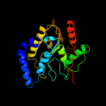
| 1 | d3euga_
|
|
 |
100.0 |
99 |
Fold:Uracil-DNA glycosylase-like
Superfamily:Uracil-DNA glycosylase-like
Family:Uracil-DNA glycosylase |
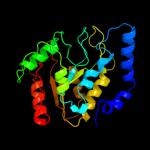
| 2 | c3tr7A_
|
|
 |
100.0 |
62 |
PDB header:hydrolase
Chain: A: PDB Molecule:uracil-dna glycosylase;
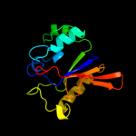
PDBTitle: structure of a uracil-dna glycosylase (ung) from coxiella burnetii
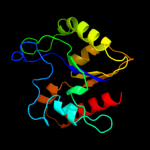
|
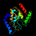
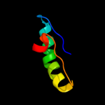
| 3 | d1okba_
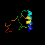
|
|
 |
100.0 |
59 |
Fold:Uracil-DNA glycosylase-like
Superfamily:Uracil-DNA glycosylase-like
Family:Uracil-DNA glycosylase |
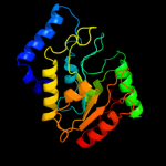
| 4 | d2hxma1
|
|
 |
100.0 |
56 |
Fold:Uracil-DNA glycosylase-like
Superfamily:Uracil-DNA glycosylase-like
Family:Uracil-DNA glycosylase |
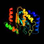
| 5 | c2booA_
|
|
 |
100.0 |
55 |
PDB header:hydrolase
Chain: A: PDB Molecule:uracil-dna glycosylase;
PDBTitle: the crystal structure of uracil-dna n-glycosylase (ung)2 from deinococcus radiodurans.
|
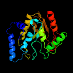
| 6 | c3cxmA_
|
|
 |
100.0 |
48 |
PDB header:hydrolase
Chain: A: PDB Molecule:uracil-dna glycosylase;
PDBTitle: leishmania naiffi uracil-dna glycosylase in complex with 5-bromouracil
|
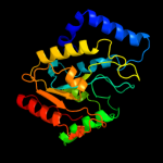
| 7 | d1laue_
|
|
 |
100.0 |
46 |
Fold:Uracil-DNA glycosylase-like
Superfamily:Uracil-DNA glycosylase-like
Family:Uracil-DNA glycosylase |
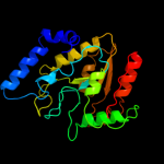
| 8 | c2zhxG_
|
|
 |
100.0 |
38 |
PDB header:hydrolase/hydrolase inhibitor
Chain: G: PDB Molecule:uracil-dna glycosylase;
PDBTitle: crystal structure of uracil-dna glycosylase from mycobacterium2 tuberculosis in complex with a proteinaceous inhibitor
|
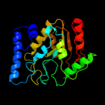
| 9 | d2j8xa1
|
|
 |
100.0 |
48 |
Fold:Uracil-DNA glycosylase-like
Superfamily:Uracil-DNA glycosylase-like
Family:Uracil-DNA glycosylase |
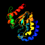
| 10 | c2owrD_
|
|
 |
100.0 |
20 |
PDB header:hydrolase
Chain: D: PDB Molecule:uracil-dna glycosylase;
PDBTitle: crystal structure of vaccinia virus uracil-dna glycosylase
|
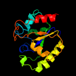
| 11 | c2rbaB_
|
|
 |
97.0 |
10 |
PDB header:hydrolase/dna
Chain: B: PDB Molecule:g/t mismatch-specific thymine dna glycosylase;
PDBTitle: structure of human thymine dna glycosylase bound to abasic and2 undamaged dna
|
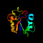
| 12 | c2c2pA_
|
|
 |
95.0 |
13 |
PDB header:hydrolase
Chain: A: PDB Molecule:g/u mismatch-specific dna glycosylase;
PDBTitle: the crystal structure of mismatch specific uracil-dna2 glycosylase (mug) from deinococcus radiodurans
|
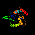
| 13 | d1oe4a_
|
|
 |
94.7 |
17 |
Fold:Uracil-DNA glycosylase-like
Superfamily:Uracil-DNA glycosylase-like
Family:Single-strand selective monofunctional uracil-DNA glycosylase SMUG1 |
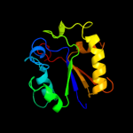
| 14 | d1muga_
|
|
 |
94.7 |
13 |
Fold:Uracil-DNA glycosylase-like
Superfamily:Uracil-DNA glycosylase-like
Family:Mug-like |
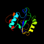
| 15 | d1ui0a_
|
|
 |
94.3 |
18 |
Fold:Uracil-DNA glycosylase-like
Superfamily:Uracil-DNA glycosylase-like
Family:Mug-like |
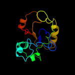
| 16 | c3ikbB_
|
|
 |
93.6 |
23 |
PDB header:structural genomics, unknown function
Chain: B: PDB Molecule:uncharacterized conserved protein;
PDBTitle: the structure of a conserved protein from streptococcus2 mutans ua159.
|
| 17 | c2d3yA_
|
|
 |
92.4 |
16 |
PDB header:hydrolase
Chain: A: PDB Molecule:uracil-dna glycosylase;
PDBTitle: crystal structure of uracil-dna glycosylase from thermus thermophilus2 hb8
|
| 18 | d1vk2a_
|
|
 |
90.4 |
20 |
Fold:Uracil-DNA glycosylase-like
Superfamily:Uracil-DNA glycosylase-like
Family:Mug-like |
| 19 | c2h2wA_
|
|
 |
50.6 |
18 |
PDB header:transferase
Chain: A: PDB Molecule:homoserine o-succinyltransferase;
PDBTitle: crystal structure of homoserine o-succinyltransferase (ec 2.3.1.46)2 (homoserine o-transsuccinylase) (hts) (tm0881) from thermotoga3 maritima at 2.52 a resolution
|
| 20 | c3p9xB_
|
|
 |
35.1 |
18 |
PDB header:transferase
Chain: B: PDB Molecule:phosphoribosylglycinamide formyltransferase;
PDBTitle: crystal structure of phosphoribosylglycinamide formyltransferase from2 bacillus halodurans
|
| 21 | d1meoa_ |
|
not modelled |
32.3 |
20 |
Fold:Formyltransferase
Superfamily:Formyltransferase
Family:Formyltransferase |
| 22 | c2l3fA_ |
|
not modelled |
30.2 |
9 |
PDB header:structural genomics, unknown function
Chain: A: PDB Molecule:uncharacterized protein;
PDBTitle: solution nmr structure of a putative uracil dna glycosylase from2 methanosarcina acetivorans, northeast structural genomics consortium3 target mvr76
|
| 23 | c2ywrA_ |
|
not modelled |
30.0 |
17 |
PDB header:transferase
Chain: A: PDB Molecule:phosphoribosylglycinamide formyltransferase;
PDBTitle: crystal structure of gar transformylase from aquifex2 aeolicus
|
| 24 | c3tqrA_ |
|
not modelled |
25.7 |
17 |
PDB header:transferase
Chain: A: PDB Molecule:phosphoribosylglycinamide formyltransferase;
PDBTitle: structure of the phosphoribosylglycinamide formyltransferase (purn) in2 complex with ches from coxiella burnetii
|
| 25 | d1jkxa_ |
|
not modelled |
22.1 |
25 |
Fold:Formyltransferase
Superfamily:Formyltransferase
Family:Formyltransferase |
| 26 | c3dcjA_ |
|
not modelled |
19.5 |
15 |
PDB header:transferase
Chain: A: PDB Molecule:probable 5'-phosphoribosylglycinamide
PDBTitle: crystal structure of glycinamide formyltransferase (purn)2 from mycobacterium tuberculosis in complex with 5-methyl-5,3 6,7,8-tetrahydrofolic acid derivative
|
| 27 | c3t7hB_ |
|
not modelled |
15.6 |
20 |
PDB header:ligase
Chain: B: PDB Molecule:ubiquitin-like modifier-activating enzyme atg7;
PDBTitle: atg8 transfer from atg7 to atg3: a distinctive e1-e2 architecture and2 mechanism in the autophagy pathway
|
| 28 | c1fmtA_ |
|
not modelled |
15.1 |
21 |
PDB header:formyltransferase
Chain: A: PDB Molecule:methionyl-trna fmet formyltransferase;
PDBTitle: methionyl-trnafmet formyltransferase from escherichia coli
|
| 29 | d2ghra1 |
|
not modelled |
14.4 |
11 |
Fold:Flavodoxin-like
Superfamily:Class I glutamine amidotransferase-like
Family:HTS-like |
| 30 | c3nrbD_ |
|
not modelled |
13.4 |
11 |
PDB header:hydrolase
Chain: D: PDB Molecule:formyltetrahydrofolate deformylase;
PDBTitle: crystal structure of a formyltetrahydrofolate deformylase (puru,2 pp_1943) from pseudomonas putida kt2440 at 2.05 a resolution
|
| 31 | d2blna2 |
|
not modelled |
12.8 |
21 |
Fold:Formyltransferase
Superfamily:Formyltransferase
Family:Formyltransferase |
| 32 | d1fmta2 |
|
not modelled |
12.3 |
18 |
Fold:Formyltransferase
Superfamily:Formyltransferase
Family:Formyltransferase |
| 33 | c2yqsA_ |
|
not modelled |
12.0 |
17 |
PDB header:transferase
Chain: A: PDB Molecule:udp-n-acetylglucosamine pyrophosphorylase;
PDBTitle: crystal structure of uridine-diphospho-n-acetylglucosamine2 pyrophosphorylase from candida albicans, in the product-binding form
|
| 34 | c3o1lB_ |
|
not modelled |
11.8 |
25 |
PDB header:hydrolase
Chain: B: PDB Molecule:formyltetrahydrofolate deformylase;
PDBTitle: crystal structure of a formyltetrahydrofolate deformylase (pspto_4314)2 from pseudomonas syringae pv. tomato str. dc3000 at 2.20 a resolution
|
| 35 | d1maba2 |
|
not modelled |
11.7 |
29 |
Fold:Domain of alpha and beta subunits of F1 ATP synthase-like
Superfamily:N-terminal domain of alpha and beta subunits of F1 ATP synthase
Family:N-terminal domain of alpha and beta subunits of F1 ATP synthase |
| 36 | d2bw0a2 |
|
not modelled |
10.7 |
27 |
Fold:Formyltransferase
Superfamily:Formyltransferase
Family:Formyltransferase |
| 37 | c3n0vD_ |
|
not modelled |
10.5 |
17 |
PDB header:hydrolase
Chain: D: PDB Molecule:formyltetrahydrofolate deformylase;
PDBTitle: crystal structure of a formyltetrahydrofolate deformylase (pp_0327)2 from pseudomonas putida kt2440 at 2.25 a resolution
|
| 38 | c3oc9A_ |
|
not modelled |
10.4 |
14 |
PDB header:transferase
Chain: A: PDB Molecule:udp-n-acetylglucosamine pyrophosphorylase;
PDBTitle: crystal structure of putative udp-n-acetylglucosamine2 pyrophosphorylase from entamoeba histolytica
|
| 39 | c2p2gD_ |
|
not modelled |
10.4 |
12 |
PDB header:transferase
Chain: D: PDB Molecule:ornithine carbamoyltransferase;
PDBTitle: crystal structure of ornithine carbamoyltransferase from mycobacterium2 tuberculosis (rv1656): orthorhombic form
|
| 40 | d1skyb2 |
|
not modelled |
9.8 |
33 |
Fold:Domain of alpha and beta subunits of F1 ATP synthase-like
Superfamily:N-terminal domain of alpha and beta subunits of F1 ATP synthase
Family:N-terminal domain of alpha and beta subunits of F1 ATP synthase |
| 41 | c3ogzA_ |
|
not modelled |
9.6 |
13 |
PDB header:transferase
Chain: A: PDB Molecule:udp-sugar pyrophosphorylase;
PDBTitle: protein structure of usp from l. major in apo-form
|
| 42 | c3obiC_ |
|
not modelled |
9.4 |
15 |
PDB header:hydrolase
Chain: C: PDB Molecule:formyltetrahydrofolate deformylase;
PDBTitle: crystal structure of a formyltetrahydrofolate deformylase (np_949368)2 from rhodopseudomonas palustris cga009 at 1.95 a resolution
|
| 43 | d1fx0a2 |
|
not modelled |
9.4 |
29 |
Fold:Domain of alpha and beta subunits of F1 ATP synthase-like
Superfamily:N-terminal domain of alpha and beta subunits of F1 ATP synthase
Family:N-terminal domain of alpha and beta subunits of F1 ATP synthase |
| 44 | c1yrwA_ |
|
not modelled |
9.3 |
21 |
PDB header:transferase
Chain: A: PDB Molecule:protein arna;
PDBTitle: crystal structure of e.coli arna transformylase domain
|
| 45 | c2xrfA_ |
|
not modelled |
8.9 |
54 |
PDB header:transferase
Chain: A: PDB Molecule:uridine phosphorylase 2;
PDBTitle: crystal structure of human uridine phosphorylase 2
|
| 46 | c3e35A_ |
|
not modelled |
8.5 |
18 |
PDB header:unknown function
Chain: A: PDB Molecule:uncharacterized protein sco1997;
PDBTitle: actinobacteria-specific protein of unknown function, sco1997
|
| 47 | d1pj3a1 |
|
not modelled |
8.5 |
21 |
Fold:NAD(P)-binding Rossmann-fold domains
Superfamily:NAD(P)-binding Rossmann-fold domains
Family:Aminoacid dehydrogenase-like, C-terminal domain |
| 48 | d2jdia2 |
|
not modelled |
8.3 |
29 |
Fold:Domain of alpha and beta subunits of F1 ATP synthase-like
Superfamily:N-terminal domain of alpha and beta subunits of F1 ATP synthase
Family:N-terminal domain of alpha and beta subunits of F1 ATP synthase |
| 49 | d1q3qa2 |
|
not modelled |
8.1 |
26 |
Fold:The "swivelling" beta/beta/alpha domain
Superfamily:GroEL apical domain-like
Family:Group II chaperonin (CCT, TRIC), apical domain |
| 50 | c3kcqA_ |
|
not modelled |
8.1 |
20 |
PDB header:transferase
Chain: A: PDB Molecule:phosphoribosylglycinamide formyltransferase;
PDBTitle: crystal structure of phosphoribosylglycinamide formyltransferase from2 anaplasma phagocytophilum
|
| 51 | d1ekxa2 |
|
not modelled |
7.9 |
26 |
Fold:ATC-like
Superfamily:Aspartate/ornithine carbamoyltransferase
Family:Aspartate/ornithine carbamoyltransferase |
| 52 | d2j13a1 |
|
not modelled |
7.8 |
9 |
Fold:7-stranded beta/alpha barrel
Superfamily:Glycoside hydrolase/deacetylase
Family:NodB-like polysaccharide deacetylase |
| 53 | c1z7eC_ |
|
not modelled |
7.6 |
21 |
PDB header:hydrolase
Chain: C: PDB Molecule:protein arna;
PDBTitle: crystal structure of full length arna
|
| 54 | c2k3mA_ |
|
not modelled |
7.2 |
70 |
PDB header:membrane protein
Chain: A: PDB Molecule:rv1761c;
PDBTitle: rv1761c
|
| 55 | c2iw0A_ |
|
not modelled |
7.2 |
12 |
PDB header:hydrolase
Chain: A: PDB Molecule:chitin deacetylase;
PDBTitle: structure of the chitin deacetylase from the fungal2 pathogen colletotrichum lindemuthianum
|
| 56 | d1r2aa_ |
|
not modelled |
7.0 |
25 |
Fold:Dimerization-anchoring domain of cAMP-dependent PK regulatory subunit
Superfamily:Dimerization-anchoring domain of cAMP-dependent PK regulatory subunit
Family:Dimerization-anchoring domain of cAMP-dependent PK regulatory subunit |
| 57 | c3eufC_ |
|
not modelled |
6.7 |
54 |
PDB header:transferase
Chain: C: PDB Molecule:uridine phosphorylase 1;
PDBTitle: crystal structure of bau-bound human uridine phosphorylase 1
|
| 58 | d1jv1a_ |
|
not modelled |
6.6 |
14 |
Fold:Nucleotide-diphospho-sugar transferases
Superfamily:Nucleotide-diphospho-sugar transferases
Family:UDP-glucose pyrophosphorylase |
| 59 | d1s3ia2 |
|
not modelled |
6.6 |
26 |
Fold:Formyltransferase
Superfamily:Formyltransferase
Family:Formyltransferase |
| 60 | c1w4xA_ |
|
not modelled |
6.4 |
27 |
PDB header:oxygenase
Chain: A: PDB Molecule:phenylacetone monooxygenase;
PDBTitle: phenylacetone monooxygenase, a baeyer-villiger2 monooxygenase
|
| 61 | d2f4za1 |
|
not modelled |
6.4 |
31 |
Fold:UBC-like
Superfamily:UBC-like
Family:UBC-related |
| 62 | d1vm8a_ |
|
not modelled |
6.3 |
17 |
Fold:Nucleotide-diphospho-sugar transferases
Superfamily:Nucleotide-diphospho-sugar transferases
Family:UDP-glucose pyrophosphorylase |
| 63 | c2i5kB_ |
|
not modelled |
6.3 |
14 |
PDB header:transferase
Chain: B: PDB Molecule:utp--glucose-1-phosphate uridylyltransferase;
PDBTitle: crystal structure of ugp1p
|
| 64 | d1w4xa1 |
|
not modelled |
6.3 |
29 |
Fold:FAD/NAD(P)-binding domain
Superfamily:FAD/NAD(P)-binding domain
Family:FAD/NAD-linked reductases, N-terminal and central domains |
| 65 | c3q0iA_ |
|
not modelled |
6.2 |
14 |
PDB header:transferase
Chain: A: PDB Molecule:methionyl-trna formyltransferase;
PDBTitle: methionyl-trna formyltransferase from vibrio cholerae
|
| 66 | d2icya2 |
|
not modelled |
6.2 |
15 |
Fold:Nucleotide-diphospho-sugar transferases
Superfamily:Nucleotide-diphospho-sugar transferases
Family:UDP-glucose pyrophosphorylase |
| 67 | d1vl6a1 |
|
not modelled |
6.0 |
42 |
Fold:NAD(P)-binding Rossmann-fold domains
Superfamily:NAD(P)-binding Rossmann-fold domains
Family:Aminoacid dehydrogenase-like, C-terminal domain |
| 68 | c2yr6A_ |
|
not modelled |
6.0 |
25 |
PDB header:oxidoreductase
Chain: A: PDB Molecule:pro-enzyme of l-phenylalanine oxidase;
PDBTitle: crystal structure of l-phenylalanine oxidase from psuedomonas sp.p501
|
| 69 | d1o0sa1 |
|
not modelled |
5.9 |
21 |
Fold:NAD(P)-binding Rossmann-fold domains
Superfamily:NAD(P)-binding Rossmann-fold domains
Family:Aminoacid dehydrogenase-like, C-terminal domain |
| 70 | c1zghA_ |
|
not modelled |
5.8 |
36 |
PDB header:transferase
Chain: A: PDB Molecule:methionyl-trna formyltransferase;
PDBTitle: methionyl-trna formyltransferase from clostridium thermocellum
|
| 71 | d2vgna2 |
|
not modelled |
5.7 |
29 |
Fold:Ribonuclease H-like motif
Superfamily:Translational machinery components
Family:ERF1/Dom34 middle domain-like |
| 72 | c3rf1B_ |
|
not modelled |
5.7 |
14 |
PDB header:ligase
Chain: B: PDB Molecule:glycyl-trna synthetase alpha subunit;
PDBTitle: the crystal structure of glycyl-trna synthetase subunit alpha from2 campylobacter jejuni subsp. jejuni nctc 11168
|
| 73 | d1gq2a1 |
|
not modelled |
5.6 |
37 |
Fold:NAD(P)-binding Rossmann-fold domains
Superfamily:NAD(P)-binding Rossmann-fold domains
Family:Aminoacid dehydrogenase-like, C-terminal domain |
| 74 | c3tqqA_ |
|
not modelled |
5.6 |
17 |
PDB header:transferase
Chain: A: PDB Molecule:methionyl-trna formyltransferase;
PDBTitle: structure of the methionyl-trna formyltransferase (fmt) from coxiella2 burnetii
|
| 75 | c2vkzH_ |
|
not modelled |
5.6 |
13 |
PDB header:transferase
Chain: H: PDB Molecule:fatty acid synthase subunit beta;
PDBTitle: structure of the cerulenin-inhibited fungal fatty acid2 synthase type i multienzyme complex
|
| 76 | d1w44a_ |
|
not modelled |
5.5 |
33 |
Fold:P-loop containing nucleoside triphosphate hydrolases
Superfamily:P-loop containing nucleoside triphosphate hydrolases
Family:RecA protein-like (ATPase-domain) |
| 77 | d2igsa1 |
|
not modelled |
5.5 |
41 |
Fold:Lysozyme-like
Superfamily:Lysozyme-like
Family:PA2222-like |
| 78 | c3louB_ |
|
not modelled |
5.1 |
12 |
PDB header:hydrolase
Chain: B: PDB Molecule:formyltetrahydrofolate deformylase;
PDBTitle: crystal structure of formyltetrahydrofolate deformylase (yp_105254.1)2 from burkholderia mallei atcc 23344 at 1.90 a resolution
|


