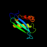
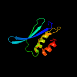
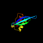
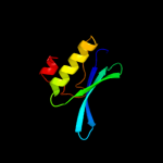
1 c1c0mA_
99.6
19
PDB header: transferaseChain: A: PDB Molecule: protein (integrase);PDBTitle: crystal structure of rsv two-domain integrase
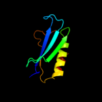
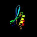
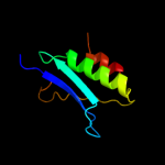
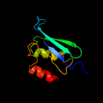
2 d1asua_
99.5
18
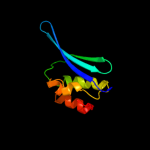
Fold: Ribonuclease H-like motifSuperfamily: Ribonuclease H-likeFamily: Retroviral integrase, catalytic domain3 d1cxqa_
99.5
18
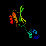
Fold: Ribonuclease H-like motifSuperfamily: Ribonuclease H-likeFamily: Retroviral integrase, catalytic domain4 d1c0ma2
99.4
18
Fold: Ribonuclease H-like motifSuperfamily: Ribonuclease H-likeFamily: Retroviral integrase, catalytic domain5 d1bcoa2
99.3
16
Fold: Ribonuclease H-like motifSuperfamily: Ribonuclease H-likeFamily: mu transposase, core domain6 d1exqa_
99.0
11
Fold: Ribonuclease H-like motifSuperfamily: Ribonuclease H-likeFamily: Retroviral integrase, catalytic domain7 c3nf9A_
99.0
11
PDB header: hydrolase/hydrolase inhibitorChain: A: PDB Molecule: integrase;PDBTitle: structural basis for a new mechanism of inhibition of hiv integrase2 identified by fragment screening and structure based design
8 d1c6va_
99.0
10
Fold: Ribonuclease H-like motifSuperfamily: Ribonuclease H-likeFamily: Retroviral integrase, catalytic domain9 c1ex4A_
98.9
11
PDB header: viral proteinChain: A: PDB Molecule: integrase;PDBTitle: hiv-1 integrase catalytic core and c-terminal domain
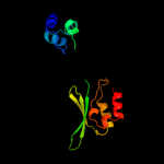
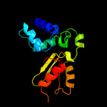
10 c3f9kV_
98.9
10
PDB header: viral protein, recombinationChain: V: PDB Molecule: integrase;PDBTitle: two domain fragment of hiv-2 integrase in complex with ledgf ibd
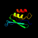
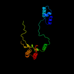
11 c1k6yB_
98.9
9
PDB header: transferaseChain: B: PDB Molecule: integrase;PDBTitle: crystal structure of a two-domain fragment of hiv-1 integrase
12 c3kksB_
98.7
11
PDB header: dna binding proteinChain: B: PDB Molecule: integrase;PDBTitle: crystal structure of catalytic core domain of biv integrase in crystal2 form ii
13 c1bcoA_
98.7
14
PDB header: transposaseChain: A: PDB Molecule: bacteriophage mu transposase;PDBTitle: bacteriophage mu transposase core domain
14 c3hpgC_
98.6
13
PDB header: transferaseChain: C: PDB Molecule: integrase;PDBTitle: visna virus integrase (residues 1-219) in complex with ledgf2 ibd: examples of open integrase dimer-dimer interfaces
15 c3dlrA_
98.5
14
PDB header: transferaseChain: A: PDB Molecule: integrase;PDBTitle: crystal structure of the catalytic core domain from pfv2 integrase
16 d1hyva_
98.5
10
Fold: Ribonuclease H-like motifSuperfamily: Ribonuclease H-likeFamily: Retroviral integrase, catalytic domain17 c3l2tB_
98.3
13
PDB header: recombination/dnaChain: B: PDB Molecule: integrase;PDBTitle: crystal structure of the prototype foamy virus (pfv) intasome in2 complex with magnesium and mk0518 (raltegravir)
18 c3hosA_
97.4
11
PDB header: transferase, dna binding protein/dnaChain: A: PDB Molecule: transposable element mariner, complete cds;PDBTitle: crystal structure of the mariner mos1 paired end complex with mg
19 c3v4gA_
95.4
14
PDB header: dna binding proteinChain: A: PDB Molecule: arginine repressor;PDBTitle: 1.60 angstrom resolution crystal structure of an arginine repressor2 from vibrio vulnificus cmcp6
20 c1u78A_
94.6
11
PDB header: dna binding protein/dnaChain: A: PDB Molecule: transposable element tc3 transposase;PDBTitle: structure of the bipartite dna-binding domain of tc32 transposase bound to transposon dna
21 d1aoya_
not modelled
93.8
11
Fold: DNA/RNA-binding 3-helical bundleSuperfamily: "Winged helix" DNA-binding domainFamily: Arginine repressor (ArgR), N-terminal DNA-binding domain22 c1b4aA_
not modelled
93.2
16
PDB header: repressorChain: A: PDB Molecule: arginine repressor;PDBTitle: structure of the arginine repressor from bacillus stearothermophilus
23 c3ereD_
not modelled
92.1
17
PDB header: dna binding protein/dnaChain: D: PDB Molecule: arginine repressor;PDBTitle: crystal structure of the arginine repressor protein from mycobacterium2 tuberculosis in complex with the dna operator
24 c6paxA_
not modelled
92.0
12
PDB header: gene regulation/dnaChain: A: PDB Molecule: homeobox protein pax-6;PDBTitle: crystal structure of the human pax-6 paired domain-dna2 complex reveals a general model for pax protein-dna3 interactions
25 d1pdnc_
not modelled
87.1
14
Fold: DNA/RNA-binding 3-helical bundleSuperfamily: Homeodomain-likeFamily: Paired domain26 d1b4aa1
not modelled
86.6
15
Fold: DNA/RNA-binding 3-helical bundleSuperfamily: "Winged helix" DNA-binding domainFamily: Arginine repressor (ArgR), N-terminal DNA-binding domain27 d2p5ka1
not modelled
86.2
21
Fold: DNA/RNA-binding 3-helical bundleSuperfamily: "Winged helix" DNA-binding domainFamily: Arginine repressor (ArgR), N-terminal DNA-binding domain28 d1f9na1
not modelled
85.6
18
Fold: DNA/RNA-binding 3-helical bundleSuperfamily: "Winged helix" DNA-binding domainFamily: Arginine repressor (ArgR), N-terminal DNA-binding domain29 c3mwmA_
not modelled
84.3
19
PDB header: transcriptionChain: A: PDB Molecule: putative metal uptake regulation protein;PDBTitle: graded expression of zinc-responsive genes through two regulatory2 zinc-binding sites in zur
30 c2o03A_
not modelled
83.4
17
PDB header: gene regulationChain: A: PDB Molecule: probable zinc uptake regulation protein furb;PDBTitle: crystal structure of furb from m. tuberculosis- a zinc uptake2 regulator
31 c2o8kA_
not modelled
83.0
17
PDB header: transcription/dnaChain: A: PDB Molecule: rna polymerase sigma factor rpon;PDBTitle: nmr structure of the sigma-54 rpon domain bound to the-242 promoter element
32 c2fe3B_
not modelled
80.8
16
PDB header: dna binding proteinChain: B: PDB Molecule: peroxide operon regulator;PDBTitle: the crystal structure of bacillus subtilis perr-zn reveals a novel2 zn(cys)4 structural redox switch
33 c2xigA_
not modelled
79.6
14
PDB header: transcriptionChain: A: PDB Molecule: ferric uptake regulation protein;PDBTitle: the structure of the helicobacter pylori ferric uptake2 regulator fur reveals three functional metal binding sites
34 d1mzba_
not modelled
79.5
14
Fold: DNA/RNA-binding 3-helical bundleSuperfamily: "Winged helix" DNA-binding domainFamily: FUR-like35 c2w57A_
not modelled
72.8
16
PDB header: metal transportChain: A: PDB Molecule: ferric uptake regulation protein;PDBTitle: crystal structure of the vibrio cholerae ferric uptake2 regulator (fur) reveals structural rearrangement of the3 dna-binding domains
36 c2r0qF_
not modelled
72.8
13
PDB header: recombination/dnaChain: F: PDB Molecule: putative transposon tn552 dna-invertase bin3;PDBTitle: crystal structure of a serine recombinase- dna regulatory2 complex
37 c3f2kB_
not modelled
68.3
11
PDB header: transferaseChain: B: PDB Molecule: histone-lysine n-methyltransferase setmar;PDBTitle: structure of the transposase domain of human histone-lysine2 n-methyltransferase setmar
38 c2fu4B_
not modelled
66.8
16
PDB header: dna binding proteinChain: B: PDB Molecule: ferric uptake regulation protein;PDBTitle: crystal structure of the dna binding domain of e.coli fur (ferric2 uptake regulator)
39 d1nkua_
not modelled
64.4
23
Fold: DNA-glycosylaseSuperfamily: DNA-glycosylaseFamily: 3-Methyladenine DNA glycosylase I (Tag)40 d1stza1
not modelled
63.7
9
Fold: DNA/RNA-binding 3-helical bundleSuperfamily: "Winged helix" DNA-binding domainFamily: Heat-inducible transcription repressor HrcA, N-terminal domain41 c2jg6A_
not modelled
62.6
17
PDB header: hydrolaseChain: A: PDB Molecule: dna-3-methyladenine glycosidase;PDBTitle: crystal structure of a 3-methyladenine dna glycosylase i2 from staphylococcus aureus
42 c3eyyA_
not modelled
56.4
19
PDB header: transportChain: A: PDB Molecule: putative iron uptake regulatory protein;PDBTitle: structural basis for the specialization of nur, a nickel-2 specific fur homologue, in metal sensing and dna3 recognition
43 c2xzmO_
not modelled
55.3
13
PDB header: ribosomeChain: O: PDB Molecule: rps13e;PDBTitle: crystal structure of the eukaryotic 40s ribosomal2 subunit in complex with initiation factor 1. this file3 contains the 40s subunit and initiation factor for4 molecule 1
44 c2f7tA_
not modelled
46.5
11
PDB header: dna binding proteinChain: A: PDB Molecule: mos1 transposase;PDBTitle: crystal structure of the catalytic domain of mos1 mariner2 transposase
45 d1u17a1
not modelled
43.6
8
Fold: LipocalinsSuperfamily: LipocalinsFamily: Retinol binding protein-like46 c1hlvA_
not modelled
42.4
20
PDB header: dna binding protein/dnaChain: A: PDB Molecule: major centromere autoantigen b;PDBTitle: crystal structure of cenp-b(1-129) complexed with the cenp-2 b box dna
47 d1x8qa_
not modelled
39.6
6
Fold: LipocalinsSuperfamily: LipocalinsFamily: Retinol binding protein-like48 d2fi9a1
not modelled
35.2
5
Fold: MTH938-likeSuperfamily: MTH938-likeFamily: MTH938-like49 d2fvta1
not modelled
33.8
6
Fold: MTH938-likeSuperfamily: MTH938-likeFamily: MTH938-like50 d1nhpa3
not modelled
22.6
16
Fold: CO dehydrogenase flavoprotein C-domain-likeSuperfamily: FAD/NAD-linked reductases, dimerisation (C-terminal) domainFamily: FAD/NAD-linked reductases, dimerisation (C-terminal) domain51 c2i8bB_
not modelled
22.3
12
PDB header: viral proteinChain: B: PDB Molecule: minor nucleoprotein vp30;PDBTitle: crystal structure of the c-terminal domain of ebola virus vp30
52 c3r1fO_
not modelled
20.8
11
PDB header: transcriptionChain: O: PDB Molecule: esx-1 secretion-associated regulator espr;PDBTitle: crystal structure of a key regulator of virulence in mycobacterium2 tuberculosis
53 d1dxla3
not modelled
20.0
10
Fold: CO dehydrogenase flavoprotein C-domain-likeSuperfamily: FAD/NAD-linked reductases, dimerisation (C-terminal) domainFamily: FAD/NAD-linked reductases, dimerisation (C-terminal) domain54 d1ebda3
not modelled
19.2
17
Fold: CO dehydrogenase flavoprotein C-domain-likeSuperfamily: FAD/NAD-linked reductases, dimerisation (C-terminal) domainFamily: FAD/NAD-linked reductases, dimerisation (C-terminal) domain55 c2rpiA_
not modelled
18.7
11
PDB header: hydrolaseChain: A: PDB Molecule: ribonuclease h;PDBTitle: the nmr structure of the submillisecond folding2 intermediate of the thermus thermophilus ribonuclease h
56 d3grsa3
not modelled
18.2
16
Fold: CO dehydrogenase flavoprotein C-domain-likeSuperfamily: FAD/NAD-linked reductases, dimerisation (C-terminal) domainFamily: FAD/NAD-linked reductases, dimerisation (C-terminal) domain57 d1gesa3
not modelled
15.2
12
Fold: CO dehydrogenase flavoprotein C-domain-likeSuperfamily: FAD/NAD-linked reductases, dimerisation (C-terminal) domainFamily: FAD/NAD-linked reductases, dimerisation (C-terminal) domain58 d1ekja_
not modelled
15.0
16
Fold: Resolvase-likeSuperfamily: beta-carbonic anhydrase, cabFamily: beta-carbonic anhydrase, cab59 d1pm1x_
not modelled
14.4
9
Fold: LipocalinsSuperfamily: LipocalinsFamily: Retinol binding protein-like60 d1lvla3
not modelled
13.3
14
Fold: CO dehydrogenase flavoprotein C-domain-likeSuperfamily: FAD/NAD-linked reductases, dimerisation (C-terminal) domainFamily: FAD/NAD-linked reductases, dimerisation (C-terminal) domain61 c3p9kD_
not modelled
12.5
8
PDB header: transferaseChain: D: PDB Molecule: caffeic acid o-methyltransferase;PDBTitle: crystal structure of perennial ryegrass lpomt1 complexed with s-2 adenosyl-l-homocysteine and coniferaldehyde
62 d1y1xa_
not modelled
12.4
9
Fold: EF Hand-likeSuperfamily: EF-handFamily: Penta-EF-hand proteins63 c2jsoA_
not modelled
11.6
8
PDB header: signaling proteinChain: A: PDB Molecule: polymyxin resistance protein pmrd;PDBTitle: antimicrobial resistance protein
64 c3izbO_
not modelled
11.6
16
PDB header: ribosomeChain: O: PDB Molecule: 40s ribosomal protein rps13 (s15p);PDBTitle: localization of the small subunit ribosomal proteins into a 6.1 a2 cryo-em map of saccharomyces cerevisiae translating 80s ribosome
65 d1lpfa3
not modelled
10.6
12
Fold: CO dehydrogenase flavoprotein C-domain-likeSuperfamily: FAD/NAD-linked reductases, dimerisation (C-terminal) domainFamily: FAD/NAD-linked reductases, dimerisation (C-terminal) domain66 d2i52a1
not modelled
10.4
10
Fold: MK0786-likeSuperfamily: MK0786-likeFamily: MK0786-like67 c3lasA_
not modelled
10.2
11
PDB header: lyaseChain: A: PDB Molecule: putative carbonic anhydrase;PDBTitle: crystal structure of carbonic anhydrase from streptococcus mutans to2 1.4 angstrom resolution
68 d2coba1
not modelled
9.9
9
Fold: DNA/RNA-binding 3-helical bundleSuperfamily: Homeodomain-likeFamily: Psq domain69 d1v59a3
not modelled
9.9
12
Fold: CO dehydrogenase flavoprotein C-domain-likeSuperfamily: FAD/NAD-linked reductases, dimerisation (C-terminal) domainFamily: FAD/NAD-linked reductases, dimerisation (C-terminal) domain70 d1ixsb1
not modelled
9.3
22
Fold: DNA/RNA-binding 3-helical bundleSuperfamily: "Winged helix" DNA-binding domainFamily: Helicase DNA-binding domain71 d1aoga3
not modelled
9.3
3
Fold: CO dehydrogenase flavoprotein C-domain-likeSuperfamily: FAD/NAD-linked reductases, dimerisation (C-terminal) domainFamily: FAD/NAD-linked reductases, dimerisation (C-terminal) domain72 d1u3em2
not modelled
8.9
11
Fold: DNA-binding domain of intron-encoded endonucleasesSuperfamily: DNA-binding domain of intron-encoded endonucleasesFamily: DNA-binding domain of intron-encoded endonucleases73 c2qv5A_
not modelled
8.9
16
PDB header: structural genomics, unknown functionChain: A: PDB Molecule: uncharacterized protein atu2773;PDBTitle: crystal structure of uncharacterized protein atu2773 from2 agrobacterium tumefaciens c58
74 c1fpqA_
not modelled
8.3
5
PDB header: transferaseChain: A: PDB Molecule: isoliquiritigenin 2'-o-methyltransferase;PDBTitle: crystal structure analysis of selenomethionine substituted chalcone o-2 methyltransferase
75 d1onfa3
not modelled
8.3
14
Fold: CO dehydrogenase flavoprotein C-domain-likeSuperfamily: FAD/NAD-linked reductases, dimerisation (C-terminal) domainFamily: FAD/NAD-linked reductases, dimerisation (C-terminal) domain76 d1ojta3
not modelled
8.1
12
Fold: CO dehydrogenase flavoprotein C-domain-likeSuperfamily: FAD/NAD-linked reductases, dimerisation (C-terminal) domainFamily: FAD/NAD-linked reductases, dimerisation (C-terminal) domain77 c2a5vB_
not modelled
8.0
8
PDB header: lyaseChain: B: PDB Molecule: carbonic anhydrase (carbonate dehydratase) (carbonicPDBTitle: crystal structure of m. tuberculosis beta carbonic anhydrase, rv3588c,2 tetrameric form
78 c2j89A_
not modelled
8.0
7
PDB header: oxidoreductaseChain: A: PDB Molecule: methionine sulfoxide reductase a;PDBTitle: functional and structural aspects of poplar cytosolic and2 plastidial type a methionine sulfoxide reductases
79 d1ej5a_
not modelled
7.9
20
Fold: Wiscott-Aldrich syndrome protein, WASP, C-terminal domainSuperfamily: Wiscott-Aldrich syndrome protein, WASP, C-terminal domainFamily: Wiscott-Aldrich syndrome protein, WASP, C-terminal domain80 d1ixrc1
not modelled
7.8
23
Fold: DNA/RNA-binding 3-helical bundleSuperfamily: "Winged helix" DNA-binding domainFamily: Helicase DNA-binding domain81 d3lada3
not modelled
7.6
14
Fold: CO dehydrogenase flavoprotein C-domain-likeSuperfamily: FAD/NAD-linked reductases, dimerisation (C-terminal) domainFamily: FAD/NAD-linked reductases, dimerisation (C-terminal) domain82 d1ff3a_
not modelled
7.4
18
Fold: Ferredoxin-likeSuperfamily: Peptide methionine sulfoxide reductaseFamily: Peptide methionine sulfoxide reductase83 d1feca3
not modelled
7.4
10
Fold: CO dehydrogenase flavoprotein C-domain-likeSuperfamily: FAD/NAD-linked reductases, dimerisation (C-terminal) domainFamily: FAD/NAD-linked reductases, dimerisation (C-terminal) domain84 d1gkab_
not modelled
7.3
16
Fold: LipocalinsSuperfamily: LipocalinsFamily: Retinol binding protein-like85 d1in4a1
not modelled
7.2
18
Fold: DNA/RNA-binding 3-helical bundleSuperfamily: "Winged helix" DNA-binding domainFamily: Helicase DNA-binding domain86 c2k27A_
not modelled
7.2
13
PDB header: transcription regulatorChain: A: PDB Molecule: paired box protein pax-8;PDBTitle: solution structure of human pax8 paired box domain
87 c1ylkA_
not modelled
7.2
4
PDB header: unknown functionChain: A: PDB Molecule: hypothetical protein rv1284/mt1322;PDBTitle: crystal structure of rv1284 from mycobacterium tuberculosis in complex2 with thiocyanate
88 c1fvaA_
not modelled
7.1
18
PDB header: oxidoreductaseChain: A: PDB Molecule: peptide methionine sulfoxide reductase;PDBTitle: crystal structure of bovine methionine sulfoxide reductase
89 c2e4jA_
not modelled
7.0
10
PDB header: isomeraseChain: A: PDB Molecule: prostaglandin-h2 d-isomerase;PDBTitle: solution structure of mouse lipocalin-type prostaglandin d2 synthase
90 c1zgaA_
not modelled
6.9
9
PDB header: plant protein, transferaseChain: A: PDB Molecule: isoflavanone 4'-o-methyltransferase';PDBTitle: crystal structure of isoflavanone 4'-o-methyltransferase complexed2 with (+)-6a-hydroxymaackiain
91 c3g43F_
not modelled
6.9
12
PDB header: metal binding proteinChain: F: PDB Molecule: voltage-dependent l-type calcium channel subunitPDBTitle: crystal structure of the calmodulin-bound cav1.2 c-terminal2 regulatory domain dimer
92 c3elkA_
not modelled
6.8
10
PDB header: transcription regulatorChain: A: PDB Molecule: putative transcriptional regulator ta0346;PDBTitle: crystal structure of putative transcriptional regulator ta0346 from2 thermoplasma acidophilum
93 c3klbA_
not modelled
6.8
17
PDB header: flavoproteinChain: A: PDB Molecule: putative flavoprotein;PDBTitle: crystal structure of putative flavoprotein in complex with fmn2 (yp_213683.1) from bacteroides fragilis nctc 9343 at 1.75 a3 resolution
94 d1fp2a2
not modelled
6.6
5
Fold: S-adenosyl-L-methionine-dependent methyltransferasesSuperfamily: S-adenosyl-L-methionine-dependent methyltransferasesFamily: Plant O-methyltransferase, C-terminal domain95 c3icrA_
not modelled
6.4
6
PDB header: oxidoreductaseChain: A: PDB Molecule: coenzyme a-disulfide reductase;PDBTitle: crystal structure of oxidized bacillus anthracis coadr-rhd
96 c3eyxB_
not modelled
6.3
14
PDB header: lyaseChain: B: PDB Molecule: carbonic anhydrase;PDBTitle: crystal structure of carbonic anhydrase nce103 from2 saccharomyces cerevisiae
97 c2a8cE_
not modelled
6.2
5
PDB header: lyaseChain: E: PDB Molecule: carbonic anhydrase 2;PDBTitle: haemophilus influenzae beta-carbonic anhydrase
98 c3ucoB_
not modelled
6.1
13
PDB header: lyase/lyase inhibitorChain: B: PDB Molecule: carbonic anhydrase;PDBTitle: coccomyxa beta-carbonic anhydrase in complex with iodide
99 c3hhhA_
not modelled
5.9
14
PDB header: transcription regulatorChain: A: PDB Molecule: transcriptional regulator, padr family;PDBTitle: crystal structure of transcriptional regulator, a member of padr2 family, from enterococcus faecalis v583