Phyre Home Retrieve Phyre Job Id
Emaill.a.kelley@imperial.ac.uk
DescriptionP0AFV4
DateThu Jan 5 11:27:22 GMT 2012
Unique Job ID7983263d7c10d3a4

Summary 

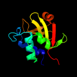
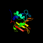
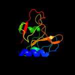
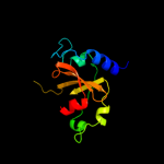
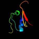
Top model
Image coloured by rainbow N → C terminus
Model (left) based on template c2k1gA_
Top template information
PDB header:lipoprotein
Chain: A: PDB Molecule:lipoprotein spr;
PDBTitle: solution nmr structure of lipoprotein spr from escherichia coli k12.2 northeast structural genomics target er541-37-162
Confidence and coverage
Confidence:100.0% Coverage: 67%
126 residues ( 67% of your sequence) have been modelled with 100.0% confidence by the single highest scoring template.
3D viewing
Interactive 3D view in Jmol

Sequence analysis 

Secondary structure and disorder prediction 

   1........10.........20.........30.........40.........50.........60
Sequence  MVKSQPILRYILRGIPAIAVAVLLSACSANNTAKNMHPETRAVGSETSSLQASQDEFENL
Secondary structure 


SS confidence 


Disorder  ??????


???????????????????????????????????
Disorder confidence 


   .........70.........80.........90.........100.........110.........120
Sequence  VRNVDVKSRIMDQYADWKGVRYRLGGSTKKGIDCSGFVQRTFREQFGLELPRSTYEQQEM
Secondary structure 


SS confidence 


Disorder  ?????


?
Disorder confidence 


   .........130.........140.........150.........160.........170.........180
Sequence  GKSVSRSNLRTGDLVLFRAGSTGRHVGIYIGNNQFVHASTSSGVIISSMNEPYWKKRYNE
Secondary structure 


SS confidence 


Disorder  ?
???


Disorder confidence 


   ........
Sequence  ARRVLSRS
Secondary structure 


SS confidence 


Disorder 


?????
Disorder confidence 


Confidence Key
High(9)                    Low (0)
?Disordered
Alpha helix
Beta strand

Domain analysis 

Hover over an aligned region to see model and summary info

Please note, only up to the top 20 hits are modelled to reduce computer load

RankAligned region

PDB 2k1g chain A

3D model

Region: 63 - 188
Aligned: 126
Modelled: 126
Confidence: 100.0%
Identity: 100%
PDB header:lipoprotein
Chain: A: PDB Molecule:lipoprotein spr;
PDBTitle: solution nmr structure of lipoprotein spr from escherichia coli k12.2 northeast structural genomics target er541-37-162

Phyre2

PDB 2fg0 chain B

3D model

Region: 39 - 185
Aligned: 146
Modelled: 147
Confidence: 100.0%
Identity: 24%
PDB header:hydrolase
Chain: B: PDB Molecule:cog0791: cell wall-associated hydrolases (invasion-
PDBTitle: crystal structure of a putative gamma-d-glutamyl-l-diamino acid2 endopeptidase (npun_r0659) from nostoc punctiforme pcc 73102 at 1.793 a resolution

Phyre2

PDB 2evr chain A domain 2

3D model

Region: 64 - 185
Aligned: 121
Modelled: 122
Confidence: 100.0%
Identity: 27%
Fold: Cysteine proteinases
Superfamily: Cysteine proteinases
Family: NlpC/P60

Phyre2

PDB 3h41 chain A

3D model

Region: 57 - 186
Aligned: 128
Modelled: 130
Confidence: 100.0%
Identity: 27%
PDB header:hydrolase
Chain: A: PDB Molecule:nlp/p60 family protein;
PDBTitle: crystal structure of a nlpc/p60 family protein (bce_2878) from2 bacillus cereus atcc 10987 at 1.79 a resolution

Phyre2

PDB 3npf chain B

3D model

Region: 62 - 188
Aligned: 123
Modelled: 127
Confidence: 100.0%
Identity: 33%
PDB header:hydrolase
Chain: B: PDB Molecule:putative dipeptidyl-peptidase vi;
PDBTitle: crystal structure of a putative dipeptidyl-peptidase vi (bacova_00612)2 from bacteroides ovatus at 1.72 a resolution

Phyre2

PDB 2xiv chain A

3D model

Region: 37 - 186
Aligned: 145
Modelled: 142
Confidence: 100.0%
Identity: 24%
PDB header:structural protein
Chain: A: PDB Molecule:hypothetical invasion protein;
PDBTitle: structure of rv1477, hypothetical invasion protein of2 mycobacterium tuberculosis

Phyre2

PDB 3pbi chain A

3D model

Region: 45 - 179
Aligned: 133
Modelled: 135
Confidence: 100.0%
Identity: 27%
PDB header:hydrolase
Chain: A: PDB Molecule:invasion protein;
PDBTitle: structure of the peptidoglycan hydrolase ripb (rv1478) from2 mycobacterium tuberculosis at 1.6 resolution

Phyre2

PDB 3i86 chain A

3D model

Region: 64 - 185
Aligned: 117
Modelled: 122
Confidence: 100.0%
Identity: 30%
PDB header:hydrolase
Chain: A: PDB Molecule:putative uncharacterized protein;
PDBTitle: crystal structure of the p60 domain from m. avium subspecies2 paratuberculosis antigen map1204

Phyre2

PDB 3gt2 chain A

3D model

Region: 64 - 185
Aligned: 115
Modelled: 122
Confidence: 100.0%
Identity: 29%
PDB header:unknown function
Chain: A: PDB Molecule:putative uncharacterized protein;
PDBTitle: crystal structure of the p60 domain from m. avium2 paratuberculosis antigen map1272c

Phyre2

PDB 3m1u chain B

3D model

Region: 67 - 187
Aligned: 114
Modelled: 121
Confidence: 99.9%
Identity: 28%
PDB header:hydrolase
Chain: B: PDB Molecule:putative gamma-d-glutamyl-l-diamino acid endopeptidase;
PDBTitle: crystal structure of a putative gamma-d-glutamyl-l-diamino acid2 endopeptidase (dvu_0896) from desulfovibrio vulgaris hildenborough at3 1.75 a resolution

Phyre2

PDB 2p1g chain A

3D model

Region: 70 - 185
Aligned: 115
Modelled: 116
Confidence: 99.6%
Identity: 22%
PDB header:structural genomics, unknown function
Chain: A: PDB Molecule:putative xylanase;
PDBTitle: crystal structure of a putative xylanase from bacteroides fragilis

Phyre2

PDB 2im9 chain A

3D model

Region: 62 - 177
Aligned: 116
Modelled: 116
Confidence: 97.8%
Identity: 33%
PDB header:structural genomics, unknown function
Chain: A: PDB Molecule:hypothetical protein;
PDBTitle: crystal structure of protein lpg0564 from legionella pneumophila str.2 philadelphia 1, pfam duf1460

Phyre2

PDB 2im9 chain A domain 1

3D model

Region: 62 - 177
Aligned: 116
Modelled: 116
Confidence: 97.8%
Identity: 33%
Fold: Cysteine proteinases
Superfamily: Cysteine proteinases
Family: Lpg0564-like

Phyre2

PDB 2kyt chain A

3D model

Region: 123 - 179
Aligned: 56
Modelled: 57
Confidence: 97.8%
Identity: 32%
PDB header:hydrolase
Chain: A: PDB Molecule:group xvi phospholipase a2;
PDBTitle: solution struture of the h-rev107 n-terminal domain

Phyre2

PDB 2if6 chain A domain 1

3D model

Region: 63 - 110
Aligned: 48
Modelled: 48
Confidence: 97.5%
Identity: 17%
Fold: Cysteine proteinases
Superfamily: Cysteine proteinases
Family: YiiX-like

Phyre2

PDB 3kw0 chain D

3D model

Region: 126 - 169
Aligned: 44
Modelled: 44
Confidence: 97.4%
Identity: 32%
PDB header:hydrolase
Chain: D: PDB Molecule:cysteine peptidase;
PDBTitle: crystal structure of cysteine peptidase (np_982244.1) from bacillus2 cereus atcc 10987 at 2.50 a resolution

Phyre2

PDB 2io8 chain A domain 2

3D model

Region: 94 - 151
Aligned: 58
Modelled: 58
Confidence: 88.2%
Identity: 24%
Fold: Cysteine proteinases
Superfamily: Cysteine proteinases
Family: CHAP domain

Phyre2

PDB 2k3a chain A

3D model

Region: 94 - 188
Aligned: 81
Modelled: 95
Confidence: 59.7%
Identity: 21%
PDB header:hydrolase
Chain: A: PDB Molecule:chap domain protein;
PDBTitle: nmr solution structure of staphylococcus saprophyticus chap2 (cysteine, histidine-dependent amidohydrolases/peptidases)3 domain protein. northeast structural genomics consortium4 target syr11

Phyre2

PDB 3kop chain B

3D model

Region: 129 - 169
Aligned: 41
Modelled: 41
Confidence: 24.5%
Identity: 20%
PDB header:structural genomics, unknown function
Chain: B: PDB Molecule:uncharacterized protein;
PDBTitle: crystal structure of protein with a cyclophilin-like fold2 (yp_831253.1) from arthrobacter sp. fb24 at 1.90 a resolution

Phyre2

PDB 2vpm chain B

3D model

Region: 94 - 151
Aligned: 58
Modelled: 58
Confidence: 21.8%
Identity: 21%
PDB header:ligase
Chain: B: PDB Molecule:trypanothione synthetase;
PDBTitle: trypanothione synthetase

Phyre2
1

c2k1gA_
2

c2fg0B_
3

d2evra2
4

c3h41A_
5

c3npfB_
6

c2xivA_
7

c3pbiA_
8

c3i86A_
9

c3gt2A_
10

c3m1uB_
11

c2p1gA_
12

c2im9A_
13

d2im9a1
14

c2kytA_
15

d2if6a1
16

c3kw0D_
17

d2io8a2
18

c2k3aA_
19

c3kopB_
20

c2vpmB_
21


22


23


24


25


26


27


28


29


30


31


32


33


34


35


36


Detailed template information 

#
Template Alignment Coverage3D Model Confidence
% i.d. Template Information
1c2k1gA_


100.0 100 PDB header:lipoprotein
Chain: A: PDB Molecule:lipoprotein spr;
PDBTitle: solution nmr structure of lipoprotein spr from escherichia coli k12.2 northeast structural genomics target er541-37-162
2c2fg0B_


100.0 24 PDB header:hydrolase
Chain: B: PDB Molecule:cog0791: cell wall-associated hydrolases (invasion-
PDBTitle: crystal structure of a putative gamma-d-glutamyl-l-diamino acid2 endopeptidase (npun_r0659) from nostoc punctiforme pcc 73102 at 1.793 a resolution
3d2evra2


100.0 27 Fold:Cysteine proteinases
Superfamily:Cysteine proteinases
Family:NlpC/P60
4c3h41A_


100.0 27 PDB header:hydrolase
Chain: A: PDB Molecule:nlp/p60 family protein;
PDBTitle: crystal structure of a nlpc/p60 family protein (bce_2878) from2 bacillus cereus atcc 10987 at 1.79 a resolution
5c3npfB_


100.0 33 PDB header:hydrolase
Chain: B: PDB Molecule:putative dipeptidyl-peptidase vi;
PDBTitle: crystal structure of a putative dipeptidyl-peptidase vi (bacova_00612)2 from bacteroides ovatus at 1.72 a resolution
6c2xivA_


100.0 24 PDB header:structural protein
Chain: A: PDB Molecule:hypothetical invasion protein;
PDBTitle: structure of rv1477, hypothetical invasion protein of2 mycobacterium tuberculosis
7c3pbiA_


100.0 27 PDB header:hydrolase
Chain: A: PDB Molecule:invasion protein;
PDBTitle: structure of the peptidoglycan hydrolase ripb (rv1478) from2 mycobacterium tuberculosis at 1.6 resolution
8c3i86A_


100.0 30 PDB header:hydrolase
Chain: A: PDB Molecule:putative uncharacterized protein;
PDBTitle: crystal structure of the p60 domain from m. avium subspecies2 paratuberculosis antigen map1204
9c3gt2A_


100.0 29 PDB header:unknown function
Chain: A: PDB Molecule:putative uncharacterized protein;
PDBTitle: crystal structure of the p60 domain from m. avium2 paratuberculosis antigen map1272c
10c3m1uB_


99.9 28 PDB header:hydrolase
Chain: B: PDB Molecule:putative gamma-d-glutamyl-l-diamino acid endopeptidase;
PDBTitle: crystal structure of a putative gamma-d-glutamyl-l-diamino acid2 endopeptidase (dvu_0896) from desulfovibrio vulgaris hildenborough at3 1.75 a resolution
11c2p1gA_


99.6 22 PDB header:structural genomics, unknown function
Chain: A: PDB Molecule:putative xylanase;
PDBTitle: crystal structure of a putative xylanase from bacteroides fragilis
12c2im9A_


97.8 33 PDB header:structural genomics, unknown function
Chain: A: PDB Molecule:hypothetical protein;
PDBTitle: crystal structure of protein lpg0564 from legionella pneumophila str.2 philadelphia 1, pfam duf1460
13d2im9a1


97.8 33 Fold:Cysteine proteinases
Superfamily:Cysteine proteinases
Family:Lpg0564-like
14c2kytA_


97.8 32 PDB header:hydrolase
Chain: A: PDB Molecule:group xvi phospholipase a2;
PDBTitle: solution struture of the h-rev107 n-terminal domain
15d2if6a1


97.5 17 Fold:Cysteine proteinases
Superfamily:Cysteine proteinases
Family:YiiX-like
16c3kw0D_


97.4 32 PDB header:hydrolase
Chain: D: PDB Molecule:cysteine peptidase;
PDBTitle: crystal structure of cysteine peptidase (np_982244.1) from bacillus2 cereus atcc 10987 at 2.50 a resolution
17d2io8a2


88.2 24 Fold:Cysteine proteinases
Superfamily:Cysteine proteinases
Family:CHAP domain
18c2k3aA_


59.7 21 PDB header:hydrolase
Chain: A: PDB Molecule:chap domain protein;
PDBTitle: nmr solution structure of staphylococcus saprophyticus chap2 (cysteine, histidine-dependent amidohydrolases/peptidases)3 domain protein. northeast structural genomics consortium4 target syr11
19c3kopB_


24.5 20 PDB header:structural genomics, unknown function
Chain: B: PDB Molecule:uncharacterized protein;
PDBTitle: crystal structure of protein with a cyclophilin-like fold2 (yp_831253.1) from arthrobacter sp. fb24 at 1.90 a resolution
20c2vpmB_


21.8 21 PDB header:ligase
Chain: B: PDB Molecule:trypanothione synthetase;
PDBTitle: trypanothione synthetase
21c2ioaA_


not modelled 18.0 24 PDB header:ligase, hydrolase
Chain: A: PDB Molecule:bifunctional glutathionylspermidine
PDBTitle: e. coli bifunctional glutathionylspermidine2 synthetase/amidase incomplex with mg2+ and adp and3 phosphinate inhibitor
22d1bg6a1


not modelled 15.2 17 Fold:6-phosphogluconate dehydrogenase C-terminal domain-like
Superfamily:6-phosphogluconate dehydrogenase C-terminal domain-like
Family:N-(1-D-carboxylethyl)-L-norvaline dehydrogenase
23d2gbsa1


not modelled 14.0 25 Fold:PUA domain-like
Superfamily:PUA domain-like
Family:Atu2648/PH1033-like
24c3eopB_


not modelled 13.0 14 PDB header:unknown function
Chain: B: PDB Molecule:thymocyte nuclear protein 1;
PDBTitle: crystal structure of the duf55 domain of human thymocyte nuclear2 protein 1
25d2g2xa1


not modelled 12.5 27 Fold:PUA domain-like
Superfamily:PUA domain-like
Family:Atu2648/PH1033-like
26d2ar1a1


not modelled 12.5 27 Fold:PUA domain-like
Superfamily:PUA domain-like
Family:Atu2648/PH1033-like
27d1zcea1


not modelled 12.1 27 Fold:PUA domain-like
Superfamily:PUA domain-like
Family:Atu2648/PH1033-like
28d1sr9a1


not modelled 10.2 40 Fold:RuvA C-terminal domain-like
Superfamily:post-HMGL domain-like
Family:DmpG/LeuA communication domain-like
29d2evea1


not modelled 9.9 25 Fold:PUA domain-like
Superfamily:PUA domain-like
Family:Atu2648/PH1033-like
30c1yy3A_


not modelled 9.2 19 PDB header:isomerase
Chain: A: PDB Molecule:s-adenosylmethionine:trna ribosyltransferase-
PDBTitle: structure of s-adenosylmethionine:trna ribosyltransferase-2 isomerase (quea)
31d2ba0a2


not modelled 8.6 25 Fold:Barrel-sandwich hybrid
Superfamily:Ribosomal L27 protein-like
Family:ECR1 N-terminal domain-like
32d1wi9a_


not modelled 7.0 19 Fold:DNA/RNA-binding 3-helical bundle
Superfamily:"Winged helix" DNA-binding domain
Family:PCI domain (PINT motif)
33d1aono_


not modelled 6.7 28 Fold:GroES-like
Superfamily:GroES-like
Family:GroES
34c3iz5G_


not modelled 6.7 24 PDB header:ribosome
Chain: G: PDB Molecule:60s ribosomal protein l6 (l6e);
PDBTitle: localization of the large subunit ribosomal proteins into a 5.5 a2 cryo-em map of triticum aestivum translating 80s ribosome
35d2hd9a1


not modelled 5.8 24 Fold:PUA domain-like
Superfamily:PUA domain-like
Family:Atu2648/PH1033-like
36d1iyca_


not modelled 5.3 60 Fold:Invertebrate chitin-binding proteins
Superfamily:Invertebrate chitin-binding proteins
Family:Antifungal peptide scarabaecin

Binding site prediction 

Due to computational demand, binding site predictions are not run for batch jobs

If you want to predict binding sites, please manually submit your model of choice to 3DLigandSite


Phyre is for academic use only

Please cite: Protein structure prediction on the web: a case study using the Phyre server
Kelley LA and Sternberg MJE. Nature Protocols 4, 363 - 371 (2009) [pdf] [Import into BibTeX]
 
If you use the binding site predictions from 3DLigandSite, please also cite:
3DLigandSite: predicting ligand-binding sites using similar structures.
Wass MN, Kelley LA and Sternberg MJ Nucleic Acids Research 38, W469-73 (2010) [PubMed]
 
© Structural Bioinformatics Group
Imperial College London
Lawrence Kelley, Benjamin Jefferys
Disclaimer
Terms and Conditions
Component software
Template detection: HHpred 1.51
Secondary structure prediction: Psi-pred 2.5
Disorder prediction: Disopred 2.4
Transmembrane prediction: Memsat_SVM
Multi-template modelling and ab initio: Poing 1.0