1 c2y4oA_
100.0
67
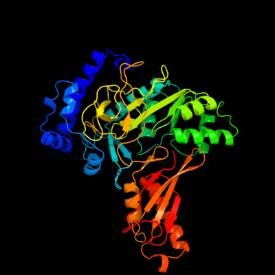
PDB header: ligaseChain: A: PDB Molecule: phenylacetate-coenzyme a ligase;PDBTitle: crystal structure of paak2 in complex with phenylacetyl adenylate
2 c2y27B_
100.0
63
PDB header: ligaseChain: B: PDB Molecule: phenylacetate-coenzyme a ligase;PDBTitle: crystal structure of paak1 in complex with atp from2 burkholderia cenocepacia
3 c3qovD_
100.0
41
PDB header: ligaseChain: D: PDB Molecule: phenylacetate-coenzyme a ligase;PDBTitle: crystal structure of a hypothetical acyl-coa ligase (bt_0428) from2 bacteroides thetaiotaomicron vpi-5482 at 2.20 a resolution
4 d1pg4a_
100.0
20
Fold: Acetyl-CoA synthetase-likeSuperfamily: Acetyl-CoA synthetase-likeFamily: Acetyl-CoA synthetase-like5 c2vsqA_
100.0
19
PDB header: ligaseChain: A: PDB Molecule: surfactin synthetase subunit 3;PDBTitle: structure of surfactin a synthetase c (srfa-c), a2 nonribosomal peptide synthetase termination module
6 d1ry2a_
100.0
19
Fold: Acetyl-CoA synthetase-likeSuperfamily: Acetyl-CoA synthetase-likeFamily: Acetyl-CoA synthetase-like7 c3hguB_
100.0
19
PDB header: biosynthetic proteinChain: B: PDB Molecule: ehpf;PDBTitle: structure of phenazine antibiotic biosynthesis protein
8 c3tsyA_
100.0
17
PDB header: ligase, transferaseChain: A: PDB Molecule: fusion protein 4-coumarate--coa ligase 1, resveratrolPDBTitle: 4-coumaroyl-coa ligase::stilbene synthase fusion protein
9 d3cw9a1
100.0
18
Fold: Acetyl-CoA synthetase-likeSuperfamily: Acetyl-CoA synthetase-likeFamily: Acetyl-CoA synthetase-like10 c3e7wA_
100.0
12
PDB header: ligaseChain: A: PDB Molecule: d-alanine--poly(phosphoribitol) ligase subunit 1;PDBTitle: crystal structure of dlta: implications for the reaction2 mechanism of non-ribosomal peptide synthetase (nrps)3 adenylation domains
11 c3gqwB_
100.0
18
PDB header: ligaseChain: B: PDB Molecule: fatty acid amp ligase;PDBTitle: crystal structure of a fatty acid amp ligase from e. coli with an acyl2 adenylate product bound
12 d1mdba_
100.0
16
Fold: Acetyl-CoA synthetase-likeSuperfamily: Acetyl-CoA synthetase-likeFamily: Acetyl-CoA synthetase-like13 c3etcB_
100.0
16
PDB header: ligaseChain: B: PDB Molecule: amp-binding protein;PDBTitle: 2.1 a structure of acyl-adenylate synthetase from methanosarcina2 acetivorans containing a link between lys256 and cys298
14 c3ni2A_
100.0
17
PDB header: ligaseChain: A: PDB Molecule: 4-coumarate:coa ligase;PDBTitle: crystal structures and enzymatic mechanisms of a populus tomentosa 4-2 coumarate:coa ligase
15 c3kxwA_
100.0
13
PDB header: ligaseChain: A: PDB Molecule: saframycin mx1 synthetase b;PDBTitle: the crystal structure of fatty acid amp ligase from legionella2 pneumophila
16 c3iteB_
100.0
15
PDB header: ligaseChain: B: PDB Molecule: sidn siderophore synthetase;PDBTitle: the third adenylation domain of the fungal sidn non-2 ribosomal peptide synthetase
17 c3eynB_
100.0
15
PDB header: ligaseChain: B: PDB Molecule: acyl-coenzyme a synthetase acsm2a;PDBTitle: crystal structure of human acyl-coa synthetase medium-chain2 family member 2a (l64p mutation) in a complex with coa
18 c3nyrA_
100.0
13
PDB header: ligaseChain: A: PDB Molecule: malonyl-coa ligase;PDBTitle: malonyl-coa ligase ternary product complex with malonyl-coa and amp2 bound
19 c2d1tA_
100.0
17
PDB header: oxidoreductaseChain: A: PDB Molecule: luciferin 4-monooxygenase;PDBTitle: crystal structure of the thermostable japanese firefly2 luciferase red-color emission s286n mutant complexed with3 high-energy intermediate analogue
20 c2v7bB_
100.0
20
PDB header: ligaseChain: B: PDB Molecule: benzoate-coenzyme a ligase;PDBTitle: crystal structures of a benzoate coa ligase from2 burkholderia xenovorans lb400
21 c3iplB_
not modelled
100.0
19
PDB header: ligaseChain: B: PDB Molecule: 2-succinylbenzoate--coa ligase;PDBTitle: crystal structure of o-succinylbenzoic acid-coa ligase from2 staphylococcus aureus subsp. aureus mu50
22 d1v25a_
not modelled
100.0
18
Fold: Acetyl-CoA synthetase-likeSuperfamily: Acetyl-CoA synthetase-likeFamily: Acetyl-CoA synthetase-like23 c3g7sA_
not modelled
100.0
19
PDB header: ligaseChain: A: PDB Molecule: long-chain-fatty-acid--coa ligase (fadd-1);PDBTitle: crystal structure of a long-chain-fatty-acid-coa ligase2 (fadd1) from archaeoglobus fulgidus
24 c3dhvA_
not modelled
100.0
16
PDB header: ligaseChain: A: PDB Molecule: d-alanine-poly(phosphoribitol) ligase;PDBTitle: crystal structure of dlta protein in complex with d-alanine2 adenylate
25 d1lcia_
not modelled
100.0
13
Fold: Acetyl-CoA synthetase-likeSuperfamily: Acetyl-CoA synthetase-likeFamily: Acetyl-CoA synthetase-like26 c1amuB_
not modelled
100.0
15
PDB header: peptide synthetaseChain: B: PDB Molecule: gramicidin synthetase 1;PDBTitle: phenylalanine activating domain of gramicidin synthetase 12 in a complex with amp and phenylalanine
27 d1amua_
not modelled
100.0
16
Fold: Acetyl-CoA synthetase-likeSuperfamily: Acetyl-CoA synthetase-likeFamily: Acetyl-CoA synthetase-like28 c3l8cA_
not modelled
100.0
17
PDB header: ligaseChain: A: PDB Molecule: d-alanine--poly(phosphoribitol) ligase subunit 1;PDBTitle: structure of probable d-alanine--poly(phosphoribitol) ligase2 subunit-1 from streptococcus pyogenes
29 c3o82B_
not modelled
99.9
18
PDB header: ligaseChain: B: PDB Molecule: peptide arylation enzyme;PDBTitle: structure of base n-terminal domain from acinetobacter baumannii bound2 to 5'-o-[n-(2,3-dihydroxybenzoyl)sulfamoyl] adenosine
30 c3ivrA_
not modelled
99.9
13
PDB header: ligaseChain: A: PDB Molecule: putative long-chain-fatty-acid coa ligase;PDBTitle: crystal structure of putative long-chain-fatty-acid coa ligase from2 rhodopseudomonas palustris cga009
31 c3e53A_
not modelled
99.9
14
PDB header: ligaseChain: A: PDB Molecule: fatty-acid-coa ligase fadd28;PDBTitle: crystal structure of n-terminal domain of a fatty acyl amp2 ligase faal28 from mycobacterium tuberculosis
32 c3o82A_
not modelled
99.8
18
PDB header: ligaseChain: A: PDB Molecule: peptide arylation enzyme;PDBTitle: structure of base n-terminal domain from acinetobacter baumannii bound2 to 5'-o-[n-(2,3-dihydroxybenzoyl)sulfamoyl] adenosine
33 c3laxA_
not modelled
99.6
28
PDB header: ligaseChain: A: PDB Molecule: phenylacetate-coenzyme a ligase;PDBTitle: the crystal structure of a domain of phenylacetate-coenzyme2 a ligase from bacteroides vulgatus atcc 8482
34 c2wh7A_
not modelled
34.7
44
PDB header: hydrolaseChain: A: PDB Molecule: hyaluronidase-phage associated;PDBTitle: the partial structure of a group a streptpcoccal phage-2 encoded tail fibre hyaluronate lyase hylp2
35 d1whua_
not modelled
34.0
27
Fold: DNA/RNA-binding 3-helical bundleSuperfamily: Polynucleotide phosphorylase/guanosine pentaphosphate synthase (PNPase/GPSI), domain 3Family: Polynucleotide phosphorylase/guanosine pentaphosphate synthase (PNPase/GPSI), domain 336 c2i2xD_
not modelled
29.9
11
PDB header: transferaseChain: D: PDB Molecule: methyltransferase 1;PDBTitle: crystal structure of methanol:cobalamin methyltransferase complex2 mtabc from methanosarcina barkeri
37 d3bula2
not modelled
29.2
11
Fold: Flavodoxin-likeSuperfamily: Cobalamin (vitamin B12)-binding domainFamily: Cobalamin (vitamin B12)-binding domain38 d1hska2
not modelled
29.1
18
Fold: Uridine diphospho-N-Acetylenolpyruvylglucosamine reductase, MurB, C-terminal domainSuperfamily: Uridine diphospho-N-Acetylenolpyruvylglucosamine reductase, MurB, C-terminal domainFamily: Uridine diphospho-N-Acetylenolpyruvylglucosamine reductase, MurB, C-terminal domain39 d1pk1c1
not modelled
27.0
21
Fold: SAM domain-likeSuperfamily: SAM/Pointed domainFamily: SAM (sterile alpha motif) domain40 d1wwva1
not modelled
23.6
15
Fold: SAM domain-likeSuperfamily: SAM/Pointed domainFamily: SAM (sterile alpha motif) domain41 c2c3fA_
not modelled
19.1
44
PDB header: lyaseChain: A: PDB Molecule: hyaluronidase, phage associated;PDBTitle: the structure of a group a streptococcal phage-encoded2 tail-fibre showing hyaluronan lyase activity.
42 d2c3fa1
not modelled
19.1
44
Fold: Triple-stranded beta-helixSuperfamily: Phage fibre proteinsFamily: HylP-like43 c1bmtB_
not modelled
16.1
13
PDB header: methyltransferaseChain: B: PDB Molecule: methionine synthase;PDBTitle: how a protein binds b12: a 3.o angstrom x-ray structure of2 the b12-binding domains of methionine synthase
44 d1dpta_
not modelled
15.1
6
Fold: Tautomerase/MIFSuperfamily: Tautomerase/MIFFamily: MIF-related45 c1pk1A_
not modelled
14.5
28
PDB header: transcription repressionChain: A: PDB Molecule: polyhomeotic-proximal chromatin protein;PDBTitle: hetero sam domain structure of ph and scm.
46 c2ormA_
not modelled
14.4
11
PDB header: isomeraseChain: A: PDB Molecule: probable tautomerase hp0924;PDBTitle: crystal structure of the 4-oxalocrotonate tautomerase homologue dmpi2 from helicobacter pylori.
47 d2o70a1
not modelled
13.8
13
Fold: UraD-likeSuperfamily: UraD-LikeFamily: UraD-like48 c3ry0A_
not modelled
13.7
14
PDB header: isomeraseChain: A: PDB Molecule: putative tautomerase;PDBTitle: crystal structure of tomn, a 4-oxalocrotonate tautomerase homologue in2 tomaymycin biosynthetic pathway
49 d1bjpa_
not modelled
13.3
10
Fold: Tautomerase/MIFSuperfamily: Tautomerase/MIFFamily: 4-oxalocrotonate tautomerase-like50 c3ezxA_
not modelled
12.5
12
PDB header: transferaseChain: A: PDB Molecule: monomethylamine corrinoid protein 1;PDBTitle: structure of methanosarcina barkeri monomethylamine2 corrinoid protein
51 d1otfa_
not modelled
12.5
16
Fold: Tautomerase/MIFSuperfamily: Tautomerase/MIFFamily: 4-oxalocrotonate tautomerase-like52 c3kk1B_
not modelled
11.6
12
PDB header: transferase/dnaChain: B: PDB Molecule: reverse transcriptase p51 subunit;PDBTitle: hiv-1 reverse transcriptase-dna complex with nuceotide inhibitor gs-2 9148-diphosphate bound in nucleotide site
53 c3ebrA_
not modelled
11.5
19
PDB header: unknown functionChain: A: PDB Molecule: uncharacterized rmlc-like cupin;PDBTitle: crystal structure of an rmlc-like cupin protein (reut_a0381) from2 ralstonia eutropha jmp134 at 2.60 a resolution
54 c1hskA_
not modelled
11.2
18
PDB header: oxidoreductaseChain: A: PDB Molecule: udp-n-acetylenolpyruvoylglucosamine reductase;PDBTitle: crystal structure of s. aureus murb
55 c3abfB_
not modelled
11.1
18
PDB header: isomeraseChain: B: PDB Molecule: 4-oxalocrotonate tautomerase;PDBTitle: crystal structure of a 4-oxalocrotonate tautomerase homologue2 (tthb242)
56 c2op8A_
not modelled
11.0
22
PDB header: isomeraseChain: A: PDB Molecule: probable tautomerase ywhb;PDBTitle: crystal structure of ywhb- homologue of 4-oxalocrotonate tautomerase
57 c2x4kB_
not modelled
10.5
16
PDB header: isomeraseChain: B: PDB Molecule: 4-oxalocrotonate tautomerase;PDBTitle: crystal structure of sar1376, a putative 4-oxalocrotonate2 tautomerase from the methicillin-resistant staphylococcus3 aureus (mrsa)
58 d1kw4a_
not modelled
10.2
22
Fold: SAM domain-likeSuperfamily: SAM/Pointed domainFamily: SAM (sterile alpha motif) domain59 d1iwga3
not modelled
10.0
8
Fold: Ferredoxin-likeSuperfamily: Multidrug efflux transporter AcrB pore domain; PN1, PN2, PC1 and PC2 subdomainsFamily: Multidrug efflux transporter AcrB pore domain; PN1, PN2, PC1 and PC2 subdomains60 d1h80a_
not modelled
10.0
31
Fold: Single-stranded right-handed beta-helixSuperfamily: Pectin lyase-likeFamily: iota-carrageenase61 d2zd1b1
not modelled
9.3
12
Fold: DNA/RNA polymerasesSuperfamily: DNA/RNA polymerasesFamily: Reverse transcriptase62 c1rthA_
not modelled
8.6
12
PDB header: nucleotidyltransferaseChain: A: PDB Molecule: hiv-1 reverse transcriptase;PDBTitle: high resolution structures of hiv-1 rt from four rt-2 inhibitor complexes
63 d1ucra_
not modelled
8.3
57
Fold: DNA/RNA-binding 3-helical bundleSuperfamily: "Winged helix" DNA-binding domainFamily: Dissimilatory sulfite reductase DsvD64 d2qfia1
not modelled
8.3
13
Fold: Alpha-lytic protease prodomain-likeSuperfamily: Cation efflux protein cytoplasmic domain-likeFamily: Cation efflux protein cytoplasmic domain-like65 c1y80A_
not modelled
8.1
17
PDB header: structural genomics, unknown functionChain: A: PDB Molecule: predicted cobalamin binding protein;PDBTitle: structure of a corrinoid (factor iiim)-binding protein from2 moorella thermoacetica
66 c1x4rA_
not modelled
7.9
71
PDB header: apoptosisChain: A: PDB Molecule: parp14 protein;PDBTitle: solution structure of wwe domain in parp14 protein
67 c3mb2G_
not modelled
7.7
12
PDB header: isomeraseChain: G: PDB Molecule: 4-oxalocrotonate tautomerase family enzyme - alpha subunit;PDBTitle: kinetic and structural characterization of a heterohexamer 4-2 oxalocrotonate tautomerase from chloroflexus aurantiacus j-10-fl:3 implications for functional and structural diversity in the4 tautomerase superfamily
68 c2pmzV_
not modelled
7.6
22
PDB header: translation, transferaseChain: V: PDB Molecule: dna-directed rna polymerase subunit h;PDBTitle: archaeal rna polymerase from sulfolobus solfataricus
69 d2icwg1
not modelled
7.5
31
Fold: Superantigen MAMSuperfamily: Superantigen MAMFamily: Superantigen MAM70 c2do9A_
not modelled
6.6
27
PDB header: signaling proteinChain: A: PDB Molecule: nacht-, lrr- and pyd-containing protein 10;PDBTitle: solution structure of the pyrin/paad-dapin domain in mouse2 nalp10 (nacht, leucine rich repeat and pyd containing 10)
71 c2pjkA_
not modelled
6.4
14
PDB header: structural genomics, unknown functionChain: A: PDB Molecule: 178aa long hypothetical molybdenum cofactorPDBTitle: structure of hypothetical molybdenum cofactor biosynthesis2 protein b from sulfolobus tokodaii
72 c2bcmB_
not modelled
6.2
43
PDB header: cell adhesionChain: B: PDB Molecule: f1845 fimbrial protein;PDBTitle: daae adhesin
73 c2epbA_
not modelled
6.2
63
PDB header: transcriptionChain: A: PDB Molecule: chromodomain-helicase-dna-binding protein 6;PDBTitle: solution structure of chromo domain 2 in chromodomain-2 helicase-dna-binding protein 6
74 d1uhra_
not modelled
6.2
36
Fold: SWIB/MDM2 domainSuperfamily: SWIB/MDM2 domainFamily: SWIB/MDM2 domain75 c2qfiB_
not modelled
6.2
14
PDB header: transport proteinChain: B: PDB Molecule: ferrous-iron efflux pump fief;PDBTitle: structure of the zinc transporter yiip
76 c3idwA_
not modelled
6.1
18
PDB header: endocytosisChain: A: PDB Molecule: actin cytoskeleton-regulatory complex protein sla1;PDBTitle: crystal structure of sla1 homology domain 2
77 d2edua1
not modelled
6.1
22
Fold: SAM domain-likeSuperfamily: RuvA domain 2-likeFamily: ComEA-like78 d1mwwa_
not modelled
6.0
11
Fold: Tautomerase/MIFSuperfamily: Tautomerase/MIFFamily: Hypothetical protein HI1388.179 c2zztA_
not modelled
6.0
8
PDB header: transport proteinChain: A: PDB Molecule: putative uncharacterized protein;PDBTitle: crystal structure of the cytosolic domain of the cation2 diffusion facilitator family protein
80 c1pk1B_
not modelled
6.0
15
PDB header: transcription repressionChain: B: PDB Molecule: sex comb on midleg cg9495-pa;PDBTitle: hetero sam domain structure of ph and scm.
81 d2f3na1
not modelled
6.0
12
Fold: SAM domain-likeSuperfamily: SAM/Pointed domainFamily: SAM (sterile alpha motif) domain82 c1k98A_
not modelled
5.9
13
PDB header: transferaseChain: A: PDB Molecule: methionine synthase;PDBTitle: adomet complex of meth c-terminal fragment
83 d1gyxa_
not modelled
5.9
6
Fold: Tautomerase/MIFSuperfamily: Tautomerase/MIFFamily: 4-oxalocrotonate tautomerase-like84 c2fvnA_
not modelled
5.8
36
PDB header: cell adhesionChain: A: PDB Molecule: protein afad;PDBTitle: the fibrillar tip complex of the afa/dr adhesins from2 pathogen e. coli displays synergistic binding to 5 1 and v3 3 integrins
85 c3bs7A_
not modelled
5.6
15
PDB header: signaling proteinChain: A: PDB Molecule: protein aveugle;PDBTitle: crystal structure of the sterile alpha motif (sam) domain2 of hyphen/aveugle
86 d2aala1
not modelled
5.6
26
Fold: Tautomerase/MIFSuperfamily: Tautomerase/MIFFamily: MSAD-like87 c3bs5A_
not modelled
5.6
15
PDB header: signaling protein/membrane proteinChain: A: PDB Molecule: protein aveugle;PDBTitle: crystal structure of hcnk2-sam/dhyp-sam complex
88 c2dn5A_
not modelled
5.4
45
PDB header: transcriptionChain: A: PDB Molecule: general transcription factor ii-i repeat domain-PDBTitle: solution structure of rsgi ruh-057, a gtf2i domain in human2 cdna
89 c3cmqA_
not modelled
5.1
10
PDB header: ligaseChain: A: PDB Molecule: phenylalanyl-trna synthetase, mitochondrial;PDBTitle: crystal structure of human mitochondrial phenylalanine trna2 synthetase
90 d1f06a2
not modelled
5.1
9
Fold: FwdE/GAPDH domain-likeSuperfamily: Glyceraldehyde-3-phosphate dehydrogenase-like, C-terminal domainFamily: Dihydrodipicolinate reductase-like91 d7reqa2
not modelled
5.0
14
Fold: Flavodoxin-likeSuperfamily: Cobalamin (vitamin B12)-binding domainFamily: Cobalamin (vitamin B12)-binding domain