Phyre Home Retrieve Phyre Job Id
Emaill.a.kelley@imperial.ac.uk
DescriptionP77733
DateThu Jan 5 12:32:15 GMT 2012
Unique Job ID7215ad166acd60f8

Summary 

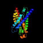
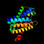
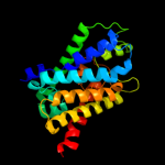
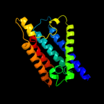
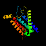
Top model
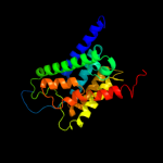
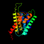
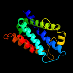
Image coloured by rainbow N → C terminus
Model (left) based on template c3klzE_
Top template information
PDB header:membrane protein
Chain: E: PDB Molecule:putative formate transporter 1;
PDBTitle: pentameric formate channel with formate bound
Confidence and coverage
Confidence:100.0% Coverage: 91%
256 residues ( 91% of your sequence) have been modelled with 100.0% confidence by the single highest scoring template.
3D viewing
Interactive 3D view in Jmol

Sequence analysis 

Secondary structure and disorder prediction 

   1........10.........20.........30.........40.........50.........60
Sequence  MRNKLSFDLQLSARKAAIAERIAAHKIARSKVSVFLMAMSAGVFMAIGFTFYLSVIADAP
Secondary structure 


SS confidence 


Disorder  ????????


?

?
Disorder confidence 


   .........70.........80.........90.........100.........110.........120
Sequence  SSQALTHLVGGLCFTLGFILLAVCGTSLFTSSVMTVMAKSRGVISWRTWLINALLVACGN
Secondary structure 


SS confidence 


Disorder 


Disorder confidence 


   .........130.........140.........150.........160.........170.........180
Sequence  LAGIACFSLLIWFSGLVMSENAMWGVAVLHCAEGKMHHTFTESVSLGIMCNLMVCLALWM
Secondary structure 


SS confidence 


Disorder 


?

?
?


Disorder confidence 


   .........190.........200.........210.........220.........230.........240
Sequence  SYCGRSLCDKIVAMILPITLFVASGFEHCIANLFVIPFAIAIRHFAPPPFWQLAHSSADN
Secondary structure 


SS confidence 


Disorder  ?
??


?


???
Disorder confidence 


   .........250.........260.........270.........280..
Sequence  FPALTVSHFITANLLPVMLGNIIGGAVLVSMCYRAIYLRQEP
Secondary structure 


SS confidence 


Disorder 


????
Disorder confidence 


Confidence Key
High(9)                    Low (0)
?Disordered
Alpha helix
Beta strand

Domain analysis 

Hover over an aligned region to see model and summary info

Please note, only up to the top 20 hits are modelled to reduce computer load

RankAligned region

PDB 3klz chain E

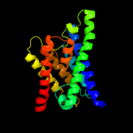
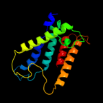
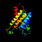
3D model

Region: 24 - 279
Aligned: 256
Modelled: 256
Confidence: 100.0%
Identity: 43%
PDB header:membrane protein
Chain: E: PDB Molecule:putative formate transporter 1;
PDBTitle: pentameric formate channel with formate bound

Phyre2

PDB 3kcv chain G

3D model

Region: 29 - 279
Aligned: 251
Modelled: 251
Confidence: 100.0%
Identity: 53%
PDB header:transport protein
Chain: G: PDB Molecule:probable formate transporter 1;
PDBTitle: structure of formate channel

Phyre2

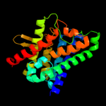
PDB 1rc2 chain A

3D model

Region: 32 - 281
Aligned: 224
Modelled: 232
Confidence: 92.4%
Identity: 9%
Fold: Aquaporin-like
Superfamily: Aquaporin-like
Family: Aquaporin-like

Phyre2

PDB 3llq chain B

3D model

Region: 28 - 279
Aligned: 224
Modelled: 234
Confidence: 90.7%
Identity: 10%
PDB header:membrane protein
Chain: B: PDB Molecule:aquaporin z 2;
PDBTitle: aquaporin structure from plant pathogen agrobacterium tumerfaciens

Phyre2

PDB 1fx8 chain A

3D model

Region: 30 - 280
Aligned: 218
Modelled: 224
Confidence: 88.8%
Identity: 18%
Fold: Aquaporin-like
Superfamily: Aquaporin-like
Family: Aquaporin-like

Phyre2

PDB 1lda chain A

3D model

Region: 30 - 280
Aligned: 218
Modelled: 224
Confidence: 88.8%
Identity: 18%
PDB header:transport protein
Chain: A: PDB Molecule:glycerol uptake facilitator protein;
PDBTitle: crystal structure of the e. coli glycerol facilitator (glpf) without2 substrate glycerol

Phyre2

PDB 2d57 chain A

3D model

Region: 28 - 209
Aligned: 172
Modelled: 182
Confidence: 77.7%
Identity: 10%
PDB header:transport protein
Chain: A: PDB Molecule:aquaporin-4;
PDBTitle: double layered 2d crystal structure of aquaporin-4 (aqp4m23) at 3.2 a2 resolution by electron crystallography

Phyre2

PDB 3gd8 chain A

3D model

Region: 29 - 209
Aligned: 171
Modelled: 181
Confidence: 76.0%
Identity: 11%
PDB header:membrane protein
Chain: A: PDB Molecule:aquaporin-4;
PDBTitle: crystal structure of human aquaporin 4 at 1.8 and its mechanism of2 conductance

Phyre2

PDB 1j4n chain A

3D model

Region: 27 - 282
Aligned: 220
Modelled: 222
Confidence: 73.4%
Identity: 15%
Fold: Aquaporin-like
Superfamily: Aquaporin-like
Family: Aquaporin-like

Phyre2

PDB 2w2e chain A

3D model

Region: 28 - 204
Aligned: 164
Modelled: 164
Confidence: 71.1%
Identity: 16%
PDB header:membrane protein
Chain: A: PDB Molecule:aquaporin;
PDBTitle: 1.15 angstrom crystal structure of p.pastoris aquaporin,2 aqy1, in a closed conformation at ph 3.5

Phyre2

PDB 3iyz chain A

3D model

Region: 28 - 277
Aligned: 212
Modelled: 225
Confidence: 67.9%
Identity: 13%
PDB header:transport protein
Chain: A: PDB Molecule:aquaporin-4;
PDBTitle: structure of aquaporin-4 s180d mutant at 10.0 a resolution from2 electron micrograph

Phyre2

PDB 1ymg chain A domain 1

3D model

Region: 44 - 282
Aligned: 204
Modelled: 215
Confidence: 57.2%
Identity: 14%
Fold: Aquaporin-like
Superfamily: Aquaporin-like
Family: Aquaporin-like

Phyre2

PDB 1ymg chain A

3D model

Region: 44 - 282
Aligned: 204
Modelled: 215
Confidence: 57.2%
Identity: 14%
PDB header:membrane protein
Chain: A: PDB Molecule:lens fiber major intrinsic protein;
PDBTitle: the channel architecture of aquaporin o at 2.2 angstrom resolution

Phyre2

PDB 2b5f chain D

3D model

Region: 28 - 276
Aligned: 216
Modelled: 226
Confidence: 49.4%
Identity: 9%
PDB header:transport protein,membrane protein
Chain: D: PDB Molecule:aquaporin;
PDBTitle: crystal structure of the spinach aquaporin sopip2;1 in an2 open conformation to 3.9 resolution

Phyre2

PDB 1h6i chain A

3D model

Region: 31 - 206
Aligned: 166
Modelled: 166
Confidence: 38.2%
Identity: 13%
Fold: Aquaporin-like
Superfamily: Aquaporin-like
Family: Aquaporin-like

Phyre2

PDB 3d9s chain B

3D model

Region: 33 - 282
Aligned: 214
Modelled: 232
Confidence: 35.6%
Identity: 15%
PDB header:membrane protein
Chain: B: PDB Molecule:aquaporin-5;
PDBTitle: human aquaporin 5 (aqp5) - high resolution x-ray structure

Phyre2

PDB 2f2b chain A

3D model

Region: 30 - 204
Aligned: 167
Modelled: 175
Confidence: 32.0%
Identity: 11%
PDB header:membrane protein
Chain: A: PDB Molecule:aquaporin aqpm;
PDBTitle: crystal structure of integral membrane protein aquaporin aqpm at 1.68a2 resolution

Phyre2

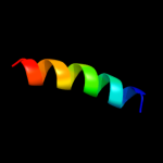
PDB 1c17 chain M

3D model

Region: 258 - 275
Aligned: 18
Modelled: 18
Confidence: 21.4%
Identity: 17%
Fold: F1F0 ATP synthase subunit A
Superfamily: F1F0 ATP synthase subunit A
Family: F1F0 ATP synthase subunit A

Phyre2

PDB 3c02 chain A

3D model

Region: 30 - 278
Aligned: 232
Modelled: 244
Confidence: 17.5%
Identity: 9%
PDB header:membrane protein
Chain: A: PDB Molecule:aquaglyceroporin;
PDBTitle: x-ray structure of the aquaglyceroporin from plasmodium falciparum

Phyre2

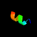
PDB 2jy0 chain A

3D model

Region: 264 - 273
Aligned: 10
Modelled: 10
Confidence: 11.4%
Identity: 50%
PDB header:membrane protein, viral protein
Chain: A: PDB Molecule:protease ns2-3;
PDBTitle: solution nmr structure of hcv ns2 protein, membrane segment2 (1-27)

Phyre2
1

c3klzE_
2

c3kcvG_
3

d1rc2a_
4

c3llqB_
5

d1fx8a_
6

c1ldaA_
7

c2d57A_
8

c3gd8A_
9

d1j4na_
10

c2w2eA_
11

c3iyzA_
12

d1ymga1
13

c1ymgA_
14

c2b5fD_
15

d1h6ia_
16

c3d9sB_
17

c2f2bA_
18

d1c17m_
19

c3c02A_
20

c2jy0A_
21


22


23


24


25


26


27


28


Detailed template information 

#
Template Alignment Coverage3D Model Confidence
% i.d. Template Information
1c3klzE_


100.0 43 PDB header:membrane protein
Chain: E: PDB Molecule:putative formate transporter 1;
PDBTitle: pentameric formate channel with formate bound
2c3kcvG_


100.0 53 PDB header:transport protein
Chain: G: PDB Molecule:probable formate transporter 1;
PDBTitle: structure of formate channel
3d1rc2a_


92.4 9 Fold:Aquaporin-like
Superfamily:Aquaporin-like
Family:Aquaporin-like
4c3llqB_


90.7 10 PDB header:membrane protein
Chain: B: PDB Molecule:aquaporin z 2;
PDBTitle: aquaporin structure from plant pathogen agrobacterium tumerfaciens
5d1fx8a_


88.8 18 Fold:Aquaporin-like
Superfamily:Aquaporin-like
Family:Aquaporin-like
6c1ldaA_


88.8 18 PDB header:transport protein
Chain: A: PDB Molecule:glycerol uptake facilitator protein;
PDBTitle: crystal structure of the e. coli glycerol facilitator (glpf) without2 substrate glycerol
7c2d57A_


77.7 10 PDB header:transport protein
Chain: A: PDB Molecule:aquaporin-4;
PDBTitle: double layered 2d crystal structure of aquaporin-4 (aqp4m23) at 3.2 a2 resolution by electron crystallography
8c3gd8A_


76.0 11 PDB header:membrane protein
Chain: A: PDB Molecule:aquaporin-4;
PDBTitle: crystal structure of human aquaporin 4 at 1.8 and its mechanism of2 conductance
9d1j4na_


73.4 15 Fold:Aquaporin-like
Superfamily:Aquaporin-like
Family:Aquaporin-like
10c2w2eA_


71.1 16 PDB header:membrane protein
Chain: A: PDB Molecule:aquaporin;
PDBTitle: 1.15 angstrom crystal structure of p.pastoris aquaporin,2 aqy1, in a closed conformation at ph 3.5
11c3iyzA_


67.9 13 PDB header:transport protein
Chain: A: PDB Molecule:aquaporin-4;
PDBTitle: structure of aquaporin-4 s180d mutant at 10.0 a resolution from2 electron micrograph
12d1ymga1


57.2 14 Fold:Aquaporin-like
Superfamily:Aquaporin-like
Family:Aquaporin-like
13c1ymgA_


57.2 14 PDB header:membrane protein
Chain: A: PDB Molecule:lens fiber major intrinsic protein;
PDBTitle: the channel architecture of aquaporin o at 2.2 angstrom resolution
14c2b5fD_


49.4 9 PDB header:transport protein,membrane protein
Chain: D: PDB Molecule:aquaporin;
PDBTitle: crystal structure of the spinach aquaporin sopip2;1 in an2 open conformation to 3.9 resolution
15d1h6ia_


38.2 13 Fold:Aquaporin-like
Superfamily:Aquaporin-like
Family:Aquaporin-like
16c3d9sB_


35.6 15 PDB header:membrane protein
Chain: B: PDB Molecule:aquaporin-5;
PDBTitle: human aquaporin 5 (aqp5) - high resolution x-ray structure
17c2f2bA_


32.0 11 PDB header:membrane protein
Chain: A: PDB Molecule:aquaporin aqpm;
PDBTitle: crystal structure of integral membrane protein aquaporin aqpm at 1.68a2 resolution
18d1c17m_


21.4 17 Fold:F1F0 ATP synthase subunit A
Superfamily:F1F0 ATP synthase subunit A
Family:F1F0 ATP synthase subunit A
19c3c02A_


17.5 9 PDB header:membrane protein
Chain: A: PDB Molecule:aquaglyceroporin;
PDBTitle: x-ray structure of the aquaglyceroporin from plasmodium falciparum
20c2jy0A_


11.4 50 PDB header:membrane protein, viral protein
Chain: A: PDB Molecule:protease ns2-3;
PDBTitle: solution nmr structure of hcv ns2 protein, membrane segment2 (1-27)
21d2c1wa1


not modelled 10.6 67 Fold:EndoU-like
Superfamily:EndoU-like
Family:Eukaryotic EndoU ribonuclease
22d2foka2


not modelled 8.7 10 Fold:DNA/RNA-binding 3-helical bundle
Superfamily:"Winged helix" DNA-binding domain
Family:Restriction endonuclease FokI, N-terminal (recognition) domain
23c2kncA_


not modelled 6.9 21 PDB header:cell adhesion
Chain: A: PDB Molecule:integrin alpha-iib;
PDBTitle: platelet integrin alfaiib-beta3 transmembrane-cytoplasmic2 heterocomplex
24c3if8B_


not modelled 6.3 11 PDB header:cell cycle
Chain: B: PDB Molecule:protein zwilch homolog;
PDBTitle: crystal structure of zwilch, a member of the rzz kinetochore complex
25c2h3oA_


not modelled 5.9 19 PDB header:membrane protein
Chain: A: PDB Molecule:merf;
PDBTitle: structure of merft, a membrane protein with two trans-2 membrane helices
26c2voyG_


not modelled 5.8 39 PDB header:hydrolase
Chain: G: PDB Molecule:sarcoplasmic/endoplasmic reticulum calcium
PDBTitle: cryoem model of copa, the copper transporting atpase from2 archaeoglobus fulgidus
27c2kncB_


not modelled 5.8 21 PDB header:cell adhesion
Chain: B: PDB Molecule:integrin beta-3;
PDBTitle: platelet integrin alfaiib-beta3 transmembrane-cytoplasmic2 heterocomplex
28d1o5wa1


not modelled 5.7 25 Fold:FAD/NAD(P)-binding domain
Superfamily:FAD/NAD(P)-binding domain
Family:FAD-linked reductases, N-terminal domain

Binding site prediction 

Due to computational demand, binding site predictions are not run for batch jobs

If you want to predict binding sites, please manually submit your model of choice to 3DLigandSite


Phyre is for academic use only

Please cite: Protein structure prediction on the web: a case study using the Phyre server
Kelley LA and Sternberg MJE. Nature Protocols 4, 363 - 371 (2009) [pdf] [Import into BibTeX]
 
If you use the binding site predictions from 3DLigandSite, please also cite:
3DLigandSite: predicting ligand-binding sites using similar structures.
Wass MN, Kelley LA and Sternberg MJ Nucleic Acids Research 38, W469-73 (2010) [PubMed]
 
© Structural Bioinformatics Group
Imperial College London
Lawrence Kelley, Benjamin Jefferys
Disclaimer
Terms and Conditions
Component software
Template detection: HHpred 1.51
Secondary structure prediction: Psi-pred 2.5
Disorder prediction: Disopred 2.4
Transmembrane prediction: Memsat_SVM
Multi-template modelling and ab initio: Poing 1.0