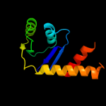
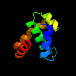
1 d1musa_
98.3
13
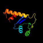
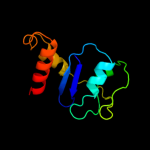
Fold: Ribonuclease H-like motifSuperfamily: Ribonuclease H-likeFamily: Transposase inhibitor (Tn5 transposase)2 d1b7ea_
97.9
14
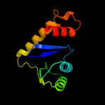
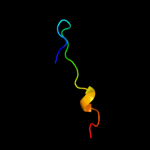
Fold: Ribonuclease H-like motifSuperfamily: Ribonuclease H-likeFamily: Transposase inhibitor (Tn5 transposase)3 d1cxqa_
76.7
22
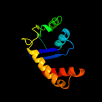
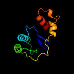
Fold: Ribonuclease H-like motifSuperfamily: Ribonuclease H-likeFamily: Retroviral integrase, catalytic domain4 d1asua_
75.9
20
Fold: Ribonuclease H-like motifSuperfamily: Ribonuclease H-likeFamily: Retroviral integrase, catalytic domain5 c3nf9A_
53.5
14
PDB header: hydrolase/hydrolase inhibitorChain: A: PDB Molecule: integrase;PDBTitle: structural basis for a new mechanism of inhibition of hiv integrase2 identified by fragment screening and structure based design
6 d1c0ma2
49.1
15
Fold: Ribonuclease H-like motifSuperfamily: Ribonuclease H-likeFamily: Retroviral integrase, catalytic domain7 c3hefB_
44.3
15
PDB header: viral proteinChain: B: PDB Molecule: gene 1 protein;PDBTitle: crystal structure of the bacteriophage sf6 terminase small2 subunit
8 d2htsa_
43.2
10
Fold: DNA/RNA-binding 3-helical bundleSuperfamily: "Winged helix" DNA-binding domainFamily: Heat-shock transcription factor9 d1hksa_
40.6
19
Fold: DNA/RNA-binding 3-helical bundleSuperfamily: "Winged helix" DNA-binding domainFamily: Heat-shock transcription factor10 d1a9xa1
39.8
14
Fold: Carbamoyl phosphate synthetase, large subunit connection domainSuperfamily: Carbamoyl phosphate synthetase, large subunit connection domainFamily: Carbamoyl phosphate synthetase, large subunit connection domain11 c1c0mA_
34.5
18
PDB header: transferaseChain: A: PDB Molecule: protein (integrase);PDBTitle: crystal structure of rsv two-domain integrase
12 d1d5ra1
29.4
26
Fold: C2 domain-likeSuperfamily: C2 domain (Calcium/lipid-binding domain, CaLB)Family: PLC-like (P variant)13 c2lduA_
28.7
21
PDB header: chaperoneChain: A: PDB Molecule: heat shock factor protein 1;PDBTitle: solution nmr structure of heat shock factor protein 1 dna binding2 domain from homo sapiens, northeast structural genomics consortium3 target hr3023c
14 c2e0yB_
28.2
20
PDB header: transferaseChain: B: PDB Molecule: gamma-glutamyltranspeptidase;PDBTitle: crystal structure of the samarium derivative of mature gamma-2 glutamyltranspeptidase from escherichia coli
15 c3pvpA_
24.2
14
PDB header: dna binding protein/dnaChain: A: PDB Molecule: chromosomal replication initiator protein dnaa;PDBTitle: structure of mycobacterium tuberculosis dnaa-dbd in complex with box22 dna
16 d1hyva_
23.1
16
Fold: Ribonuclease H-like motifSuperfamily: Ribonuclease H-likeFamily: Retroviral integrase, catalytic domain17 c2nqoB_
22.2
15
PDB header: transferaseChain: B: PDB Molecule: gamma-glutamyltranspeptidase;PDBTitle: crystal structure of helicobacter pylori gamma-glutamyltranspeptidase
18 d1exqa_
18.8
16
Fold: Ribonuclease H-like motifSuperfamily: Ribonuclease H-likeFamily: Retroviral integrase, catalytic domain19 c3f9kV_
18.4
12
PDB header: viral protein, recombinationChain: V: PDB Molecule: integrase;PDBTitle: two domain fragment of hiv-2 integrase in complex with ledgf ibd
20 c3eplA_
15.3
10
PDB header: transferase/rnaChain: A: PDB Molecule: trna isopentenyltransferase;PDBTitle: crystallographic snapshots of eukaryotic2 dimethylallyltransferase acting on trna: insight into trna3 recognition and reaction mechanism
21 c2v36D_
not modelled
15.1
30
PDB header: transferaseChain: D: PDB Molecule: gamma-glutamyltranspeptidase small chain;PDBTitle: crystal structure of gamma-glutamyl transferase from2 bacillus subtilis
22 c2o8kA_
not modelled
15.0
27
PDB header: transcription/dnaChain: A: PDB Molecule: rna polymerase sigma factor rpon;PDBTitle: nmr structure of the sigma-54 rpon domain bound to the-242 promoter element
23 d1mnga2
not modelled
14.3
18
Fold: Fe,Mn superoxide dismutase (SOD), C-terminal domainSuperfamily: Fe,Mn superoxide dismutase (SOD), C-terminal domainFamily: Fe,Mn superoxide dismutase (SOD), C-terminal domain24 c1p7gL_
not modelled
13.5
5
PDB header: oxidoreductaseChain: L: PDB Molecule: superoxide dismutase;PDBTitle: crystal structure of superoxide dismutase from pyrobaculum2 aerophilum
25 d1bsma2
not modelled
13.4
11
Fold: Fe,Mn superoxide dismutase (SOD), C-terminal domainSuperfamily: Fe,Mn superoxide dismutase (SOD), C-terminal domainFamily: Fe,Mn superoxide dismutase (SOD), C-terminal domain26 d1slma1
not modelled
13.2
27
Fold: PGBD-likeSuperfamily: PGBD-likeFamily: MMP N-terminal domain27 d1c6va_
not modelled
12.7
16
Fold: Ribonuclease H-like motifSuperfamily: Ribonuclease H-likeFamily: Retroviral integrase, catalytic domain28 d1p7ga2
not modelled
12.6
5
Fold: Fe,Mn superoxide dismutase (SOD), C-terminal domainSuperfamily: Fe,Mn superoxide dismutase (SOD), C-terminal domainFamily: Fe,Mn superoxide dismutase (SOD), C-terminal domain29 d1kkca2
not modelled
12.5
5
Fold: Fe,Mn superoxide dismutase (SOD), C-terminal domainSuperfamily: Fe,Mn superoxide dismutase (SOD), C-terminal domainFamily: Fe,Mn superoxide dismutase (SOD), C-terminal domain30 d1b06a2
not modelled
11.9
5
Fold: Fe,Mn superoxide dismutase (SOD), C-terminal domainSuperfamily: Fe,Mn superoxide dismutase (SOD), C-terminal domainFamily: Fe,Mn superoxide dismutase (SOD), C-terminal domain31 d1ma1a2
not modelled
11.6
13
Fold: Fe,Mn superoxide dismutase (SOD), C-terminal domainSuperfamily: Fe,Mn superoxide dismutase (SOD), C-terminal domainFamily: Fe,Mn superoxide dismutase (SOD), C-terminal domain32 d1idsa2
not modelled
11.1
13
Fold: Fe,Mn superoxide dismutase (SOD), C-terminal domainSuperfamily: Fe,Mn superoxide dismutase (SOD), C-terminal domainFamily: Fe,Mn superoxide dismutase (SOD), C-terminal domain33 c3g7pA_
not modelled
10.9
17
PDB header: unknown functionChain: A: PDB Molecule: nitrogen fixation protein;PDBTitle: crystal structure of a nifx-associated protein of unknown function2 (afe_1514) from acidithiobacillus ferrooxidans atcc at 2.00 a3 resolution
34 d2vo1a1
not modelled
9.9
18
Fold: P-loop containing nucleoside triphosphate hydrolasesSuperfamily: P-loop containing nucleoside triphosphate hydrolasesFamily: Nitrogenase iron protein-like35 c3nj2B_
not modelled
9.8
17
PDB header: unknown functionChain: B: PDB Molecule: duf269-containing protein;PDBTitle: crystal structure of cce_0566 from the cyanobacterium cyanothece2 51142, a protein associated with nitrogen fixation from the duf2693 family
36 d1jr9a2
not modelled
9.7
13
Fold: Fe,Mn superoxide dismutase (SOD), C-terminal domainSuperfamily: Fe,Mn superoxide dismutase (SOD), C-terminal domainFamily: Fe,Mn superoxide dismutase (SOD), C-terminal domain37 d1wb8a2
not modelled
9.7
5
Fold: Fe,Mn superoxide dismutase (SOD), C-terminal domainSuperfamily: Fe,Mn superoxide dismutase (SOD), C-terminal domainFamily: Fe,Mn superoxide dismutase (SOD), C-terminal domain38 c3d3kD_
not modelled
9.2
12
PDB header: protein bindingChain: D: PDB Molecule: enhancer of mrna-decapping protein 3;PDBTitle: crystal structure of human edc3p
39 d2i3oa1
not modelled
9.2
25
Fold: Ntn hydrolase-likeSuperfamily: N-terminal nucleophile aminohydrolases (Ntn hydrolases)Family: Gamma-glutamyltranspeptidase-like40 d1gv3a2
not modelled
9.0
11
Fold: Fe,Mn superoxide dismutase (SOD), C-terminal domainSuperfamily: Fe,Mn superoxide dismutase (SOD), C-terminal domainFamily: Fe,Mn superoxide dismutase (SOD), C-terminal domain41 d2nyba2
not modelled
8.9
8
Fold: Fe,Mn superoxide dismutase (SOD), C-terminal domainSuperfamily: Fe,Mn superoxide dismutase (SOD), C-terminal domainFamily: Fe,Mn superoxide dismutase (SOD), C-terminal domain42 c3ceiA_
not modelled
8.8
9
PDB header: oxidoreductaseChain: A: PDB Molecule: superoxide dismutase;PDBTitle: crystal structure of superoxide dismutase from helicobacter2 pylori
43 d2p4ka2
not modelled
8.8
11
Fold: Fe,Mn superoxide dismutase (SOD), C-terminal domainSuperfamily: Fe,Mn superoxide dismutase (SOD), C-terminal domainFamily: Fe,Mn superoxide dismutase (SOD), C-terminal domain44 d2nlza1
not modelled
8.7
20
Fold: Ntn hydrolase-likeSuperfamily: N-terminal nucleophile aminohydrolases (Ntn hydrolases)Family: Gamma-glutamyltranspeptidase-like45 c3n0aA_
not modelled
8.6
29
PDB header: hydrolaseChain: A: PDB Molecule: tyrosine-protein phosphatase auxilin;PDBTitle: crystal structure of auxilin (40-400)
46 c1y67D_
not modelled
8.5
11
PDB header: oxidoreductaseChain: D: PDB Molecule: manganese superoxide dismutase;PDBTitle: crystal structure of manganese superoxide dismutase from2 deinococcus radiodurans
47 c2kvcA_
not modelled
8.1
20
PDB header: unknown functionChain: A: PDB Molecule: putative uncharacterized protein;PDBTitle: solution structure of the mycobacterium tuberculosis protein rv0543c,2 a member of the duf3349 superfamily. seattle structural genomics3 center for infectious disease target mytud.17112.a
48 d1y67a2
not modelled
7.9
11
Fold: Fe,Mn superoxide dismutase (SOD), C-terminal domainSuperfamily: Fe,Mn superoxide dismutase (SOD), C-terminal domainFamily: Fe,Mn superoxide dismutase (SOD), C-terminal domain49 c3d3jA_
not modelled
7.4
12
PDB header: protein bindingChain: A: PDB Molecule: enhancer of mrna-decapping protein 3;PDBTitle: crystal structure of human edc3p
50 d1s1ma2
not modelled
7.3
24
Fold: P-loop containing nucleoside triphosphate hydrolasesSuperfamily: P-loop containing nucleoside triphosphate hydrolasesFamily: Nitrogenase iron protein-like51 c3ga9S_
not modelled
7.2
36
PDB header: hydrolaseChain: S: PDB Molecule: capsule biosynthesis protein capd;PDBTitle: crystal structure of bacillus anthracis transpeptidase enzyme capd,2 crystal form ii
52 c1k6yB_
not modelled
7.1
15
PDB header: transferaseChain: B: PDB Molecule: integrase;PDBTitle: crystal structure of a two-domain fragment of hiv-1 integrase
53 d1j1va_
not modelled
6.7
17
Fold: DNA/RNA-binding 3-helical bundleSuperfamily: TrpR-likeFamily: Chromosomal replication initiation factor DnaA C-terminal domain IV54 d1sfka_
not modelled
6.4
24
Fold: Flavivirus capsid protein CSuperfamily: Flavivirus capsid protein CFamily: Flavivirus capsid protein C55 d1vcoa2
not modelled
6.3
18
Fold: P-loop containing nucleoside triphosphate hydrolasesSuperfamily: P-loop containing nucleoside triphosphate hydrolasesFamily: Nitrogenase iron protein-like56 c1cojA_
not modelled
6.2
8
PDB header: oxidoreductaseChain: A: PDB Molecule: protein (superoxide dismutase);PDBTitle: fe-sod from aquifex pyrophilus, a hyperthermophilic bacterium
57 d1su3a1
not modelled
6.1
28
Fold: PGBD-likeSuperfamily: PGBD-likeFamily: MMP N-terminal domain58 d2fug11
not modelled
5.9
19
Fold: Bromodomain-likeSuperfamily: Nqo1C-terminal domain-likeFamily: Nqo1C-terminal domain-like59 d1b48a1
not modelled
5.9
44
Fold: GST C-terminal domain-likeSuperfamily: GST C-terminal domain-likeFamily: Glutathione S-transferase (GST), C-terminal domain60 d1coja2
not modelled
5.8
8
Fold: Fe,Mn superoxide dismutase (SOD), C-terminal domainSuperfamily: Fe,Mn superoxide dismutase (SOD), C-terminal domainFamily: Fe,Mn superoxide dismutase (SOD), C-terminal domain61 c1m6vE_
not modelled
5.8
14
PDB header: ligaseChain: E: PDB Molecule: carbamoyl phosphate synthetase large chain;PDBTitle: crystal structure of the g359f (small subunit) point mutant of2 carbamoyl phosphate synthetase
62 c1dt0A_
not modelled
5.8
12
PDB header: oxidoreductaseChain: A: PDB Molecule: superoxide dismutase;PDBTitle: cloning, sequence, and crystallographic structure of2 recombinant iron superoxide dismutase from pseudomonas3 ovalis
63 c2jg6A_
not modelled
5.7
10
PDB header: hydrolaseChain: A: PDB Molecule: dna-3-methyladenine glycosidase;PDBTitle: crystal structure of a 3-methyladenine dna glycosylase i2 from staphylococcus aureus
64 c3kksB_
not modelled
5.6
14
PDB header: dna binding proteinChain: B: PDB Molecule: integrase;PDBTitle: crystal structure of catalytic core domain of biv integrase in crystal2 form ii
65 c3eusB_
not modelled
5.6
16
PDB header: dna binding proteinChain: B: PDB Molecule: dna-binding protein;PDBTitle: the crystal structure of the dna binding protein from silicibacter2 pomeroyi
66 c2vkpA_
not modelled
5.5
12
PDB header: protein-bindingChain: A: PDB Molecule: btb/poz domain-containing protein 6;PDBTitle: crystal structure of btb domain from btbd6
67 d1uera2
not modelled
5.2
15
Fold: Fe,Mn superoxide dismutase (SOD), C-terminal domainSuperfamily: Fe,Mn superoxide dismutase (SOD), C-terminal domainFamily: Fe,Mn superoxide dismutase (SOD), C-terminal domain68 d1ix9a2
not modelled
5.1
13
Fold: Fe,Mn superoxide dismutase (SOD), C-terminal domainSuperfamily: Fe,Mn superoxide dismutase (SOD), C-terminal domainFamily: Fe,Mn superoxide dismutase (SOD), C-terminal domain69 d1v77a_
not modelled
5.1
7
Fold: 7-stranded beta/alpha barrelSuperfamily: PHP domain-likeFamily: RNase P subunit p30