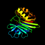
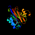
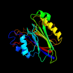
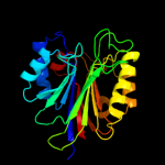
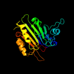
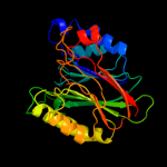
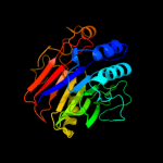
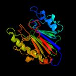
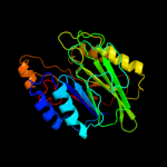
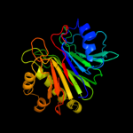
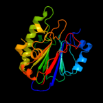
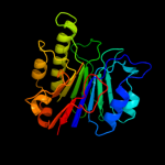
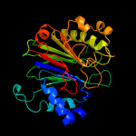
1 c3tebA_
100.0
17
PDB header: hydrolaseChain: A: PDB Molecule: endonuclease/exonuclease/phosphatase;PDBTitle: endonuclease/exonuclease/phosphatase family protein from leptotrichia2 buccalis c-1013-b
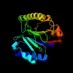
2 c3ngoA_
100.0
16
PDB header: hydrolase/dnaChain: A: PDB Molecule: ccr4-not transcription complex subunit 6-like;PDBTitle: crystal structure of the human cnot6l nuclease domain in complex with2 poly(a) dna
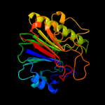
3 c3g6sA_
100.0
16
PDB header: hydrolaseChain: A: PDB Molecule: putative endonuclease/exonuclease/phosphatasePDBTitle: crystal structure of the2 endonuclease/exonuclease/phosphatase (bvu_0621) from3 bacteroides vulgatus. northeast structural genomics4 consortium target bvr56d
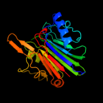
4 c3mprB_
100.0
20
PDB header: hydrolaseChain: B: PDB Molecule: putative endonuclease/exonuclease/phosphatase familyPDBTitle: crystal structure of endonuclease/exonuclease/phosphatase family2 protein from bacteroides thetaiotaomicron, northeast structural3 genomics consortium target btr318a
5 c2j63B_
100.0
20
PDB header: lyaseChain: B: PDB Molecule: ap-endonuclease;PDBTitle: crystal structure of ap endonuclease lmap from leishmania2 major
6 c3l1wE_
100.0
21
PDB header: structural genomics, unknown functionChain: E: PDB Molecule: uncharacterized protein;PDBTitle: the crystal structure of a functionally unknown conserved2 protein from enterococcus faecalis v583
7 d2ddra1
100.0
19
Fold: DNase I-likeSuperfamily: DNase I-likeFamily: Sphingomyelin phosphodiesterase-like8 c1e9nB_
100.0
21
PDB header: dna repairChain: B: PDB Molecule: dna-(apurinic or apyrimidinic site) lyase;PDBTitle: a second divalent metal ion in the active site of a new2 crystal form of human apurinic/apyrimidinic endonuclease,3 ape1, and its implications for the catalytic mechanism
9 d1sr4b_
100.0
17
Fold: DNase I-likeSuperfamily: DNase I-likeFamily: DNase I-like10 d1zwxa1
100.0
19
Fold: DNase I-likeSuperfamily: DNase I-likeFamily: Sphingomyelin phosphodiesterase-like11 d1akoa_
100.0
21
Fold: DNase I-likeSuperfamily: DNase I-likeFamily: DNase I-like12 c2jc5A_
100.0
16
PDB header: hydrolaseChain: A: PDB Molecule: exodeoxyribonuclease;PDBTitle: apurinic apyrimidinic (ap) endonuclease (nape) from2 neisseria meningitidis
13 c3i46B_
100.0
16
PDB header: toxinChain: B: PDB Molecule: beta-hemolysin;PDBTitle: crystal structure of beta toxin from staphylococcus aureus f277a,2 p278a mutant with bound calcium ions
14 c2voaB_
100.0
19
PDB header: lyaseChain: B: PDB Molecule: exodeoxyribonuclease iii;PDBTitle: structure of an ap endonuclease from archaeoglobus fulgidus
15 c3g0rA_
100.0
20
PDB header: hydrolase/dnaChain: A: PDB Molecule: exodeoxyribonuclease;PDBTitle: complex of mth0212 and an 8bp dsdna with distorted ends
16 d1vyba_
99.9
16
Fold: DNase I-likeSuperfamily: DNase I-likeFamily: DNase I-like17 d2f1na1
99.9
16
Fold: DNase I-likeSuperfamily: DNase I-likeFamily: DNase I-like18 d1hd7a_
99.9
19
Fold: DNase I-likeSuperfamily: DNase I-likeFamily: DNase I-like19 c2jc4A_
99.9
23
PDB header: hydrolaseChain: A: PDB Molecule: exodeoxyribonuclease iii;PDBTitle: 3'-5' exonuclease (nexo) from neisseria meningitidis
20 d2a40b1
99.9
15
Fold: DNase I-likeSuperfamily: DNase I-likeFamily: DNase I-like21 d1wdua_
not modelled
99.9
13
Fold: DNase I-likeSuperfamily: DNase I-likeFamily: DNase I-like22 d2imqx1
not modelled
99.9
16
Fold: DNase I-likeSuperfamily: DNase I-likeFamily: Inositol polyphosphate 5-phosphatase (IPP5)23 c2ei9A_
not modelled
99.9
15
PDB header: gene regulationChain: A: PDB Molecule: non-ltr retrotransposon r1bmks orf2 protein;PDBTitle: crystal structure of r1bm endonuclease domain
24 c3mtcA_
not modelled
99.9
18
PDB header: hydrolase/hydrolase inhibitorChain: A: PDB Molecule: type ii inositol-1,4,5-trisphosphate 5-phosphatase;PDBTitle: crystal structure of inpp5b in complex with phosphatidylinositol 4-2 phosphate
25 c3nr8A_
not modelled
99.9
17
PDB header: hydrolaseChain: A: PDB Molecule: phosphatidylinositol-3,4,5-trisphosphate 5-phosphatase 2;PDBTitle: crystal structure of human ship2
26 c2xswB_
not modelled
99.7
13
PDB header: hydrolaseChain: B: PDB Molecule: 72 kda inositol polyphosphate 5-phosphatase;PDBTitle: crystal structure of human inpp5e
27 d1i9za_
not modelled
99.6
14
Fold: DNase I-likeSuperfamily: DNase I-likeFamily: Inositol polyphosphate 5-phosphatase (IPP5)28 d1emsa2
not modelled
83.9
7
Fold: Carbon-nitrogen hydrolaseSuperfamily: Carbon-nitrogen hydrolaseFamily: Nitrilase29 c2w1vA_
not modelled
61.9
19
PDB header: hydrolaseChain: A: PDB Molecule: nitrilase homolog 2;PDBTitle: crystal structure of mouse nitrilase-2 at 1.4a resolution
30 c2vhiG_
not modelled
61.7
20
PDB header: hydrolaseChain: G: PDB Molecule: cg3027-pa;PDBTitle: crystal structure of a pyrimidine degrading enzyme from2 drosophila melanogaster
31 d1f89a_
not modelled
60.7
14
Fold: Carbon-nitrogen hydrolaseSuperfamily: Carbon-nitrogen hydrolaseFamily: Nitrilase32 c3hkxA_
not modelled
53.4
14
PDB header: hydrolaseChain: A: PDB Molecule: amidase;PDBTitle: crystal structure analysis of an amidase from nesterenkonia sp.
33 c2plqA_
not modelled
47.0
15
PDB header: hydrolaseChain: A: PDB Molecule: aliphatic amidase;PDBTitle: crystal structure of the amidase from geobacillus pallidus rapc8
34 c1emsB_
not modelled
45.2
7
PDB header: antitumor proteinChain: B: PDB Molecule: nit-fragile histidine triad fusion protein;PDBTitle: crystal structure of the c. elegans nitfhit protein
35 c3ilvA_
not modelled
31.7
17
PDB header: ligaseChain: A: PDB Molecule: glutamine-dependent nad(+) synthetase;PDBTitle: crystal structure of a glutamine-dependent nad(+) synthetase2 from cytophaga hutchinsonii
36 c2e2kC_
not modelled
29.2
5
PDB header: hydrolaseChain: C: PDB Molecule: formamidase;PDBTitle: helicobacter pylori formamidase amif contains a fine-tuned cysteine-2 glutamate-lysine catalytic triad
37 c3n05B_
not modelled
26.6
18
PDB header: ligaseChain: B: PDB Molecule: nh(3)-dependent nad(+) synthetase;PDBTitle: crystal structure of nh3-dependent nad+ synthetase from streptomyces2 avermitilis
38 d1uf5a_
not modelled
22.8
14
Fold: Carbon-nitrogen hydrolaseSuperfamily: Carbon-nitrogen hydrolaseFamily: Carbamilase39 d3bula2
not modelled
20.4
21
Fold: Flavodoxin-likeSuperfamily: Cobalamin (vitamin B12)-binding domainFamily: Cobalamin (vitamin B12)-binding domain40 c2e11B_
not modelled
16.1
10
PDB header: hydrolaseChain: B: PDB Molecule: hydrolase;PDBTitle: the crystal structure of xc1258 from xanthomonas campestris: a cn-2 hydrolase superfamily protein with an arsenic adduct in the active3 site
41 d1xrsb1
not modelled
14.3
11
Fold: Flavodoxin-likeSuperfamily: Cobalamin (vitamin B12)-binding domainFamily: Cobalamin (vitamin B12)-binding domain42 c1chmA_
not modelled
13.6
12
PDB header: creatinaseChain: A: PDB Molecule: creatine amidinohydrolase;PDBTitle: enzymatic mechanism of creatine amidinohydrolase as deduced2 from crystal structures
43 c1y80A_
not modelled
12.9
28
PDB header: structural genomics, unknown functionChain: A: PDB Molecule: predicted cobalamin binding protein;PDBTitle: structure of a corrinoid (factor iiim)-binding protein from2 moorella thermoacetica
44 c1xrsB_
not modelled
12.3
11
PDB header: isomeraseChain: B: PDB Molecule: d-lysine 5,6-aminomutase beta subunit;PDBTitle: crystal structure of lysine 5,6-aminomutase in complex with plp,2 cobalamin, and 5'-deoxyadenosine
45 c3t1iC_
not modelled
10.4
17
PDB header: hydrolaseChain: C: PDB Molecule: double-strand break repair protein mre11a;PDBTitle: crystal structure of human mre11: understanding tumorigenic mutations
46 c3jvvA_
not modelled
10.4
11
PDB header: atp binding proteinChain: A: PDB Molecule: twitching mobility protein;PDBTitle: crystal structure of p. aeruginosa pilt with bound amp-pcp
47 d7reqa2
not modelled
10.1
11
Fold: Flavodoxin-likeSuperfamily: Cobalamin (vitamin B12)-binding domainFamily: Cobalamin (vitamin B12)-binding domain48 d1edqa1
not modelled
9.8
20
Fold: Immunoglobulin-like beta-sandwichSuperfamily: E set domainsFamily: E-set domains of sugar-utilizing enzymes49 d1ccwa_
not modelled
9.3
16
Fold: Flavodoxin-likeSuperfamily: Cobalamin (vitamin B12)-binding domainFamily: Cobalamin (vitamin B12)-binding domain50 d1j31a_
not modelled
9.2
18
Fold: Carbon-nitrogen hydrolaseSuperfamily: Carbon-nitrogen hydrolaseFamily: Carbamilase51 c1bmtB_
not modelled
8.5
21
PDB header: methyltransferaseChain: B: PDB Molecule: methionine synthase;PDBTitle: how a protein binds b12: a 3.o angstrom x-ray structure of2 the b12-binding domains of methionine synthase
52 c2yxbA_
not modelled
8.4
11
PDB header: isomeraseChain: A: PDB Molecule: coenzyme b12-dependent mutase;PDBTitle: crystal structure of the methylmalonyl-coa mutase alpha-subunit from2 aeropyrum pernix
53 c3nrbD_
not modelled
8.2
24
PDB header: hydrolaseChain: D: PDB Molecule: formyltetrahydrofolate deformylase;PDBTitle: crystal structure of a formyltetrahydrofolate deformylase (puru,2 pp_1943) from pseudomonas putida kt2440 at 2.05 a resolution
54 c1k98A_
not modelled
8.2
21
PDB header: transferaseChain: A: PDB Molecule: methionine synthase;PDBTitle: adomet complex of meth c-terminal fragment
55 c3menC_
not modelled
8.1
14
PDB header: hydrolaseChain: C: PDB Molecule: acetylpolyamine aminohydrolase;PDBTitle: crystal structure of acetylpolyamine aminohydrolase from burkholderia2 pseudomallei, iodide soak
56 c2eyuA_
not modelled
7.3
11
PDB header: protein transportChain: A: PDB Molecule: twitching motility protein pilt;PDBTitle: the crystal structure of the c-terminal domain of aquifex2 aeolicus pilt
57 c3n0vD_
not modelled
7.1
18
PDB header: hydrolaseChain: D: PDB Molecule: formyltetrahydrofolate deformylase;PDBTitle: crystal structure of a formyltetrahydrofolate deformylase (pp_0327)2 from pseudomonas putida kt2440 at 2.25 a resolution
58 c2qlvB_
not modelled
7.0
39
PDB header: transferase/protein bindingChain: B: PDB Molecule: protein sip2;PDBTitle: crystal structure of the heterotrimer core of the s.2 cerevisiae ampk homolog snf1
59 c3ezxA_
not modelled
6.8
11
PDB header: transferaseChain: A: PDB Molecule: monomethylamine corrinoid protein 1;PDBTitle: structure of methanosarcina barkeri monomethylamine2 corrinoid protein
60 c3obiC_
not modelled
6.6
18
PDB header: hydrolaseChain: C: PDB Molecule: formyltetrahydrofolate deformylase;PDBTitle: crystal structure of a formyltetrahydrofolate deformylase (np_949368)2 from rhodopseudomonas palustris cga009 at 1.95 a resolution
61 c3o1lB_
not modelled
6.6
35
PDB header: hydrolaseChain: B: PDB Molecule: formyltetrahydrofolate deformylase;PDBTitle: crystal structure of a formyltetrahydrofolate deformylase (pspto_4314)2 from pseudomonas syringae pv. tomato str. dc3000 at 2.20 a resolution
62 d3bzka5
not modelled
6.5
6
Fold: Ribonuclease H-like motifSuperfamily: Ribonuclease H-likeFamily: Tex RuvX-like domain-like63 c3louB_
not modelled
6.5
29
PDB header: hydrolaseChain: B: PDB Molecule: formyltetrahydrofolate deformylase;PDBTitle: crystal structure of formyltetrahydrofolate deformylase (yp_105254.1)2 from burkholderia mallei atcc 23344 at 1.90 a resolution
64 d2qlvb1
not modelled
6.5
39
Fold: Immunoglobulin-like beta-sandwichSuperfamily: E set domainsFamily: AMPK-beta glycogen binding domain-like65 d1g6oa_
not modelled
6.5
10
Fold: P-loop containing nucleoside triphosphate hydrolasesSuperfamily: P-loop containing nucleoside triphosphate hydrolasesFamily: RecA protein-like (ATPase-domain)66 c3p9xB_
not modelled
6.4
13
PDB header: transferaseChain: B: PDB Molecule: phosphoribosylglycinamide formyltransferase;PDBTitle: crystal structure of phosphoribosylglycinamide formyltransferase from2 bacillus halodurans
67 c2i2xD_
not modelled
6.3
17
PDB header: transferaseChain: D: PDB Molecule: methyltransferase 1;PDBTitle: crystal structure of methanol:cobalamin methyltransferase complex2 mtabc from methanosarcina barkeri
68 d1p9ra_
not modelled
6.1
11
Fold: P-loop containing nucleoside triphosphate hydrolasesSuperfamily: P-loop containing nucleoside triphosphate hydrolasesFamily: RecA protein-like (ATPase-domain)69 d2eg6a1
not modelled
5.6
50
Fold: TIM beta/alpha-barrelSuperfamily: Metallo-dependent hydrolasesFamily: Dihydroorotase70 d1fmfa_
not modelled
5.5
22
Fold: Flavodoxin-likeSuperfamily: Cobalamin (vitamin B12)-binding domainFamily: Cobalamin (vitamin B12)-binding domain71 c3pn9C_
not modelled
5.4
10
PDB header: hydrolaseChain: C: PDB Molecule: proline dipeptidase;PDBTitle: crystal structure of a proline dipeptidase from streptococcus2 pneumoniae tigr4
72 d1hjra_
not modelled
5.2
13
Fold: Ribonuclease H-like motifSuperfamily: Ribonuclease H-likeFamily: RuvC resolvase73 c3o5vA_
not modelled
5.2
14
PDB header: hydrolaseChain: A: PDB Molecule: x-pro dipeptidase;PDBTitle: the crystal structure of the creatinase/prolidase n-terminal domain of2 an x-pro dipeptidase from streptococcus pyogenes to 1.85a
74 d1z0na1
not modelled
5.2
40
Fold: Immunoglobulin-like beta-sandwichSuperfamily: E set domainsFamily: AMPK-beta glycogen binding domain-like75 d1q8ia1
not modelled
5.1
18
Fold: Ribonuclease H-like motifSuperfamily: Ribonuclease H-likeFamily: DnaQ-like 3'-5' exonuclease76 c3i7mA_
not modelled
5.1
15
PDB header: hydrolaseChain: A: PDB Molecule: xaa-pro dipeptidase;PDBTitle: n-terminal domain of xaa-pro dipeptidase from lactobacillus brevis.


