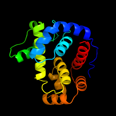
Phyre Home Retrieve Phyre Job Id
Emaill.a.kelley@imperial.ac.uk
DescriptionP0C960
DateThu Jan 5 11:30:08 GMT 2012
Unique Job ID67b2e1c85b2cb0ac

Summary 

Top model
Image coloured by rainbow N → C terminus
Model (left) based on template c2y8pA_
Top template information
PDB header:lyase
Chain: A: PDB Molecule:endo-type membrane-bound lytic murein transglycosylase a;
PDBTitle: crystal structure of an outer membrane-anchored endolytic2 peptidoglycan lytic transglycosylase (mlte) from3 escherichia coli
Confidence and coverage
Confidence:100.0% Coverage: 91%
185 residues ( 91% of your sequence) have been modelled with 100.0% confidence by the single highest scoring template.
3D viewing
Interactive 3D view in Jmol

Sequence analysis 

Secondary structure and disorder prediction 

   1........10.........20.........30.........40.........50.........60
Sequence  MKLRWFAFLIVLLAGCSSKHDYTNPPWNAKVPVQRAMQWMPISQKAGAAWGVDPQLITAI
Secondary structure 


SS confidence 


Disorder  ???


??


?????
??


?


Disorder confidence 


   .........70.........80.........90.........100.........110.........120
Sequence  IAIESGGNPNAVSKSNAIGLMQLKASTSGRDVYRRMGWSGEPTTSELKNPERNISMGAAY
Secondary structure 


SS confidence 


Disorder 


?????
?


Disorder confidence 


   .........130.........140.........150.........160.........170.........180
Sequence  LNILETGPLAGIEDPKVLQYALVVSYANGAGALLRTFSSDRKKAISKINDLDADEFLEHV
Secondary structure 


SS confidence 


Disorder 


?
??


???
??


Disorder confidence 


   .........190.........200...
Sequence  ARNHPAPQAPRYIYKLEQALDAM
Secondary structure 


SS confidence 


Disorder 


??
Disorder confidence 


Confidence Key
High(9)                    Low (0)
?Disordered
Alpha helix
Beta strand

Domain analysis 

Hover over an aligned region to see model and summary info

Please note, only up to the top 20 hits are modelled to reduce computer load

RankAligned region

PDB 2y8p chain A

3D model

Region: 19 - 203
Aligned: 185
Modelled: 185
Confidence: 100.0%
Identity: 100%
PDB header:lyase
Chain: A: PDB Molecule:endo-type membrane-bound lytic murein transglycosylase a;
PDBTitle: crystal structure of an outer membrane-anchored endolytic2 peptidoglycan lytic transglycosylase (mlte) from3 escherichia coli

Phyre2

PDB 1sly chain A

3D model

Region: 39 - 200
Aligned: 143
Modelled: 155
Confidence: 100.0%
Identity: 22%
PDB header:glycosyltransferase
Chain: A: PDB Molecule:70-kda soluble lytic transglycosylase;
PDBTitle: complex of the 70-kda soluble lytic transglycosylase with2 bulgecin a

Phyre2

PDB 1qsa chain A domain 2

3D model

Region: 38 - 201
Aligned: 145
Modelled: 157
Confidence: 100.0%
Identity: 21%
Fold: Lysozyme-like
Superfamily: Lysozyme-like
Family: Bacterial muramidase, catalytic domain

Phyre2

PDB 3mgw chain A

3D model

Region: 29 - 201
Aligned: 135
Modelled: 135
Confidence: 100.0%
Identity: 19%
PDB header:hydrolase
Chain: A: PDB Molecule:lysozyme g;
PDBTitle: thermodynamics and structure of a salmon cold-active goose-type2 lysozyme

Phyre2

PDB 1gbs chain A

3D model

Region: 28 - 201
Aligned: 136
Modelled: 136
Confidence: 100.0%
Identity: 21%
Fold: Lysozyme-like
Superfamily: Lysozyme-like
Family: G-type lysozyme

Phyre2

PDB 3gxk chain B

3D model

Region: 29 - 201
Aligned: 135
Modelled: 135
Confidence: 100.0%
Identity: 18%
PDB header:hydrolase
Chain: B: PDB Molecule:goose-type lysozyme 1;
PDBTitle: the crystal structure of g-type lysozyme from atlantic cod2 (gadus morhua l.) in complex with nag oligomers sheds new3 light on substrate binding and the catalytic mechanism.4 native structure to 1.9

Phyre2

PDB 1qus chain A

3D model

Region: 25 - 123
Aligned: 95
Modelled: 99
Confidence: 99.1%
Identity: 24%
Fold: Lysozyme-like
Superfamily: Lysozyme-like
Family: Bacterial muramidase, catalytic domain

Phyre2

PDB 3bkh chain A

3D model

Region: 41 - 168
Aligned: 127
Modelled: 128
Confidence: 99.0%
Identity: 20%
PDB header:hydrolase
Chain: A: PDB Molecule:lytic transglycosylase;
PDBTitle: crystal structure of the bacteriophage phikz lytic2 transglycosylase, gp144

Phyre2

PDB 1xsf chain A

3D model

Region: 51 - 122
Aligned: 67
Modelled: 72
Confidence: 97.4%
Identity: 21%
PDB header:cell cycle, hydrolase
Chain: A: PDB Molecule:probable resuscitation-promoting factor rpfb;
PDBTitle: solution structure of a resuscitation promoting factor2 domain from mycobacterium tuberculosis

Phyre2

PDB 1xsf chain A domain 1

3D model

Region: 52 - 122
Aligned: 66
Modelled: 71
Confidence: 97.2%
Identity: 21%
Fold: Lysozyme-like
Superfamily: Lysozyme-like
Family: RPF-like

Phyre2

PDB 3eo5 chain A

3D model

Region: 58 - 122
Aligned: 60
Modelled: 65
Confidence: 96.6%
Identity: 22%
PDB header:cell adhesion
Chain: A: PDB Molecule:resuscitation-promoting factor rpfb;
PDBTitle: crystal structure of the resuscitation promoting factor rpfb

Phyre2

PDB 2fbd chain B

3D model

Region: 43 - 83
Aligned: 41
Modelled: 41
Confidence: 96.2%
Identity: 32%
PDB header:hydrolase
Chain: B: PDB Molecule:lysozyme 1;
PDBTitle: the crystallographic structure of the digestive lysozyme 1 from musca2 domestica at 1.90 ang.

Phyre2

PDB 1gd6 chain A

3D model

Region: 43 - 83
Aligned: 41
Modelled: 41
Confidence: 96.1%
Identity: 20%
Fold: Lysozyme-like
Superfamily: Lysozyme-like
Family: C-type lysozyme

Phyre2

PDB 1iiz chain A

3D model

Region: 43 - 83
Aligned: 41
Modelled: 41
Confidence: 96.1%
Identity: 24%
Fold: Lysozyme-like
Superfamily: Lysozyme-like
Family: C-type lysozyme

Phyre2

PDB 1hhl chain A

3D model

Region: 43 - 83
Aligned: 40
Modelled: 41
Confidence: 95.6%
Identity: 25%
Fold: Lysozyme-like
Superfamily: Lysozyme-like
Family: C-type lysozyme

Phyre2

PDB 2vb1 chain A domain 1

3D model

Region: 43 - 83
Aligned: 40
Modelled: 41
Confidence: 95.6%
Identity: 25%
Fold: Lysozyme-like
Superfamily: Lysozyme-like
Family: C-type lysozyme

Phyre2

PDB 1ghl chain A

3D model

Region: 43 - 83
Aligned: 40
Modelled: 41
Confidence: 95.5%
Identity: 25%
Fold: Lysozyme-like
Superfamily: Lysozyme-like
Family: C-type lysozyme

Phyre2

PDB 3fi7 chain A

3D model

Region: 34 - 67
Aligned: 34
Modelled: 34
Confidence: 95.3%
Identity: 15%
PDB header:hydrolase
Chain: A: PDB Molecule:lmo1076 protein;
PDBTitle: crystal structure of the autolysin auto (lmo1076) from listeria2 monocytogenes, catalytic domain

Phyre2

PDB 2zyc chain A

3D model

Region: 33 - 73
Aligned: 41
Modelled: 41
Confidence: 95.2%
Identity: 22%
PDB header:hydrolase
Chain: A: PDB Molecule:peptidoglycan hydrolase flgj;
PDBTitle: crystal structure of peptidoglycan hydrolase from2 sphingomonas sp. a1

Phyre2

PDB 1jse chain A

3D model

Region: 43 - 83
Aligned: 40
Modelled: 41
Confidence: 95.1%
Identity: 23%
Fold: Lysozyme-like
Superfamily: Lysozyme-like
Family: C-type lysozyme

Phyre2
1

c2y8pA_
2

c1slyA_
3

d1qsaa2
4

c3mgwA_
5

d1gbsa_
6

c3gxkB_
7

d1qusa_
8

c3bkhA_
9

c1xsfA_
10

d1xsfa1
11

c3eo5A_
12

c2fbdB_
13

d1gd6a_
14

d1iiza_
15

d1hhla_
16

d2vb1a1
17

d1ghla_
18

c3fi7A_
19

c2zycA_
20

d1jsea_
21


22


23


24


25


26


27


28


29


30


31


32


33


34


35


36


37


38


39


40


41


42


43


44


45


46


47


48


49


50


Detailed template information 

#
Template Alignment Coverage3D Model Confidence
% i.d. Template Information
1c2y8pA_


100.0 100 PDB header:lyase
Chain: A: PDB Molecule:endo-type membrane-bound lytic murein transglycosylase a;
PDBTitle: crystal structure of an outer membrane-anchored endolytic2 peptidoglycan lytic transglycosylase (mlte) from3 escherichia coli
2c1slyA_


100.0 22 PDB header:glycosyltransferase
Chain: A: PDB Molecule:70-kda soluble lytic transglycosylase;
PDBTitle: complex of the 70-kda soluble lytic transglycosylase with2 bulgecin a
3d1qsaa2


100.0 21 Fold:Lysozyme-like
Superfamily:Lysozyme-like
Family:Bacterial muramidase, catalytic domain
4c3mgwA_


100.0 19 PDB header:hydrolase
Chain: A: PDB Molecule:lysozyme g;
PDBTitle: thermodynamics and structure of a salmon cold-active goose-type2 lysozyme
5d1gbsa_


100.0 21 Fold:Lysozyme-like
Superfamily:Lysozyme-like
Family:G-type lysozyme
6c3gxkB_


100.0 18 PDB header:hydrolase
Chain: B: PDB Molecule:goose-type lysozyme 1;
PDBTitle: the crystal structure of g-type lysozyme from atlantic cod2 (gadus morhua l.) in complex with nag oligomers sheds new3 light on substrate binding and the catalytic mechanism.4 native structure to 1.9
7d1qusa_


99.1 24 Fold:Lysozyme-like
Superfamily:Lysozyme-like
Family:Bacterial muramidase, catalytic domain
8c3bkhA_


99.0 20 PDB header:hydrolase
Chain: A: PDB Molecule:lytic transglycosylase;
PDBTitle: crystal structure of the bacteriophage phikz lytic2 transglycosylase, gp144
9c1xsfA_


97.4 21 PDB header:cell cycle, hydrolase
Chain: A: PDB Molecule:probable resuscitation-promoting factor rpfb;
PDBTitle: solution structure of a resuscitation promoting factor2 domain from mycobacterium tuberculosis
10d1xsfa1


97.2 21 Fold:Lysozyme-like
Superfamily:Lysozyme-like
Family:RPF-like
11c3eo5A_


96.6 22 PDB header:cell adhesion
Chain: A: PDB Molecule:resuscitation-promoting factor rpfb;
PDBTitle: crystal structure of the resuscitation promoting factor rpfb
12c2fbdB_


96.2 32 PDB header:hydrolase
Chain: B: PDB Molecule:lysozyme 1;
PDBTitle: the crystallographic structure of the digestive lysozyme 1 from musca2 domestica at 1.90 ang.
13d1gd6a_


96.1 20 Fold:Lysozyme-like
Superfamily:Lysozyme-like
Family:C-type lysozyme
14d1iiza_


96.1 24 Fold:Lysozyme-like
Superfamily:Lysozyme-like
Family:C-type lysozyme
15d1hhla_


95.6 25 Fold:Lysozyme-like
Superfamily:Lysozyme-like
Family:C-type lysozyme
16d2vb1a1


95.6 25 Fold:Lysozyme-like
Superfamily:Lysozyme-like
Family:C-type lysozyme
17d1ghla_


95.5 25 Fold:Lysozyme-like
Superfamily:Lysozyme-like
Family:C-type lysozyme
18c3fi7A_


95.3 15 PDB header:hydrolase
Chain: A: PDB Molecule:lmo1076 protein;
PDBTitle: crystal structure of the autolysin auto (lmo1076) from listeria2 monocytogenes, catalytic domain
19c2zycA_


95.2 22 PDB header:hydrolase
Chain: A: PDB Molecule:peptidoglycan hydrolase flgj;
PDBTitle: crystal structure of peptidoglycan hydrolase from2 sphingomonas sp. a1
20d1jsea_


95.1 23 Fold:Lysozyme-like
Superfamily:Lysozyme-like
Family:C-type lysozyme
21c3ct5A_


not modelled 95.1 17 PDB header:hydrolase
Chain: A: PDB Molecule:morphogenesis protein 1;
PDBTitle: crystal and cryoem structural studies of a cell wall degrading enzyme2 in the bacteriophage phi29 tail
22d1lmqa_


not modelled 95.1 30 Fold:Lysozyme-like
Superfamily:Lysozyme-like
Family:C-type lysozyme
23c2goiC_


not modelled 95.0 30 PDB header:cell adhesion, sugar binding protein
Chain: C: PDB Molecule:sperm lysozyme-like protein 1;
PDBTitle: crystal structure of mouse sperm c-type lysozyme-like2 protein 1
24d1qqya_


not modelled 94.9 25 Fold:Lysozyme-like
Superfamily:Lysozyme-like
Family:C-type lysozyme
25d1juga_


not modelled 94.8 28 Fold:Lysozyme-like
Superfamily:Lysozyme-like
Family:C-type lysozyme
26c2z2fA_


not modelled 94.7 20 PDB header:hydrolase
Chain: A: PDB Molecule:lysozyme c-2;
PDBTitle: x-ray crystal structure of bovine stomach lysozyme
27d1lsga1


not modelled 94.5 24 Fold:Lysozyme-like
Superfamily:Lysozyme-like
Family:C-type lysozyme
28d1ivma_


not modelled 94.5 18 Fold:Lysozyme-like
Superfamily:Lysozyme-like
Family:C-type lysozyme
29d2nwdx1


not modelled 93.8 23 Fold:Lysozyme-like
Superfamily:Lysozyme-like
Family:C-type lysozyme
30d1f6sa_


not modelled 92.5 28 Fold:Lysozyme-like
Superfamily:Lysozyme-like
Family:C-type lysozyme
31d2eqla_


not modelled 92.5 34 Fold:Lysozyme-like
Superfamily:Lysozyme-like
Family:C-type lysozyme
32d1b9oa_


not modelled 92.3 31 Fold:Lysozyme-like
Superfamily:Lysozyme-like
Family:C-type lysozyme
33d1yroa1


not modelled 91.9 31 Fold:Lysozyme-like
Superfamily:Lysozyme-like
Family:C-type lysozyme
34d1hfxa_


not modelled 91.9 38 Fold:Lysozyme-like
Superfamily:Lysozyme-like
Family:C-type lysozyme
35d1fkqa_


not modelled 91.3 28 Fold:Lysozyme-like
Superfamily:Lysozyme-like
Family:C-type lysozyme
36d1alca_


not modelled 90.3 31 Fold:Lysozyme-like
Superfamily:Lysozyme-like
Family:C-type lysozyme
37c3csqC_


not modelled 80.4 18 PDB header:hydrolase
Chain: C: PDB Molecule:morphogenesis protein 1;
PDBTitle: crystal and cryoem structural studies of a cell wall2 degrading enzyme in the bacteriophage phi29 tail
38d1nvma1


not modelled 34.0 15 Fold:RuvA C-terminal domain-like
Superfamily:post-HMGL domain-like
Family:DmpG/LeuA communication domain-like
39d1idra_


not modelled 29.8 16 Fold:Globin-like
Superfamily:Globin-like
Family:Truncated hemoglobin
40d1yt3a2


not modelled 21.8 4 Fold:SAM domain-like
Superfamily:HRDC-like
Family:RNase D C-terminal domains
41d2coba1


not modelled 15.0 17 Fold:DNA/RNA-binding 3-helical bundle
Superfamily:Homeodomain-like
Family:Psq domain
42c3aq8A_


not modelled 9.2 14 PDB header:oxygen binding
Chain: A: PDB Molecule:group 1 truncated hemoglobin;
PDBTitle: crystal structure of truncated hemoglobin from tetrahymena pyriformis,2 q46e mutant, fe(iii) form
43c2l5qA_


not modelled 9.0 23 PDB header:structural genomics, unknown function
Chain: A: PDB Molecule:uncharacterized protein;
PDBTitle: solution nmr structure of bvu_3817 from bacteroides vulgatus,2 northeast structural genomics consortium target bvr159
44d2cvza1


not modelled 8.7 22 Fold:6-phosphogluconate dehydrogenase C-terminal domain-like
Superfamily:6-phosphogluconate dehydrogenase C-terminal domain-like
Family:Hydroxyisobutyrate and 6-phosphogluconate dehydrogenase domain
45d1dlwa_


not modelled 8.3 19 Fold:Globin-like
Superfamily:Globin-like
Family:Truncated hemoglobin
46c1mgtA_


not modelled 7.1 16 PDB header:transferase
Chain: A: PDB Molecule:protein (o6-methylguanine-dna methyltransferase);
PDBTitle: crystal structure of o6-methylguanine-dna methyltransferase from2 hyperthermophilic archaeon pyrococcus kodakaraensis strain kod1
47d1a5za1


not modelled 6.7 13 Fold:NAD(P)-binding Rossmann-fold domains
Superfamily:NAD(P)-binding Rossmann-fold domains
Family:LDH N-terminal domain-like
48d2fnoa1


not modelled 6.2 32 Fold:GST C-terminal domain-like
Superfamily:GST C-terminal domain-like
Family:Glutathione S-transferase (GST), C-terminal domain
49d2fp1a1


not modelled 5.8 17 Fold:Chorismate mutase II
Superfamily:Chorismate mutase II
Family:Secreted chorismate mutase-like
50d1bw6a_


not modelled 5.3 10 Fold:DNA/RNA-binding 3-helical bundle
Superfamily:Homeodomain-like
Family:Centromere-binding

Binding site prediction 

Due to computational demand, binding site predictions are not run for batch jobs

If you want to predict binding sites, please manually submit your model of choice to 3DLigandSite


Phyre is for academic use only

Please cite: Protein structure prediction on the web: a case study using the Phyre server
Kelley LA and Sternberg MJE. Nature Protocols 4, 363 - 371 (2009) [pdf] [Import into BibTeX]
 
If you use the binding site predictions from 3DLigandSite, please also cite:
3DLigandSite: predicting ligand-binding sites using similar structures.
Wass MN, Kelley LA and Sternberg MJ Nucleic Acids Research 38, W469-73 (2010) [PubMed]
 
© Structural Bioinformatics Group
Imperial College London
Lawrence Kelley, Benjamin Jefferys
Disclaimer
Terms and Conditions
Component software
Template detection: HHpred 1.51
Secondary structure prediction: Psi-pred 2.5
Disorder prediction: Disopred 2.4
Transmembrane prediction: Memsat_SVM
Multi-template modelling and ab initio: Poing 1.0