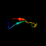
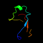
1 c2kluA_
87.9
20
PDB header: immune system, membrane proteinChain: A: PDB Molecule: t-cell surface glycoprotein cd4;PDBTitle: nmr structure of the transmembrane and cytoplasmic domains2 of human cd4
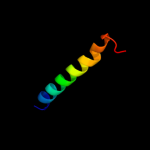
2 c3db3A_
86.2
17
PDB header: ligaseChain: A: PDB Molecule: e3 ubiquitin-protein ligase uhrf1;PDBTitle: crystal structure of the tandem tudor domains of the e3 ubiquitin-2 protein ligase uhrf1 in complex with trimethylated histone h3-k93 peptide
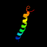
3 d1bcoa1
79.3
19
Fold: mu transposase, C-terminal domainSuperfamily: mu transposase, C-terminal domainFamily: mu transposase, C-terminal domain4 c2jp3A_
76.4
14
PDB header: transcriptionChain: A: PDB Molecule: fxyd domain-containing ion transport regulator 4;PDBTitle: solution structure of the human fxyd4 (chif) protein in sds2 micelles
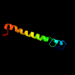
5 c2l2tA_
50.6
28
PDB header: membrane proteinChain: A: PDB Molecule: receptor tyrosine-protein kinase erbb-4;PDBTitle: solution nmr structure of the erbb4 dimeric membrane domain
6 c1bcoA_
50.2
19
PDB header: transposaseChain: A: PDB Molecule: bacteriophage mu transposase;PDBTitle: bacteriophage mu transposase core domain
7 c2jqoA_
32.2
19
PDB header: structural genomicsChain: A: PDB Molecule: hypothetical protein yoba;PDBTitle: nmr solution structure of bacillus subtilis yoba 21-120:2 northeast structural genomics consortium target sr547
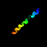
8 c1afoB_
31.9
18
PDB header: integral membrane proteinChain: B: PDB Molecule: glycophorin a;PDBTitle: dimeric transmembrane domain of human glycophorin a, nmr,2 20 structures
9 c2jo1A_
29.2
17
PDB header: hydrolase regulatorChain: A: PDB Molecule: phospholemman;PDBTitle: structure of the na,k-atpase regulatory protein fxyd1 in2 micelles
10 c1kqfB_
25.9
12
PDB header: oxidoreductaseChain: B: PDB Molecule: formate dehydrogenase, nitrate-inducible, iron-sulfurPDBTitle: formate dehydrogenase n from e. coli
11 c2k52A_
22.6
18
PDB header: structural genomics, unknown functionChain: A: PDB Molecule: uncharacterized protein mj1198;PDBTitle: structure of uncharacterized protein mj1198 from2 methanocaldococcus jannaschii. northeast structural3 genomics target mjr117b
12 d1v43a2
21.8
13
Fold: OB-foldSuperfamily: MOP-likeFamily: ABC-transporter additional domain13 c3f1zF_
20.6
31
PDB header: dna binding proteinChain: F: PDB Molecule: putative nucleic acid-binding lipoprotein;PDBTitle: crystal structure of putative nucleic acid-binding lipoprotein2 (yp_001337197.1) from klebsiella pneumoniae subsp. pneumoniae mgh3 78578 at 2.46 a resolution
14 d1ugpb_
19.5
18
Fold: SH3-like barrelSuperfamily: Electron transport accessory proteinsFamily: Nitrile hydratase beta chain15 c3dwqD_
16.5
43
PDB header: toxinChain: D: PDB Molecule: subtilase cytotoxin, subunit b;PDBTitle: crystal structure of the a-subunit of the ab5 toxin from e.2 coli with neu5gc-2,3gal-1,3glcnac
16 c2l0cA_
16.5
13
PDB header: structural genomics, unknown functionChain: A: PDB Molecule: putative membrane protein;PDBTitle: solution nmr structure of protein sty4237 (residues 36-120) from2 salmonella enterica, northeast structural genomics consortium target3 slr115
17 d1y5ic1
15.7
18
Fold: Heme-binding four-helical bundleSuperfamily: Respiratory nitrate reductase 1 gamma chainFamily: Respiratory nitrate reductase 1 gamma chain18 c3qz9D_
15.6
18
PDB header: lyaseChain: D: PDB Molecule: co-type nitrile hydratase beta subunit;PDBTitle: crystal structure of co-type nitrile hydratase beta-y215f from2 pseudomonas putida.
19 d2ahob2
14.8
13
Fold: OB-foldSuperfamily: Nucleic acid-binding proteinsFamily: Cold shock DNA-binding domain-like20 c2dxcG_
14.3
11
PDB header: hydrolaseChain: G: PDB Molecule: thiocyanate hydrolase subunit alpha;PDBTitle: recombinant thiocyanate hydrolase, fully-matured form
21 c3kdpH_
not modelled
14.1
26
PDB header: hydrolaseChain: H: PDB Molecule: na+/k+ atpase gamma subunit transcript variant a;PDBTitle: crystal structure of the sodium-potassium pump
22 c3kdpG_
not modelled
14.1
26
PDB header: hydrolaseChain: G: PDB Molecule: na+/k+ atpase gamma subunit transcript variant a;PDBTitle: crystal structure of the sodium-potassium pump
23 d1v29b_
not modelled
13.5
7
Fold: SH3-like barrelSuperfamily: Electron transport accessory proteinsFamily: Nitrile hydratase beta chain24 c2ky9A_
not modelled
13.3
19
PDB header: structural genomics, unknown functionChain: A: PDB Molecule: uncharacterized protein ydhk;PDBTitle: solution nmr structure of ydhk c-terminal domain from b.subtilis,2 northeast structural genomics consortium target target sr518
25 d2qdyb1
not modelled
12.6
15
Fold: SH3-like barrelSuperfamily: Electron transport accessory proteinsFamily: Nitrile hydratase beta chain26 d1vqoq1
not modelled
12.2
31
Fold: SH3-like barrelSuperfamily: Translation proteins SH3-like domainFamily: Ribosomal proteins L24p and L21e27 d2e74f1
not modelled
11.9
20
Fold: Single transmembrane helixSuperfamily: PetM subunit of the cytochrome b6f complexFamily: PetM subunit of the cytochrome b6f complex28 c2kpeB_
not modelled
11.0
17
PDB header: membrane proteinChain: B: PDB Molecule: glycophorin-a;PDBTitle: refined structure of glycophorin a transmembrane segment dimer in dpc2 micelles
29 c2kpeA_
not modelled
11.0
17
PDB header: membrane proteinChain: A: PDB Molecule: glycophorin-a;PDBTitle: refined structure of glycophorin a transmembrane segment dimer in dpc2 micelles
30 c1s1iQ_
not modelled
10.9
14
PDB header: ribosomeChain: Q: PDB Molecule: 60s ribosomal protein l21-a;PDBTitle: structure of the ribosomal 80s-eef2-sordarin complex from2 yeast obtained by docking atomic models for rna and protein3 components into a 11.7 a cryo-em map. this file, 1s1i,4 contains 60s subunit. the 40s ribosomal subunit is in file5 1s1h.
31 c3i4oA_
not modelled
10.3
32
PDB header: translationChain: A: PDB Molecule: translation initiation factor if-1;PDBTitle: crystal structure of translation initiation factor 1 from2 mycobacterium tuberculosis
32 d1wjsa_
not modelled
10.3
19
Fold: SH3-like barrelSuperfamily: Tudor/PWWP/MBTFamily: MBT repeat33 c2qtsA_
not modelled
10.3
12
PDB header: membrane proteinChain: A: PDB Molecule: acid-sensing ion channel;PDBTitle: structure of an acid-sensing ion channel 1 at 1.9 a resolution and low2 ph
34 d3d31a1
not modelled
10.3
9
Fold: OB-foldSuperfamily: MOP-likeFamily: ABC-transporter additional domain35 c2zkrq_
not modelled
9.3
20
PDB header: ribosomal protein/rnaChain: Q: PDB Molecule: rna expansion segment es31 part ii;PDBTitle: structure of a mammalian ribosomal 60s subunit within an2 80s complex obtained by docking homology models of the rna3 and proteins into an 8.7 a cryo-em map
36 d1hr0w_
not modelled
9.2
37
Fold: OB-foldSuperfamily: Nucleic acid-binding proteinsFamily: Cold shock DNA-binding domain-like37 c2kncA_
not modelled
9.0
23
PDB header: cell adhesionChain: A: PDB Molecule: integrin alpha-iib;PDBTitle: platelet integrin alfaiib-beta3 transmembrane-cytoplasmic2 heterocomplex
38 c3cm1C_
not modelled
8.4
19
PDB header: cell cycleChain: C: PDB Molecule: ssga-like sporulation-specific cell division protein;PDBTitle: crystal structure of ssga-like sporulation-specific cell division2 protein (yp_290167.1) from thermobifida fusca yx-er1 at 2.60 a3 resolution
39 d2k5qa1
not modelled
8.3
9
Fold: OB-foldSuperfamily: BC4932-likeFamily: BC4932-like40 d1fvia1
not modelled
8.0
16
Fold: OB-foldSuperfamily: Nucleic acid-binding proteinsFamily: DNA ligase/mRNA capping enzyme postcatalytic domain41 d1fftb2
not modelled
7.6
11
Fold: Transmembrane helix hairpinSuperfamily: Cytochrome c oxidase subunit II-like, transmembrane regionFamily: Cytochrome c oxidase subunit II-like, transmembrane region42 c2ahoB_
not modelled
7.6
11
PDB header: translationChain: B: PDB Molecule: translation initiation factor 2 alpha subunit;PDBTitle: structure of the archaeal initiation factor eif2 alpha-2 gamma heterodimer from sulfolobus solfataricus complexed3 with gdpnp
43 c2k1aA_
not modelled
7.6
23
PDB header: cell adhesionChain: A: PDB Molecule: integrin alpha-iib;PDBTitle: bicelle-embedded integrin alpha(iib) transmembrane segment
44 d1h9ra2
not modelled
7.5
15
Fold: OB-foldSuperfamily: MOP-likeFamily: BiMOP, duplicated molybdate-binding domain45 c2jwaA_
not modelled
7.5
9
PDB header: transferaseChain: A: PDB Molecule: receptor tyrosine-protein kinase erbb-2;PDBTitle: erbb2 transmembrane segment dimer spatial structure
46 c3j0cH_
not modelled
7.5
11
PDB header: virusChain: H: PDB Molecule: e2 envelope glycoprotein;PDBTitle: models of e1, e2 and cp of venezuelan equine encephalitis virus tc-832 strain restrained by a near atomic resolution cryo-em map
47 d1qcsa1
not modelled
7.3
32
Fold: Double psi beta-barrelSuperfamily: ADC-likeFamily: Cdc48 N-terminal domain-like48 d1f20a1
not modelled
7.1
11
Fold: Reductase/isomerase/elongation factor common domainSuperfamily: Riboflavin synthase domain-likeFamily: NADPH-cytochrome p450 reductase FAD-binding domain-like49 c2zxeG_
not modelled
6.7
17
PDB header: hydrolase/transport proteinChain: G: PDB Molecule: phospholemman-like protein;PDBTitle: crystal structure of the sodium - potassium pump in the e2.2k+.pi2 state
50 c2e6zA_
not modelled
6.7
31
PDB header: transcriptionChain: A: PDB Molecule: transcription elongation factor spt5;PDBTitle: solution structure of the second kow motif of human2 transcription elongation factor spt5
51 c2xzn2_
not modelled
6.5
27
PDB header: ribosomeChain: 2: PDB Molecule: 40s ribosomal protein s8;PDBTitle: crystal structure of the eukaryotic 40s ribosomal2 subunit in complex with initiation factor 1. this file3 contains the 40s subunit and initiation factor for4 molecule 2
52 d1y14b1
not modelled
6.3
16
Fold: OB-foldSuperfamily: Nucleic acid-binding proteinsFamily: Cold shock DNA-binding domain-like53 c2zklA_
not modelled
6.3
17
PDB header: isomeraseChain: A: PDB Molecule: capsular polysaccharide synthesis enzyme cap5f;PDBTitle: crystal structure of capsular polysaccharide assembling protein capf2 from staphylococcus aureus
54 d1h9ma2
not modelled
6.3
6
Fold: OB-foldSuperfamily: MOP-likeFamily: BiMOP, duplicated molybdate-binding domain55 c2ks1B_
not modelled
6.2
11
PDB header: transferaseChain: B: PDB Molecule: epidermal growth factor receptor;PDBTitle: heterodimeric association of transmembrane domains of erbb1 and erbb22 receptors enabling kinase activation
56 d1h6la_
not modelled
6.2
18
Fold: 6-bladed beta-propellerSuperfamily: Thermostable phytase (3-phytase)Family: Thermostable phytase (3-phytase)57 c3u5cI_
not modelled
6.0
36
PDB header: ribosomeChain: I: PDB Molecule: 40s ribosomal protein s8-a;PDBTitle: the structure of the eukaryotic ribosome at 3.0 a resolution
58 c4a1aP_
not modelled
6.0
14
PDB header: ribosomeChain: P: PDB Molecule: 60s ribosomal protein l21;PDBTitle: t.thermophila 60s ribosomal subunit in complex with2 initiation factor 6. this file contains 5s rrna,3 5.8s rrna and proteins of molecule 3.
59 c3iz5U_
not modelled
6.0
21
PDB header: ribosomeChain: U: PDB Molecule: 60s ribosomal protein l21 (l21e);PDBTitle: localization of the large subunit ribosomal proteins into a 5.5 a2 cryo-em map of triticum aestivum translating 80s ribosome
60 c1vf5G_
not modelled
5.8
21
PDB header: photosynthesisChain: G: PDB Molecule: protein pet g;PDBTitle: crystal structure of cytochrome b6f complex from m.laminosus
61 d1vf5g_
not modelled
5.8
21
Fold: Single transmembrane helixSuperfamily: PetG subunit of the cytochrome b6f complexFamily: PetG subunit of the cytochrome b6f complex62 c1ddiA_
not modelled
5.7
8
PDB header: oxidoreductaseChain: A: PDB Molecule: sulfite reductase [nadph] flavoprotein alpha-PDBTitle: crystal structure of sir-fp60
63 c2k21A_
not modelled
5.6
13
PDB header: membrane proteinChain: A: PDB Molecule: potassium voltage-gated channel subfamily ePDBTitle: nmr structure of human kcne1 in lmpg micelles at ph 6.0 and2 40 degree c
64 d3bn0a1
not modelled
5.5
15
Fold: Ribosomal protein S16Superfamily: Ribosomal protein S16Family: Ribosomal protein S1665 c2kcyA_
not modelled
5.5
25
PDB header: ribosomal proteinChain: A: PDB Molecule: 30s ribosomal protein s8e;PDBTitle: solution structure of ribosomal protein s8e from2 methanothermobacter thermautotrophicus,3 northeaststructural genomics consortium (nesg) target tr71d
66 c2kcoA_
not modelled
5.5
18
PDB header: ribosomal proteinChain: A: PDB Molecule: 30s ribosomal protein s8e;PDBTitle: solution nmr structure of ribosomal protein sso0164 from2 sulfolobus solfataricus. northeast structural genomics3 consortium (nesg) target sst4.
67 c1fftG_
not modelled
5.4
14
PDB header: oxidoreductaseChain: G: PDB Molecule: ubiquinol oxidase;PDBTitle: the structure of ubiquinol oxidase from escherichia coli
68 c3izcU_
not modelled
5.3
21
PDB header: ribosomeChain: U: PDB Molecule: 60s ribosomal protein rpl21 (l21e);PDBTitle: localization of the large subunit ribosomal proteins into a 6.1 a2 cryo-em map of saccharomyces cerevisiae translating 80s ribosome
69 d1q46a2
not modelled
5.3
18
Fold: OB-foldSuperfamily: Nucleic acid-binding proteinsFamily: Cold shock DNA-binding domain-like70 d1oi1a2
not modelled
5.3
12
Fold: SH3-like barrelSuperfamily: Tudor/PWWP/MBTFamily: MBT repeat71 d1g2914
not modelled
5.2
16
Fold: OB-foldSuperfamily: MOP-likeFamily: ABC-transporter additional domain72 c2khjA_
not modelled
5.2
21
PDB header: ribosomal proteinChain: A: PDB Molecule: 30s ribosomal protein s1;PDBTitle: nmr structure of the domain 6 of the e. coli ribosomal2 protein s1
73 d2e74g1
not modelled
5.2
19
Fold: Single transmembrane helixSuperfamily: PetG subunit of the cytochrome b6f complexFamily: PetG subunit of the cytochrome b6f complex74 c2khiA_
not modelled
5.1
29
PDB header: ribosomal proteinChain: A: PDB Molecule: 30s ribosomal protein s1;PDBTitle: nmr structure of the domain 4 of the e. coli ribosomal2 protein s1