|
| ||||||
| Protein Homology/analogY Recognition Engine V 2.2 | |||||||
|
|
|
| ||||||
| Protein Homology/analogY Recognition Engine V 2.2 | |||||||
|
| Fold library id | PDB Header | Molecule | Title |
|---|---|---|---|
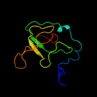
| c1s1iQ_ | PDB header: ribosome | Chain: Q: PDB Molecule: 60s ribosomal protein l21-a; | PDBTitle: structure of the ribosomal 80s-eef2-sordarin complex from2 yeast obtained by docking atomic models for rna and protein3 components into a 11.7 a cryo-em map. this file, 1s1i,4 contains 60s subunit. the 40s ribosomal subunit is in file5 1s1h. |
| Added to library: Tue Mar 16 13:16:29 2010 | |
| Links to external resources | |
|---|---|
| 1 | . | . | . | . | . | . | . | . | 10 | . | . | . | . | . | . | . | . | . | 20 | . | . | . | . | . | . | . | . | . | 30 | . | . | . | . | . | . | . | . | . | 40 | . | . | . | . | . | . | . | . | . | 50 | . | . | . | . | . | . | . | . | . | 60 | . | . | . | . | . | . | . | . | . | 70 | |
| Sequence | S | H | G | Y | R | S | R | T | R | Y | M | F | Q | R | D | F | R | K | H | G | A | V | H | L | S | T | Y | L | K | V | Y | K | V | G | D | I | V | D | I | K | A | N | G | S | I | Q | K | G | M | P | H | K | F | Y | Q | G | K | T | G | V | V | Y | N | V | T | K | S | S | V | G |
| Predicted secondary structure |  | | | | | | | | | |  | | | | |  | | | | | | | | | | | | | | | | | ||||||||||||||||||||||||||||||||||||||
| SS confidence | ||||||||||||||||||||||||||||||||||||||||||||||||||||||||||||||||||||||
| Known secondary structure (DSSP) | S | S | S | S | T | T | S | S | S | S | S | S | T | T | T | T | T | S | S | | | S | S | S | S | S | S | S | S | | | B | S | S | S | B | S | . | . | . | . | . | . | . | . | . | 80 | . | . | . | . | . | . | . | . | . | 90 | . | . | . | . | . | . | . | ||||||
| Sequence | V | I | I | N | K | M | V | G | N | R | Y | L | E | K | R | L | N | L | R | V | E | H | I | K | H | S | K | |||||||||||||||||||||||||||||||||||||||||||
| Predicted secondary structure | | | | | | | | | | | | | | | | | | | | | | | | |||||||||||||||||||||||||||||||||||||||||||||||
| SS confidence | ||||||||||||||||||||||||||||||||||||||||||||||||||||||||||||||||||||||
| Known secondary structure (DSSP) | S | S | S | S | S | S | B | T | T | S |
| Download: | PDB structure | FASTA sequence |
Phyre is now FREE for commercial users! All images and data generated by Phyre2 are free to use in any publication with acknowledgement Accessibility Statement
| ||||||||||||||||||||||||