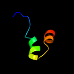
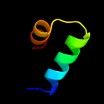
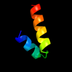
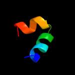
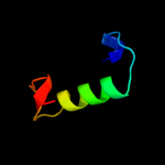
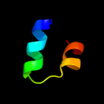
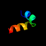
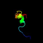
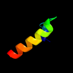
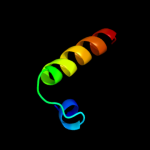
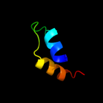
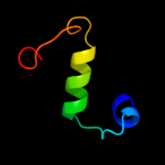
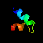
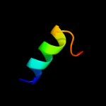
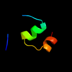
1 c1stzB_
66.9
18
PDB header: transcriptionChain: B: PDB Molecule: heat-inducible transcription repressor hrca homolog;PDBTitle: crystal structure of a hypothetical protein at 2.2 a resolution
2 d1oqya1
54.4
23
Fold: RuvA C-terminal domain-likeSuperfamily: UBA-likeFamily: UBA domain3 c3d5lA_
43.1
14
PDB header: signaling proteinChain: A: PDB Molecule: regulatory protein recx;PDBTitle: crystal structure of regulatory protein recx
4 d1ifya_
40.1
25
Fold: RuvA C-terminal domain-likeSuperfamily: UBA-likeFamily: UBA domain5 c2dakA_
39.9
22
PDB header: hydrolaseChain: A: PDB Molecule: ubiquitin carboxyl-terminal hydrolase 5;PDBTitle: solution structure of the second uba domain in the human2 ubiquitin specific protease 5 (isopeptidase 5)
6 d3e46a1
38.7
30
Fold: RuvA C-terminal domain-likeSuperfamily: UBA-likeFamily: UBA domain7 c3dfgA_
36.8
24
PDB header: recombinationChain: A: PDB Molecule: regulatory protein recx;PDBTitle: crystal structure of recx: a potent inhibitor protein of2 reca from xanthomonas campestris
8 d2cosa1
36.7
32
Fold: RuvA C-terminal domain-likeSuperfamily: UBA-likeFamily: UBA domain9 c2fu4B_
32.1
21
PDB header: dna binding proteinChain: B: PDB Molecule: ferric uptake regulation protein;PDBTitle: crystal structure of the dna binding domain of e.coli fur (ferric2 uptake regulator)
10 c2cosA_
31.8
32
PDB header: transferaseChain: A: PDB Molecule: serine/threonine protein kinase lats2;PDBTitle: solution structure of rsgi ruh-038, a uba domain from mouse2 lats2 (large tumor suppressor homolog 2)
11 c2l02B_
28.6
17
PDB header: structural genomics, unknown functionChain: B: PDB Molecule: uncharacterized protein;PDBTitle: solution nmr structure of protein bt2368 from bacteroides2 thetaiotaomicron, northeast structural genomics consortium target3 btr375
12 c2crnA_
26.4
26
PDB header: immune systemChain: A: PDB Molecule: ubash3a protein;PDBTitle: solution structure of the uba domain of human ubash3a2 protein
13 c1dpuA_
26.1
19
PDB header: dna binding proteinChain: A: PDB Molecule: replication protein a (rpa32) c-terminal domain;PDBTitle: solution structure of the c-terminal domain of human rpa322 complexed with ung2(73-88)
14 d1dpua_
26.1
19
Fold: DNA/RNA-binding 3-helical bundleSuperfamily: "Winged helix" DNA-binding domainFamily: C-terminal domain of RPA3215 d1t95a1
25.4
12
Fold: RuvA C-terminal domain-likeSuperfamily: Hypothetical protein AF0491, middle domainFamily: Hypothetical protein AF0491, middle domain16 c2w57A_
22.4
12
PDB header: metal transportChain: A: PDB Molecule: ferric uptake regulation protein;PDBTitle: crystal structure of the vibrio cholerae ferric uptake2 regulator (fur) reveals structural rearrangement of the3 dna-binding domains
17 d2g3qa1
20.6
30
Fold: RuvA C-terminal domain-likeSuperfamily: UBA-likeFamily: UBA domain18 d1whqa_
19.2
24
Fold: dsRBD-likeSuperfamily: dsRNA-binding domain-likeFamily: Double-stranded RNA-binding domain (dsRBD)19 d1whca_
19.1
30
Fold: RuvA C-terminal domain-likeSuperfamily: UBA-likeFamily: UBA domain20 c2cpwA_
18.6
27
PDB header: structural genomics, unknown functionChain: A: PDB Molecule: cbl-interacting protein sts-1 variant;PDBTitle: solution structure of rsgi ruh-031, a uba domain from human2 cdna
21 c3l9kZ_
not modelled
18.0
22
PDB header: motor proteinChain: Z: PDB Molecule: dynein intermediate chain, cytosolic;PDBTitle: insights into dynein assembly from a dynein intermediate chain-light2 chain roadblock structure
22 c3kevA_
not modelled
17.6
22
PDB header: structural genomics, unknown functionChain: A: PDB Molecule: galieria sulfuraria dcun1 domain-containing protein;PDBTitle: x-ray crystal structure of a dcun1 domain-containing protein from2 galdieria sulfuraria
23 d1wiva_
not modelled
17.0
27
Fold: RuvA C-terminal domain-likeSuperfamily: UBA-likeFamily: UBA domain24 c1fuiB_
not modelled
16.5
17
PDB header: isomeraseChain: B: PDB Molecule: l-fucose isomerase;PDBTitle: l-fucose isomerase from escherichia coli
25 d1vega_
not modelled
16.3
27
Fold: RuvA C-terminal domain-likeSuperfamily: UBA-likeFamily: UBA domain26 c3qqmD_
not modelled
16.2
23
PDB header: transferaseChain: D: PDB Molecule: mlr3007 protein;PDBTitle: crystal structure of a putative amino-acid aminotransferase2 (np_104211.1) from mesorhizobium loti at 2.30 a resolution
27 c3a1yF_
not modelled
15.5
21
PDB header: ribosomal proteinChain: F: PDB Molecule: 50s ribosomal protein p1 (l12p);PDBTitle: the structure of protein complex
28 c3e3vA_
not modelled
15.0
17
PDB header: recombinationChain: A: PDB Molecule: regulatory protein recx;PDBTitle: crystal structure of recx from lactobacillus salivarius
29 d1r4wa_
not modelled
13.8
10
Fold: Thioredoxin foldSuperfamily: Thioredoxin-likeFamily: DsbA-like30 d1veka_
not modelled
13.3
19
Fold: RuvA C-terminal domain-likeSuperfamily: UBA-likeFamily: UBA domain31 c3eyyA_
not modelled
13.3
21
PDB header: transportChain: A: PDB Molecule: putative iron uptake regulatory protein;PDBTitle: structural basis for the specialization of nur, a nickel-2 specific fur homologue, in metal sensing and dna3 recognition
32 c2wbmA_
not modelled
12.8
12
PDB header: rna-binding proteinChain: A: PDB Molecule: ribosome maturation protein sdo1 homolog;PDBTitle: crystal structure of mthsbds, the homologue of the2 shwachman-bodian-diamond syndrome protein in the3 euriarchaeon methanothermobacter thermautotrophicus
33 c1qzeA_
not modelled
12.4
13
PDB header: replicationChain: A: PDB Molecule: uv excision repair protein rad23 homolog a;PDBTitle: hhr23a protein structure based on residual dipolar coupling2 data
34 d2cpwa1
not modelled
12.3
27
Fold: RuvA C-terminal domain-likeSuperfamily: UBA-likeFamily: UBA domain35 c2dagA_
not modelled
12.1
22
PDB header: hydrolaseChain: A: PDB Molecule: ubiquitin carboxyl-terminal hydrolase 5;PDBTitle: solution structure of the first uba domain in the human2 ubiquitin specific protease 5 (isopeptidase 5)
36 c2do6A_
not modelled
11.8
28
PDB header: ligaseChain: A: PDB Molecule: e3 ubiquitin-protein ligase cbl-b;PDBTitle: solution structure of rsgi ruh-065, a uba domain from human2 cdna
37 c3itcA_
not modelled
11.8
13
PDB header: hydrolaseChain: A: PDB Molecule: renal dipeptidase;PDBTitle: crystal structure of sco3058 with bound citrate and glycerol
38 d2fi0a1
not modelled
11.7
31
Fold: SP0561-likeSuperfamily: SP0561-likeFamily: SP0561-like39 d1dhsa_
not modelled
11.3
13
Fold: DHS-like NAD/FAD-binding domainSuperfamily: DHS-like NAD/FAD-binding domainFamily: Deoxyhypusine synthase, DHS40 c3fpnB_
not modelled
11.2
23
PDB header: dna binding proteinChain: B: PDB Molecule: geobacillus stearothermophilus uvrb interactionPDBTitle: crystal structure of uvra-uvrb interaction domains
41 c2fe3B_
not modelled
11.0
20
PDB header: dna binding proteinChain: B: PDB Molecule: peroxide operon regulator;PDBTitle: the crystal structure of bacillus subtilis perr-zn reveals a novel2 zn(cys)4 structural redox switch
42 c2jnhA_
not modelled
10.4
28
PDB header: ligaseChain: A: PDB Molecule: e3 ubiquitin-protein ligase cbl-b;PDBTitle: solution structure of the uba domain from cbl-b
43 d1ntha_
not modelled
10.3
22
Fold: TIM beta/alpha-barrelSuperfamily: Monomethylamine methyltransferase MtmBFamily: Monomethylamine methyltransferase MtmB44 c2k2pA_
not modelled
9.9
24
PDB header: structural genomics, unknown functionChain: A: PDB Molecule: uncharacterized protein atu1203;PDBTitle: solution nmr structure of protein atu1203 from agrobacterium2 tumefaciens. northeast structural genomics consortium (nesg) target3 att10, ontario center for structural proteomics target atc1183
45 c2is9A_
not modelled
9.8
36
PDB header: transcriptionChain: A: PDB Molecule: defective in cullin neddylation protein 1;PDBTitle: structure of yeast dcn-1
46 c2d9sA_
not modelled
9.7
33
PDB header: ligaseChain: A: PDB Molecule: cbl e3 ubiquitin protein ligase;PDBTitle: solution structure of rsgi ruh-049, a uba domain from mouse2 cdna
47 d1oqya2
not modelled
9.6
20
Fold: RuvA C-terminal domain-likeSuperfamily: UBA-likeFamily: UBA domain48 c2v79B_
not modelled
9.6
11
PDB header: dna-binding proteinChain: B: PDB Molecule: dna replication protein dnad;PDBTitle: crystal structure of the n-terminal domain of dnad from2 bacillus subtilis
49 c3dp7B_
not modelled
8.6
26
PDB header: transferaseChain: B: PDB Molecule: sam-dependent methyltransferase;PDBTitle: crystal structure of sam-dependent methyltransferase from bacteroides2 vulgatus atcc 8482
50 c2daiA_
not modelled
8.6
37
PDB header: structural genomics, unknown functionChain: A: PDB Molecule: ubiquitin associated domain containing 1;PDBTitle: solution structure of the first uba domain in the human2 ubiquitin associated domain containing 1 (ubadc1)
51 c3ocjA_
not modelled
8.6
19
PDB header: structural genomics, unknown functionChain: A: PDB Molecule: putative exported protein;PDBTitle: the crystal structure of a possilbe exported protein from bordetella2 parapertussis
52 d1jjcb1
not modelled
8.0
23
Fold: Putative DNA-binding domainSuperfamily: Putative DNA-binding domainFamily: Domains B1 and B5 of PheRS-beta, PheT53 d1daaa_
not modelled
7.9
19
Fold: D-aminoacid aminotransferase-like PLP-dependent enzymesSuperfamily: D-aminoacid aminotransferase-like PLP-dependent enzymesFamily: D-aminoacid aminotransferase-like PLP-dependent enzymes54 c3o10D_
not modelled
7.7
18
PDB header: chaperoneChain: D: PDB Molecule: sacsin;PDBTitle: crystal structure of the hepn domain from human sacsin
55 c3nznA_
not modelled
7.5
33
PDB header: oxidoreductaseChain: A: PDB Molecule: glutaredoxin;PDBTitle: the crystal structure of the glutaredoxin from methanosarcina mazei2 go1
56 d1okra_
not modelled
7.3
17
Fold: DNA/RNA-binding 3-helical bundleSuperfamily: "Winged helix" DNA-binding domainFamily: Penicillinase repressor57 c2l01A_
not modelled
7.2
20
PDB header: structural genomics, unknown functionChain: A: PDB Molecule: uncharacterized protein;PDBTitle: solution nmr structure of protein bvu3908 from bacteroides vulgatus,2 northeast structural genomics consortium target bvr153
58 c2kdoA_
not modelled
7.1
12
PDB header: rna binding proteinChain: A: PDB Molecule: ribosome maturation protein sbds;PDBTitle: structure of the human shwachman-bodian-diamond syndrome protein, sbds
59 c3gp4B_
not modelled
7.0
9
PDB header: transcription regulatorChain: B: PDB Molecule: transcriptional regulator, merr family;PDBTitle: crystal structure of putative merr family transcriptional regulator2 from listeria monocytogenes
60 d1t5la1
not modelled
6.9
19
Fold: P-loop containing nucleoside triphosphate hydrolasesSuperfamily: P-loop containing nucleoside triphosphate hydrolasesFamily: Tandem AAA-ATPase domain61 d1sb6a_
not modelled
6.8
24
Fold: Ferredoxin-likeSuperfamily: HMA, heavy metal-associated domainFamily: HMA, heavy metal-associated domain62 d1jb0f_
not modelled
6.6
22
Fold: Single transmembrane helixSuperfamily: Subunit III of photosystem I reaction centre, PsaFFamily: Subunit III of photosystem I reaction centre, PsaF63 d2eyqa4
not modelled
6.3
24
Fold: P-loop containing nucleoside triphosphate hydrolasesSuperfamily: P-loop containing nucleoside triphosphate hydrolasesFamily: Tandem AAA-ATPase domain64 c3hjhA_
not modelled
6.3
24
PDB header: hydrolaseChain: A: PDB Molecule: transcription-repair-coupling factor;PDBTitle: a rigid n-terminal clamp restrains the motor domains of the bacterial2 transcription-repair coupling factor
65 c2e75H_
not modelled
6.2
57
PDB header: photosynthesisChain: H: PDB Molecule: cytochrome b6-f complex subunit 8;PDBTitle: crystal structure of the cytochrome b6f complex with 2-nonyl-4-2 hydroxyquinoline n-oxide (nqno) from m.laminosus
66 c2e74H_
not modelled
6.2
57
PDB header: photosynthesisChain: H: PDB Molecule: cytochrome b6-f complex subunit 8;PDBTitle: crystal structure of the cytochrome b6f complex from m.laminosus
67 c2e76H_
not modelled
6.2
57
PDB header: photosynthesisChain: H: PDB Molecule: cytochrome b6-f complex subunit 8;PDBTitle: crystal structure of the cytochrome b6f complex with tridecyl-2 stigmatellin (tds) from m.laminosus
68 c3lulA_
not modelled
6.1
13
PDB header: lyaseChain: A: PDB Molecule: 4-amino-4-deoxychorismate lyase;PDBTitle: crystal structure of putative 4-amino-4-deoxychorismate lyase.2 (yp_094631.1) from legionella pneumophila subsp. pneumophila str.3 philadelphia 1 at 1.78 a resolution
69 d1iyea_
not modelled
5.9
19
Fold: D-aminoacid aminotransferase-like PLP-dependent enzymesSuperfamily: D-aminoacid aminotransferase-like PLP-dependent enzymesFamily: D-aminoacid aminotransferase-like PLP-dependent enzymes70 d1efub3
not modelled
5.7
29
Fold: RuvA C-terminal domain-likeSuperfamily: UBA-likeFamily: TS-N domain71 c3c1dA_
not modelled
5.6
19
PDB header: recombination, dna binding proteinChain: A: PDB Molecule: regulatory protein recx;PDBTitle: x-ray crystal structure of recx
72 c2zc2A_
not modelled
5.6
18
PDB header: replicationChain: A: PDB Molecule: dnad-like replication protein;PDBTitle: crystal structure of dnad-like replication protein from2 streptococcus mutans ua159, gi 24377835, residues 127-199
73 d1w6ka1
not modelled
5.5
22
Fold: alpha/alpha toroidSuperfamily: Terpenoid cyclases/Protein prenyltransferasesFamily: Terpene synthases74 c2abjG_
not modelled
5.4
20
PDB header: transferaseChain: G: PDB Molecule: branched-chain-amino-acid aminotransferase, cytosolic;PDBTitle: crystal structure of human branched chain amino acid transaminase in a2 complex with an inhibitor, c16h10n2o4f3scl, and pyridoxal 5'3 phosphate.
75 c1t95A_
not modelled
5.4
12
PDB header: unknown functionChain: A: PDB Molecule: hypothetical protein af0491;PDBTitle: crystal structure of the shwachman-bodian-diamond syndrome2 protein orthologue from archaeoglobus fulgidus
76 d2b2na1
not modelled
5.4
24
Fold: P-loop containing nucleoside triphosphate hydrolasesSuperfamily: P-loop containing nucleoside triphosphate hydrolasesFamily: Tandem AAA-ATPase domain77 d1m2ia_
not modelled
5.3
33
Fold: Cytochrome b5-like heme/steroid binding domainSuperfamily: Cytochrome b5-like heme/steroid binding domainFamily: Cytochrome b578 c3itfA_
not modelled
5.3
21
PDB header: signaling proteinChain: A: PDB Molecule: periplasmic adaptor protein cpxp;PDBTitle: structural basis for the inhibitory function of the cpxp adaptor2 protein


