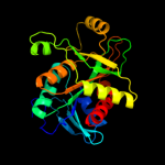
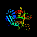
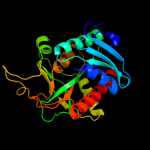
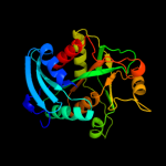
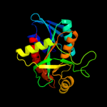
| 1 | d1k9sa_
|
|
 |
100.0 |
100 |
Fold:Phosphorylase/hydrolase-like
Superfamily:Purine and uridine phosphorylases
Family:Purine and uridine phosphorylases |
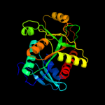
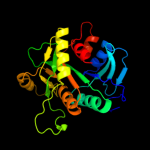
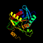
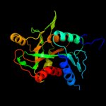
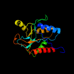
| 2 | d1vhwa_
|
|
 |
100.0 |
80 |
Fold:Phosphorylase/hydrolase-like
Superfamily:Purine and uridine phosphorylases
Family:Purine and uridine phosphorylases |
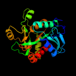
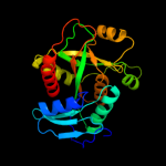
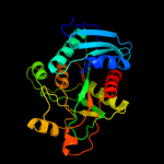
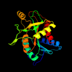
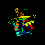
| 3 | c1z34A_
|
|
 |
100.0 |
59 |
PDB header:transferase
Chain: A: PDB Molecule:purine nucleoside phosphorylase;
PDBTitle: crystal structure of trichomonas vaginalis purine nucleoside2 phosphorylase complexed with 2-fluoro-2'-deoxyadenosine
|
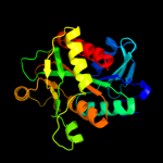
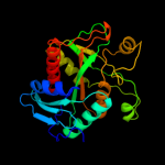
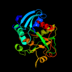
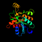
| 4 | d1odka_
|
|
 |
100.0 |
36 |
Fold:Phosphorylase/hydrolase-like
Superfamily:Purine and uridine phosphorylases
Family:Purine and uridine phosphorylases |
| 5 | c3bjeA_
|
|
 |
100.0 |
25 |
PDB header:transferase
Chain: A: PDB Molecule:nucleoside phosphorylase, putative;
PDBTitle: crystal structure of trypanosoma brucei nucleoside phosphorylase shows2 uridine phosphorylase activity
|
| 6 | c3tl6B_
|
|
 |
100.0 |
48 |
PDB header:transferase
Chain: B: PDB Molecule:purine nucleoside phosphorylase;
PDBTitle: crystal structure of purine nucleoside phosphorylase from entamoeba2 histolytica
|
| 7 | d2ac7a1
|
|
 |
100.0 |
58 |
Fold:Phosphorylase/hydrolase-like
Superfamily:Purine and uridine phosphorylases
Family:Purine and uridine phosphorylases |
| 8 | d1je0a_
|
|
 |
100.0 |
33 |
Fold:Phosphorylase/hydrolase-like
Superfamily:Purine and uridine phosphorylases
Family:Purine and uridine phosphorylases |
| 9 | c3mb8A_
|
|
 |
100.0 |
29 |
PDB header:transferase
Chain: A: PDB Molecule:purine nucleoside phosphorylase;
PDBTitle: crystal structure of purine nucleoside phosphorylase from toxoplasma2 gondii in complex with immucillin-h
|
| 10 | d1rxya_
|
|
 |
100.0 |
28 |
Fold:Phosphorylase/hydrolase-like
Superfamily:Purine and uridine phosphorylases
Family:Purine and uridine phosphorylases |
| 11 | c3qpbB_
|
|
 |
100.0 |
31 |
PDB header:transferase
Chain: B: PDB Molecule:uridine phosphorylase;
PDBTitle: crystal structure of streptococcus pyogenes uridine phosphorylase2 reveals a subclass of the np-i superfamily
|
| 12 | c1nw4C_
|
|
 |
100.0 |
27 |
PDB header:transferase
Chain: C: PDB Molecule:uridine phosphorylase, putative;
PDBTitle: crystal structure of plasmodium falciparum purine nucleoside2 phosphorylase in complex with immh and sulfate
|
| 13 | d1q1ga_
|
|
 |
100.0 |
27 |
Fold:Phosphorylase/hydrolase-like
Superfamily:Purine and uridine phosphorylases
Family:Purine and uridine phosphorylases |
| 14 | c2xrfA_
|
|
 |
100.0 |
23 |
PDB header:transferase
Chain: A: PDB Molecule:uridine phosphorylase 2;
PDBTitle: crystal structure of human uridine phosphorylase 2
|
| 15 | c3eufC_
|
|
 |
100.0 |
22 |
PDB header:transferase
Chain: C: PDB Molecule:uridine phosphorylase 1;
PDBTitle: crystal structure of bau-bound human uridine phosphorylase 1
|
| 16 | d1ybfa_
|
|
 |
100.0 |
15 |
Fold:Phosphorylase/hydrolase-like
Superfamily:Purine and uridine phosphorylases
Family:Purine and uridine phosphorylases |
| 17 | c3nm5B_
|
|
 |
100.0 |
18 |
PDB header:hydrolase
Chain: B: PDB Molecule:mta/sah nucleosidase;
PDBTitle: helicobacter pylori mtan complexed with formycin a
|
| 18 | d1t8sa_
|
|
 |
100.0 |
19 |
Fold:Phosphorylase/hydrolase-like
Superfamily:Purine and uridine phosphorylases
Family:Purine and uridine phosphorylases |
| 19 | c1zosE_
|
|
 |
100.0 |
20 |
PDB header:hydrolase
Chain: E: PDB Molecule:5'-methylthioadenosine / s-adenosylhomocysteine
PDBTitle: structure of 5'-methylthionadenosine/s-adenosylhomocysteine2 nucleosidase from s. pneumoniae with a transition-state3 inhibitor mt-imma
|
| 20 | c3bl6A_
|
|
 |
100.0 |
18 |
PDB header:hydrolase
Chain: A: PDB Molecule:5'-methylthioadenosine nucleosidase/s-
PDBTitle: crystal structure of staphylococcus aureus 5'-2 methylthioadenosine/s-adenosylhomocysteine nucleosidase in3 complex with formycin a
|
| 21 | c3eeiA_ |
|
not modelled |
100.0 |
20 |
PDB header:hydrolase
Chain: A: PDB Molecule:5-methylthioadenosine nucleosidase/s-
PDBTitle: crystal structure of 5'-methylthioadenosine/s-2 adenosylhomocysteine nucleosidase from neisseria3 meningitidis in complex with methylthio-immucillin-a
|
| 22 | c3dp9A_ |
|
not modelled |
100.0 |
20 |
PDB header:hydrolase
Chain: A: PDB Molecule:mta/sah nucleosidase;
PDBTitle: crystal structure of vibrio cholerae 5'-methylthioadenosine/s-adenosyl2 homocysteine nucleosidase (mtan) complexed with butylthio-dadme-3 immucillin a
|
| 23 | d1jysa_ |
|
not modelled |
100.0 |
18 |
Fold:Phosphorylase/hydrolase-like
Superfamily:Purine and uridine phosphorylases
Family:Purine and uridine phosphorylases |
| 24 | c2h8gA_ |
|
not modelled |
100.0 |
16 |
PDB header:hydrolase
Chain: A: PDB Molecule:5'-methylthioadenosine nucleosidase;
PDBTitle: 5'-methylthioadenosine nucleosidase from arabidopsis2 thaliana
|
| 25 | c3bsfB_ |
|
not modelled |
100.0 |
19 |
PDB header:hydrolase
Chain: B: PDB Molecule:at4g34840;
PDBTitle: crystal structure of the mta/sah nucleosidase
|
| 26 | c3ozbF_ |
|
not modelled |
100.0 |
17 |
PDB header:transferase
Chain: F: PDB Molecule:methylthioadenosine phosphorylase;
PDBTitle: crystal structure of 5'-methylthioinosine phosphorylase from2 psedomonas aeruginosa in complex with hypoxanthine
|
| 27 | c3khsB_ |
|
not modelled |
99.9 |
16 |
PDB header:hydrolase
Chain: B: PDB Molecule:purine nucleoside phosphorylase;
PDBTitle: crystal structure of grouper iridovirus purine nucleoside2 phosphorylase
|
| 28 | d1v4na_ |
|
not modelled |
99.9 |
19 |
Fold:Phosphorylase/hydrolase-like
Superfamily:Purine and uridine phosphorylases
Family:Purine and uridine phosphorylases |
| 29 | c1wtaA_ |
|
not modelled |
99.9 |
19 |
PDB header:transferase
Chain: A: PDB Molecule:5'-methylthioadenosine phosphorylase;
PDBTitle: crystal structure of 5'-deoxy-5'-methylthioadenosine from aeropyrum2 pernix (r32 form)
|
| 30 | c3la8A_ |
|
not modelled |
99.9 |
16 |
PDB header:transferase
Chain: A: PDB Molecule:putative purine nucleoside phosphorylase;
PDBTitle: the crystal structure of smu.1229 from streptococcus mutans ua159
|
| 31 | d1g2oa_ |
|
not modelled |
99.9 |
12 |
Fold:Phosphorylase/hydrolase-like
Superfamily:Purine and uridine phosphorylases
Family:Purine and uridine phosphorylases |
| 32 | d1cb0a_ |
|
not modelled |
99.9 |
16 |
Fold:Phosphorylase/hydrolase-like
Superfamily:Purine and uridine phosphorylases
Family:Purine and uridine phosphorylases |
| 33 | c2p4sA_ |
|
not modelled |
99.9 |
14 |
PDB header:transferase
Chain: A: PDB Molecule:purine nucleoside phosphorylase;
PDBTitle: structure of purine nucleoside phosphorylase from anopheles gambiae in2 complex with dadme-immh
|
| 34 | d1vmka_ |
|
not modelled |
99.9 |
17 |
Fold:Phosphorylase/hydrolase-like
Superfamily:Purine and uridine phosphorylases
Family:Purine and uridine phosphorylases |
| 35 | c1yr3A_ |
|
not modelled |
99.9 |
15 |
PDB header:transferase
Chain: A: PDB Molecule:xanthosine phosphorylase;
PDBTitle: escherichia coli purine nucleoside phosphorylase ii, the2 product of the xapa gene
|
| 36 | c1tcvB_ |
|
not modelled |
99.9 |
19 |
PDB header:transferase
Chain: B: PDB Molecule:purine-nucleoside phosphorylase;
PDBTitle: crystal structure of the purine nucleoside phosphorylase2 from schistosoma mansoni in complex with non-detergent3 sulfobetaine 195 and acetate
|
| 37 | d3bgsa1 |
|
not modelled |
99.9 |
17 |
Fold:Phosphorylase/hydrolase-like
Superfamily:Purine and uridine phosphorylases
Family:Purine and uridine phosphorylases |
| 38 | d1qe5a_ |
|
not modelled |
99.9 |
11 |
Fold:Phosphorylase/hydrolase-like
Superfamily:Purine and uridine phosphorylases
Family:Purine and uridine phosphorylases |
| 39 | c3ggsA_ |
|
not modelled |
99.9 |
17 |
PDB header:transferase
Chain: A: PDB Molecule:purine nucleoside phosphorylase;
PDBTitle: human purine nucleoside phosphorylase double mutant e201q,n243d2 complexed with 2-fluoro-2'-deoxyadenosine
|
| 40 | d3pnpa_ |
|
not modelled |
99.9 |
16 |
Fold:Phosphorylase/hydrolase-like
Superfamily:Purine and uridine phosphorylases
Family:Purine and uridine phosphorylases |
| 41 | c1tvmA_ |
|
not modelled |
71.7 |
8 |
PDB header:transferase
Chain: A: PDB Molecule:pts system, galactitol-specific iib component;
PDBTitle: nmr structure of enzyme gatb of the galactitol-specific2 phosphoenolpyruvate-dependent phosphotransferase system
|
| 42 | c3czcA_ |
|
not modelled |
45.7 |
16 |
PDB header:transferase
Chain: A: PDB Molecule:rmpb;
PDBTitle: the crystal structure of a putative pts iib(ptxb) from2 streptococcus mutans
|
| 43 | c3efhB_ |
|
not modelled |
43.7 |
16 |
PDB header:transferase
Chain: B: PDB Molecule:ribose-phosphate pyrophosphokinase 1;
PDBTitle: crystal structure of human phosphoribosyl pyrophosphate2 synthetase 1
|
| 44 | d2c4ka2 |
|
not modelled |
40.8 |
17 |
Fold:PRTase-like
Superfamily:PRTase-like
Family:Phosphoribosylpyrophosphate synthetase-like |
| 45 | c1vkrA_ |
|
not modelled |
40.7 |
25 |
PDB header:transferase
Chain: A: PDB Molecule:mannitol-specific pts system enzyme iiabc components;
PDBTitle: structure of iib domain of the mannitol-specific permease enzyme ii
|
| 46 | d1vkra_ |
|
not modelled |
40.7 |
25 |
Fold:Phosphotyrosine protein phosphatases I-like
Superfamily:PTS system IIB component-like
Family:PTS system, Lactose/Cellobiose specific IIB subunit |
| 47 | c2q1yB_ |
|
not modelled |
40.5 |
18 |
PDB header:cell cycle, signaling protein
Chain: B: PDB Molecule:cell division protein ftsz;
PDBTitle: crystal structure of cell division protein ftsz from mycobacterium2 tuberculosis in complex with gtp-gamma-s
|
| 48 | d1v58a2 |
|
not modelled |
37.3 |
20 |
Fold:Cystatin-like
Superfamily:DsbC/DsbG N-terminal domain-like
Family:DsbC/DsbG N-terminal domain-like |
| 49 | c2f9iD_ |
|
not modelled |
29.6 |
19 |
PDB header:transferase
Chain: D: PDB Molecule:acetyl-coenzyme a carboxylase carboxyl
PDBTitle: crystal structure of the carboxyltransferase subunit of acc2 from staphylococcus aureus
|
| 50 | d2csba3 |
|
not modelled |
27.7 |
46 |
Fold:SAM domain-like
Superfamily:RuvA domain 2-like
Family:Topoisomerase V repeat domain |
| 51 | d1ea9c1 |
|
not modelled |
26.0 |
27 |
Fold:Immunoglobulin-like beta-sandwich
Superfamily:E set domains
Family:E-set domains of sugar-utilizing enzymes |
| 52 | c1ii0A_ |
|
not modelled |
24.6 |
35 |
PDB header:hydrolase
Chain: A: PDB Molecule:arsenical pump-driving atpase;
PDBTitle: crystal structure of the escherichia coli arsenite-translocating2 atpase
|
| 53 | d1a4ia1 |
|
not modelled |
23.5 |
18 |
Fold:NAD(P)-binding Rossmann-fold domains
Superfamily:NAD(P)-binding Rossmann-fold domains
Family:Aminoacid dehydrogenase-like, C-terminal domain |
| 54 | d2a7sa2 |
|
not modelled |
22.4 |
18 |
Fold:ClpP/crotonase
Superfamily:ClpP/crotonase
Family:Biotin dependent carboxylase carboxyltransferase domain |
| 55 | d2bm8a1 |
|
not modelled |
21.1 |
11 |
Fold:S-adenosyl-L-methionine-dependent methyltransferases
Superfamily:S-adenosyl-L-methionine-dependent methyltransferases
Family:CmcI-like |
| 56 | c3onoA_ |
|
not modelled |
20.4 |
21 |
PDB header:isomerase
Chain: A: PDB Molecule:ribose/galactose isomerase;
PDBTitle: crystal structure of ribose-5-phosphate isomerase lacab_rpib from2 vibrio parahaemolyticus
|
| 57 | c3peiA_ |
|
not modelled |
20.3 |
17 |
PDB header:hydrolase
Chain: A: PDB Molecule:cytosol aminopeptidase;
PDBTitle: crystal structure of cytosol aminopeptidase from francisella2 tularensis
|
| 58 | c3giuA_ |
|
not modelled |
20.2 |
7 |
PDB header:hydrolase
Chain: A: PDB Molecule:pyrrolidone-carboxylate peptidase;
PDBTitle: 1.25 angstrom crystal structure of pyrrolidone-carboxylate peptidase2 (pcp) from staphylococcus aureus
|
| 59 | c3o38D_ |
|
not modelled |
20.2 |
36 |
PDB header:oxidoreductase
Chain: D: PDB Molecule:short chain dehydrogenase;
PDBTitle: crystal structure of a short chain dehydrogenase from mycobacterium2 smegmatis
|
| 60 | c1a4iB_ |
|
not modelled |
20.0 |
18 |
PDB header:oxidoreductase
Chain: B: PDB Molecule:methylenetetrahydrofolate dehydrogenase /
PDBTitle: human tetrahydrofolate dehydrogenase / cyclohydrolase
|
| 61 | d1dkua2 |
|
not modelled |
19.5 |
12 |
Fold:PRTase-like
Superfamily:PRTase-like
Family:Phosphoribosylpyrophosphate synthetase-like |
| 62 | d1wzla1 |
|
not modelled |
19.4 |
22 |
Fold:Immunoglobulin-like beta-sandwich
Superfamily:E set domains
Family:E-set domains of sugar-utilizing enzymes |
| 63 | c1dkrB_ |
|
not modelled |
19.3 |
13 |
PDB header:transferase
Chain: B: PDB Molecule:phosphoribosyl pyrophosphate synthetase;
PDBTitle: crystal structures of bacillus subtilis phosphoribosylpyrophosphate2 synthetase: molecular basis of allosteric inhibition and activation.
|
| 64 | d2bj7a2 |
|
not modelled |
19.3 |
19 |
Fold:Ferredoxin-like
Superfamily:ACT-like
Family:Nickel responsive regulator NikR, C-terminal domain |
| 65 | d1gvia1 |
|
not modelled |
18.7 |
33 |
Fold:Immunoglobulin-like beta-sandwich
Superfamily:E set domains
Family:E-set domains of sugar-utilizing enzymes |
| 66 | d1u9ya2 |
|
not modelled |
18.7 |
16 |
Fold:PRTase-like
Superfamily:PRTase-like
Family:Phosphoribosylpyrophosphate synthetase-like |
| 67 | d1x94a_ |
|
not modelled |
18.7 |
20 |
Fold:SIS domain
Superfamily:SIS domain
Family:mono-SIS domain |
| 68 | c1w5fA_ |
|
not modelled |
18.6 |
22 |
PDB header:cell division
Chain: A: PDB Molecule:cell division protein ftsz;
PDBTitle: ftsz, t7 mutated, domain swapped (t. maritima)
|
| 69 | c2qlcC_ |
|
not modelled |
18.4 |
17 |
PDB header:dna binding protein
Chain: C: PDB Molecule:dna repair protein radc homolog;
PDBTitle: the crystal structure of dna repair protein radc from chlorobium2 tepidum tls
|
| 70 | c2bj3D_ |
|
not modelled |
18.1 |
19 |
PDB header:transcription
Chain: D: PDB Molecule:nickel responsive regulator;
PDBTitle: nikr-apo
|
| 71 | c2ppwA_ |
|
not modelled |
18.1 |
21 |
PDB header:isomerase
Chain: A: PDB Molecule:conserved domain protein;
PDBTitle: the crystal structure of uncharacterized ribose 5-phosphate isomerase2 rpib from streptococcus pneumoniae
|
| 72 | c1q5vB_ |
|
not modelled |
17.8 |
22 |
PDB header:transcription
Chain: B: PDB Molecule:nickel responsive regulator;
PDBTitle: apo-nikr
|
| 73 | c1e9yB_ |
|
not modelled |
17.7 |
40 |
PDB header:hydrolase
Chain: B: PDB Molecule:urease subunit beta;
PDBTitle: crystal structure of helicobacter pylori urease in complex with2 acetohydroxamic acid
|
| 74 | c3k7pA_ |
|
not modelled |
17.7 |
21 |
PDB header:isomerase
Chain: A: PDB Molecule:ribose 5-phosphate isomerase;
PDBTitle: structure of mutant of ribose 5-phosphate isomerase type b from2 trypanosoma cruzi.
|
| 75 | c3m1pA_ |
|
not modelled |
17.7 |
21 |
PDB header:isomerase
Chain: A: PDB Molecule:ribose 5-phosphate isomerase;
PDBTitle: structure of ribose 5-phosphate isomerase type b from trypanosoma2 cruzi, soaked with allose-6-phosphate
|
| 76 | d1q5ya_ |
|
not modelled |
17.5 |
22 |
Fold:Ferredoxin-like
Superfamily:ACT-like
Family:Nickel responsive regulator NikR, C-terminal domain |
| 77 | d1j0ha1 |
|
not modelled |
17.4 |
33 |
Fold:Immunoglobulin-like beta-sandwich
Superfamily:E set domains
Family:E-set domains of sugar-utilizing enzymes |
| 78 | c2ca9B_ |
|
not modelled |
17.3 |
13 |
PDB header:transcriptional regulation
Chain: B: PDB Molecule:putative nickel-responsive regulator;
PDBTitle: apo-nikr from helicobacter pylori in closed trans-2 conformation
|
| 79 | d1id0a_ |
|
not modelled |
17.2 |
19 |
Fold:ATPase domain of HSP90 chaperone/DNA topoisomerase II/histidine kinase
Superfamily:ATPase domain of HSP90 chaperone/DNA topoisomerase II/histidine kinase
Family:Histidine kinase |
| 80 | c3s5pA_ |
|
not modelled |
17.1 |
43 |
PDB header:isomerase
Chain: A: PDB Molecule:ribose 5-phosphate isomerase;
PDBTitle: crystal structure of ribose-5-phosphate isomerase b rpib from giardia2 lamblia
|
| 81 | c3he8A_ |
|
not modelled |
16.9 |
36 |
PDB header:isomerase
Chain: A: PDB Molecule:ribose-5-phosphate isomerase;
PDBTitle: structural study of clostridium thermocellum ribose-5-phosphate2 isomerase b
|
| 82 | d2vvpa1 |
|
not modelled |
16.9 |
29 |
Fold:Ribose/Galactose isomerase RpiB/AlsB
Superfamily:Ribose/Galactose isomerase RpiB/AlsB
Family:Ribose/Galactose isomerase RpiB/AlsB |
| 83 | d1b0aa1 |
|
not modelled |
16.8 |
20 |
Fold:NAD(P)-binding Rossmann-fold domains
Superfamily:NAD(P)-binding Rossmann-fold domains
Family:Aminoacid dehydrogenase-like, C-terminal domain |
| 84 | d1nn4a_ |
|
not modelled |
16.6 |
36 |
Fold:Ribose/Galactose isomerase RpiB/AlsB
Superfamily:Ribose/Galactose isomerase RpiB/AlsB
Family:Ribose/Galactose isomerase RpiB/AlsB |
| 85 | d1sv6a_ |
|
not modelled |
16.5 |
15 |
Fold:FAH
Superfamily:FAH
Family:FAH |
| 86 | c2y3yC_ |
|
not modelled |
16.5 |
13 |
PDB header:transcription
Chain: C: PDB Molecule:putative nickel-responsive regulator;
PDBTitle: holo-ni(ii) hpnikr is a symmetric tetramer containing four2 canonic square-planar ni(ii) ions at physiological ph
|
| 87 | d1b12a_ |
|
not modelled |
16.5 |
14 |
Fold:LexA/Signal peptidase
Superfamily:LexA/Signal peptidase
Family:Type 1 signal peptidase |
| 88 | d1a2za_ |
|
not modelled |
16.4 |
9 |
Fold:Phosphorylase/hydrolase-like
Superfamily:Pyrrolidone carboxyl peptidase (pyroglutamate aminopeptidase)
Family:Pyrrolidone carboxyl peptidase (pyroglutamate aminopeptidase) |
| 89 | c2l2qA_ |
|
not modelled |
16.3 |
18 |
PDB header:transferase
Chain: A: PDB Molecule:pts system, cellobiose-specific iib component (cela);
PDBTitle: solution structure of cellobiose-specific phosphotransferase iib2 component protein from borrelia burgdorferi
|
| 90 | c2equA_ |
|
not modelled |
15.3 |
25 |
PDB header:protein binding
Chain: A: PDB Molecule:phd finger protein 20-like 1;
PDBTitle: solution structure of the tudor domain of phd finger2 protein 20-like 1
|
| 91 | c2c4kD_ |
|
not modelled |
15.1 |
16 |
PDB header:regulatory protein
Chain: D: PDB Molecule:phosphoribosyl pyrophosphate synthetase-
PDBTitle: crystal structure of human phosphoribosylpyrophosphate2 synthetase-associated protein 39 (pap39)
|
| 92 | c2jjyD_ |
|
not modelled |
14.9 |
16 |
PDB header:oxidoreductase
Chain: D: PDB Molecule:enoyl-[acyl-carrier-protein] reductase;
PDBTitle: crystal structure of francisella tularensis enoyl reductase2 (ftfabi) with bound nad
|
| 93 | d1sqsa_ |
|
not modelled |
14.8 |
13 |
Fold:Flavodoxin-like
Superfamily:Flavoproteins
Family:Hypothetical protein SP1951 |
| 94 | d2d1ya1 |
|
not modelled |
14.6 |
19 |
Fold:NAD(P)-binding Rossmann-fold domains
Superfamily:NAD(P)-binding Rossmann-fold domains
Family:Tyrosine-dependent oxidoreductases |
| 95 | c2qmoA_ |
|
not modelled |
14.3 |
21 |
PDB header:ligase
Chain: A: PDB Molecule:dethiobiotin synthetase;
PDBTitle: crystal structure of dethiobiotin synthetase (biod) from helicobacter2 pylori
|
| 96 | d2h7ma1 |
|
not modelled |
14.2 |
18 |
Fold:NAD(P)-binding Rossmann-fold domains
Superfamily:NAD(P)-binding Rossmann-fold domains
Family:Tyrosine-dependent oxidoreductases |
| 97 | d1fdra2 |
|
not modelled |
14.0 |
10 |
Fold:Ferredoxin reductase-like, C-terminal NADP-linked domain
Superfamily:Ferredoxin reductase-like, C-terminal NADP-linked domain
Family:Reductases |
| 98 | d1rq2a1 |
|
not modelled |
14.0 |
20 |
Fold:Tubulin nucleotide-binding domain-like
Superfamily:Tubulin nucleotide-binding domain-like
Family:Tubulin, GTPase domain |
| 99 | c3ngmB_ |
|
not modelled |
13.8 |
21 |
PDB header:hydrolase
Chain: B: PDB Molecule:extracellular lipase;
PDBTitle: crystal structure of lipase from gibberella zeae
|