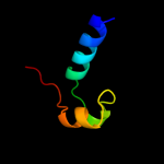
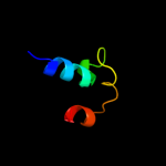
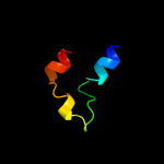
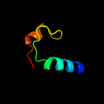
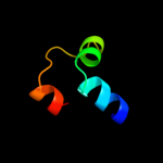
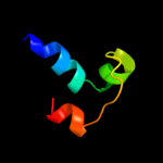
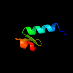
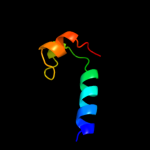
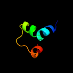
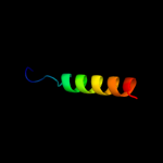
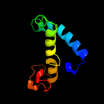
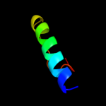
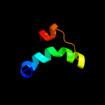
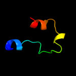
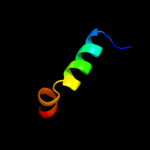
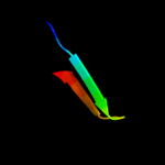
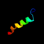
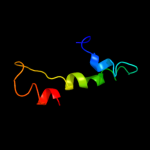
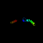
1 d2c8sa1
60.6
18
Fold: Cytochrome cSuperfamily: Cytochrome cFamily: monodomain cytochrome c2 d1fc2c_
59.7
36
Fold: immunoglobulin/albumin-binding domain-likeSuperfamily: Bacterial immunoglobulin/albumin-binding domainsFamily: Immunoglobulin-binding protein A modules3 d1lp1b_
59.3
38
Fold: immunoglobulin/albumin-binding domain-likeSuperfamily: Bacterial immunoglobulin/albumin-binding domainsFamily: Immunoglobulin-binding protein A modules4 c2d0wA_
49.7
25
PDB header: electron transportChain: A: PDB Molecule: cytochrome cl;PDBTitle: crystal structure of cytochrome cl from hyphomicrobium2 denitrificans
5 d1deeg_
47.0
43
Fold: immunoglobulin/albumin-binding domain-likeSuperfamily: Bacterial immunoglobulin/albumin-binding domainsFamily: Immunoglobulin-binding protein A modules6 c1zdbA_
47.0
25
PDB header: igg binding domainChain: A: PDB Molecule: mini protein a domain, z38;PDBTitle: phage-selected mini protein a domain, z38, nmr, minimized2 mean structure
7 d2jwda1
46.5
32
Fold: immunoglobulin/albumin-binding domain-likeSuperfamily: Bacterial immunoglobulin/albumin-binding domainsFamily: Immunoglobulin-binding protein A modules8 d2gc4d1
42.3
22
Fold: Cytochrome cSuperfamily: Cytochrome cFamily: monodomain cytochrome c9 d1lp1a_
42.3
33
Fold: immunoglobulin/albumin-binding domain-likeSuperfamily: Bacterial immunoglobulin/albumin-binding domainsFamily: Immunoglobulin-binding protein A modules10 c2g38A_
38.3
16
PDB header: structural genomics, unknown functionChain: A: PDB Molecule: pe family protein;PDBTitle: a pe/ppe protein complex from mycobacterium tuberculosis
11 d2g38a1
38.3
16
Fold: Ferritin-likeSuperfamily: PE/PPE dimer-likeFamily: PE12 c3fggA_
36.6
20
PDB header: structural genomics, unknown functionChain: A: PDB Molecule: uncharacterized protein bce2196;PDBTitle: crystal structure of putative ecf-type sigma factor negative effector2 from bacillus cereus
13 c3lueP_
31.9
24
PDB header: structural proteinChain: P: PDB Molecule: alpha-actinin-3;PDBTitle: model of alpha-actinin ch1 bound to f-actin
14 d1edla_
24.8
44
Fold: immunoglobulin/albumin-binding domain-likeSuperfamily: Bacterial immunoglobulin/albumin-binding domainsFamily: Immunoglobulin-binding protein A modules15 d2iy5a1
20.3
13
Fold: Long alpha-hairpinSuperfamily: tRNA-binding armFamily: Phenylalanyl-tRNA synthetase (PheRS)16 d1ojqa_
18.0
4
Fold: ADP-ribosylationSuperfamily: ADP-ribosylationFamily: ADP-ribosylating toxins17 c3uc2A_
15.8
27
PDB header: structural genomics, unknown functionChain: A: PDB Molecule: hypothetical protein with immunoglobulin-like fold;PDBTitle: crystal structure of a hypothetical protein with immunoglobulin-like2 fold (pa0388) from pseudomonas aeruginosa pao1 at 2.09 a resolution
18 c2xglB_
14.2
33
PDB header: antibioticChain: B: PDB Molecule: colicin-m immunity protein;PDBTitle: the x-ray structure of the escherichia coli colicin m immunity2 protein demonstrates the presence of a disulphide bridge, which is3 functionally essential
19 c1hynQ_
13.9
27
PDB header: membrane proteinChain: Q: PDB Molecule: band 3 anion transport protein;PDBTitle: crystal structure of the cytoplasmic domain of human2 erythrocyte band-3 protein
20 c1g5gA_
13.8
24
PDB header: viral proteinChain: A: PDB Molecule: fusion protein;PDBTitle: fragment of fusion protein from newcastle disease virus
21 d1r45a_
not modelled
12.1
13
Fold: ADP-ribosylationSuperfamily: ADP-ribosylationFamily: ADP-ribosylating toxins22 d1fqva1
not modelled
11.8
38
Fold: F-box domainSuperfamily: F-box domainFamily: F-box domain23 c2kv5A_
not modelled
11.7
36
PDB header: toxinChain: A: PDB Molecule: putative uncharacterized protein rnai;PDBTitle: solution structure of the par toxin fst in dpc micelles
24 c2yskA_
not modelled
10.9
12
PDB header: structural genomics, unknown functionChain: A: PDB Molecule: hypothetical protein ttha1432;PDBTitle: crystal structure of a hypothetical protein ttha1432 from thermus2 thermophilus
25 d1qn2a_
not modelled
10.6
13
Fold: Cytochrome cSuperfamily: Cytochrome cFamily: monodomain cytochrome c26 d1qhma_
not modelled
10.4
14
Fold: PFL-like glycyl radical enzymesSuperfamily: PFL-like glycyl radical enzymesFamily: PFL-like27 c2dbhA_
not modelled
10.3
22
PDB header: signaling proteinChain: A: PDB Molecule: tumor necrosis factor receptor superfamilyPDBTitle: solution structure of the carboxyl-terminal card-like2 domain in human tnfr-related death receptor-6
28 d1eiya1
not modelled
10.3
12
Fold: Long alpha-hairpinSuperfamily: tRNA-binding armFamily: Phenylalanyl-tRNA synthetase (PheRS)29 d1y9ua_
not modelled
10.1
15
Fold: Periplasmic binding protein-like IISuperfamily: Periplasmic binding protein-like IIFamily: Phosphate binding protein-like30 c3ermD_
not modelled
10.0
16
PDB header: structural genomics, unknown functionChain: D: PDB Molecule: uncharacterized conserved protein;PDBTitle: the crystal structure of a conserved protein with unknown function2 from pseudomonas syringae pv. tomato str. dc3000
31 d4icba_
not modelled
9.7
31
Fold: EF Hand-likeSuperfamily: EF-handFamily: Calbindin D9K32 c3swyB_
not modelled
8.8
18
PDB header: transport proteinChain: B: PDB Molecule: cyclic nucleotide-gated cation channel alpha-3;PDBTitle: cnga3 626-672 containing clz domain
33 d1j3sa_
not modelled
8.3
21
Fold: Cytochrome cSuperfamily: Cytochrome cFamily: monodomain cytochrome c34 c2x7aB_
not modelled
8.0
22
PDB header: immune systemChain: B: PDB Molecule: bone marrow stromal antigen 2;PDBTitle: structural basis of hiv-1 tethering to membranes by the2 bst2-tetherin ectodomain
35 c2kwhA_
not modelled
7.6
27
PDB header: transport proteinChain: A: PDB Molecule: rala-binding protein 1;PDBTitle: ral binding domain of rlip76 (ralbp1)
36 d1sh5a1
not modelled
7.6
10
Fold: CH domain-likeSuperfamily: Calponin-homology domain, CH-domainFamily: Calponin-homology domain, CH-domain37 d1hynp_
not modelled
7.2
24
Fold: Phoshotransferase/anion transport proteinSuperfamily: Phoshotransferase/anion transport proteinFamily: Anion transport protein, cytoplasmic domain38 d1y4ta_
not modelled
7.1
26
Fold: Periplasmic binding protein-like IISuperfamily: Periplasmic binding protein-like IIFamily: Phosphate binding protein-like39 c2zykA_
not modelled
7.0
14
PDB header: sugar binding proteinChain: A: PDB Molecule: solute-binding protein;PDBTitle: crystal structure of cyclo/maltodextrin-binding protein2 complexed with gamma-cyclodextrin
40 c3bw8B_
not modelled
7.0
17
PDB header: transferaseChain: B: PDB Molecule: mono-adp-ribosyltransferase c3;PDBTitle: crystal structure of the clostridium limosum c3 exoenzyme
41 c3d0wD_
not modelled
6.9
36
PDB header: structural genomics, unknown functionChain: D: PDB Molecule: yflh protein;PDBTitle: crystal structure of yflh protein from bacillus subtilis.2 northeast structural genomics consortium target sr326
42 d1lmsa_
not modelled
6.8
21
Fold: Cytochrome cSuperfamily: Cytochrome cFamily: monodomain cytochrome c43 d1ccra_
not modelled
6.8
23
Fold: Cytochrome cSuperfamily: Cytochrome cFamily: monodomain cytochrome c44 d1l8ya_
not modelled
6.7
42
Fold: HMG-boxSuperfamily: HMG-boxFamily: HMG-box45 d1nmla2
not modelled
6.4
28
Fold: Cytochrome cSuperfamily: Cytochrome cFamily: Di-heme cytochrome c peroxidase46 c3gdzA_
not modelled
6.3
18
PDB header: ligaseChain: A: PDB Molecule: arginyl-trna synthetase;PDBTitle: crystal structure of arginyl-trna synthetase from klebsiella2 pneumoniae subsp. pneumoniae
47 d3ctda1
not modelled
6.2
0
Fold: post-AAA+ oligomerization domain-likeSuperfamily: post-AAA+ oligomerization domain-likeFamily: MgsA/YrvN C-terminal domain-like48 c2pt1A_
not modelled
6.0
24
PDB header: metal transportChain: A: PDB Molecule: iron transport protein;PDBTitle: futa1 synechocystis pcc 6803
49 c3oeoD_
not modelled
6.0
78
PDB header: signaling proteinChain: D: PDB Molecule: spheroplast protein y;PDBTitle: the crystal structure e. coli spy
50 c1sj8A_
not modelled
5.9
12
PDB header: structural proteinChain: A: PDB Molecule: talin 1;PDBTitle: crystal structure of talin residues 482-789
51 c1sszA_
not modelled
5.8
33
PDB header: surface active proteinChain: A: PDB Molecule: pulmonary surfactant-associated protein b;PDBTitle: conformational mapping of mini-b: an n-terminal/c-terminal2 construct of surfactant protein b using 13c-enhanced3 fourier transform infrared (ftir) spectroscopy
52 d1aoaa1
not modelled
5.7
8
Fold: CH domain-likeSuperfamily: Calponin-homology domain, CH-domainFamily: Calponin-homology domain, CH-domain53 c3m97X_
not modelled
5.7
21
PDB header: electron transportChain: X: PDB Molecule: cytochrome c-552;PDBTitle: structure of the soluble domain of cytochrome c552 with its flexible2 linker segment from paracoccus denitrificans
54 d1ycca_
not modelled
5.6
21
Fold: Cytochrome cSuperfamily: Cytochrome cFamily: monodomain cytochrome c55 c2z6vA_
not modelled
5.6
14
PDB header: unknown functionChain: A: PDB Molecule: putative uncharacterized protein;PDBTitle: crystal structure of sulfotransferase stf9 from2 mycobacterium avium
56 d1hywa_
not modelled
5.3
36
Fold: gpW/XkdW-likeSuperfamily: Head-to-tail joining protein W, gpWFamily: Head-to-tail joining protein W, gpW57 d1cl1a_
not modelled
5.2
35
Fold: PLP-dependent transferase-likeSuperfamily: PLP-dependent transferasesFamily: Cystathionine synthase-like58 d1hroa_
not modelled
5.2
23
Fold: Cytochrome cSuperfamily: Cytochrome cFamily: monodomain cytochrome c59 c2qk1A_
not modelled
5.2
13
PDB header: protein bindingChain: A: PDB Molecule: protein stu2;PDBTitle: structural basis of microtubule plus end tracking by2 xmap215, clip-170 and eb1


