1 c1fpyE_
100.0
24
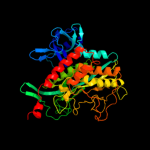
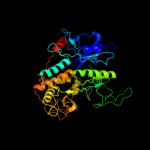
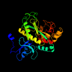
PDB header: ligaseChain: E: PDB Molecule: glutamine synthetase;PDBTitle: crystal structure of glutamine synthetase from salmonella2 typhimurium with inhibitor phosphinothricin
2 c3ng0A_
100.0
22
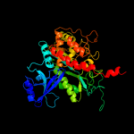
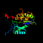
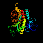
PDB header: ligaseChain: A: PDB Molecule: glutamine synthetase;PDBTitle: crystal structure of glutamine synthetase from synechocystis sp. pcc2 6803
3 c1htoB_
100.0
24
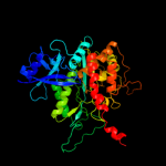
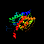
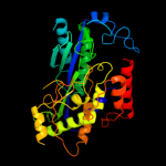
PDB header: ligaseChain: B: PDB Molecule: glutamine synthetase;PDBTitle: crystallographic structure of a relaxed glutamine synthetase from2 mycobacterium tuberculosis
4 c2j9iL_
100.0
22
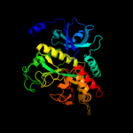
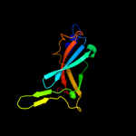
PDB header: ligaseChain: L: PDB Molecule: glutamate-ammonia ligase domain-containingPDBTitle: lengsin is a survivor of an ancient family of class i2 glutamine synthetases in eukaryotes that has undergone3 evolutionary re-engineering for a tissue-specific role4 in the vertebrate eye lens.
5 d1f52a2
100.0
24
Fold: Glutamine synthetase/guanido kinaseSuperfamily: Glutamine synthetase/guanido kinaseFamily: Glutamine synthetase catalytic domain6 d2bvca2
100.0
24
Fold: Glutamine synthetase/guanido kinaseSuperfamily: Glutamine synthetase/guanido kinaseFamily: Glutamine synthetase catalytic domain7 c2qc8J_
100.0
20
PDB header: ligaseChain: J: PDB Molecule: glutamine synthetase;PDBTitle: crystal structure of human glutamine synthetase in complex with adp2 and methionine sulfoximine phosphate
8 c2d3aJ_
100.0
16
PDB header: ligaseChain: J: PDB Molecule: glutamine synthetase;PDBTitle: crystal structure of the maize glutamine synthetase2 complexed with adp and methionine sulfoximine phosphate
9 c3fkyD_
100.0
20
PDB header: ligaseChain: D: PDB Molecule: glutamine synthetase;PDBTitle: crystal structure of the glutamine synthetase gln1deltan182 from the yeast saccharomyces cerevisiae
10 c3o6xC_
100.0
22
PDB header: ligaseChain: C: PDB Molecule: glutamine synthetase;PDBTitle: crystal structure of the type iii glutamine synthetase from2 bacteroides fragilis
11 d1f52a1
99.9
19
Fold: beta-Grasp (ubiquitin-like)Superfamily: Glutamine synthetase, N-terminal domainFamily: Glutamine synthetase, N-terminal domain12 d2bvca1
99.9
23
Fold: beta-Grasp (ubiquitin-like)Superfamily: Glutamine synthetase, N-terminal domainFamily: Glutamine synthetase, N-terminal domain13 c1tt4B_
99.0
19
PDB header: structural genomics, unknown functionChain: B: PDB Molecule: putative cytoplasmic protein;PDBTitle: structure of np459575, a predicted glutathione synthase from2 salmonella typhimurium
14 d1tt4a_
99.0
19
Fold: Glutamine synthetase/guanido kinaseSuperfamily: Glutamine synthetase/guanido kinaseFamily: Glutamate-cysteine ligase family 2 (GCS2)15 d1r8ga_
98.9
18
Fold: Glutamine synthetase/guanido kinaseSuperfamily: Glutamine synthetase/guanido kinaseFamily: Glutamate-cysteine ligase family 2 (GCS2)16 c2gwcE_
97.9
16
PDB header: ligaseChain: E: PDB Molecule: glutamate cysteine ligase;PDBTitle: crystal structure of plant glutamate cysteine ligase in complex with a2 transition state analogue
17 d1u8sa2
32.8
9
Fold: Ferredoxin-likeSuperfamily: ACT-likeFamily: Glycine cleavage system transcriptional repressor18 c2jy5A_
24.7
24
PDB header: signaling proteinChain: A: PDB Molecule: ubiquilin-1;PDBTitle: nmr structure of ubiquilin 1 uba domain
19 d1owxa_
20.2
9
Fold: Ferredoxin-likeSuperfamily: RNA-binding domain, RBDFamily: Canonical RBD20 d1nxia_
18.5
17
Fold: Ferredoxin-likeSuperfamily: Hypothetical protein VC0424Family: Hypothetical protein VC042421 c3ln7A_
not modelled
14.5
18
PDB header: ligaseChain: A: PDB Molecule: glutathione biosynthesis bifunctional protein gshab;PDBTitle: crystal structure of a bifunctional glutathione synthetase from2 pasteurella multocida
22 d1neka3
not modelled
13.6
16
Fold: Succinate dehydrogenase/fumarate reductase flavoprotein, catalytic domainSuperfamily: Succinate dehydrogenase/fumarate reductase flavoprotein, catalytic domainFamily: Succinate dehydrogenase/fumarate reductase flavoprotein, catalytic domain23 c3ns5B_
not modelled
13.1
6
PDB header: translationChain: B: PDB Molecule: eukaryotic translation initiation factor 3 subunit b;PDBTitle: crystal structure of the rna recognition motif of yeast eif3b residues2 76-161
24 c2vxeA_
not modelled
10.4
17
PDB header: transcriptionChain: A: PDB Molecule: cg10686-pa;PDBTitle: solution structure of the lsm domain of drosophila2 melanogaster tral (trailer hitch)
25 c2k7rA_
not modelled
10.4
14
PDB header: replicationChain: A: PDB Molecule: primosomal protein dnai;PDBTitle: n-terminal domain of the bacillus subtilis helicase-loading2 protein dnai
26 c3htnA_
not modelled
9.9
12
PDB header: metal binding proteinChain: A: PDB Molecule: putative dna binding protein;PDBTitle: crystal structure of a putative dna binding protein (bt_1116) from2 bacteroides thetaiotaomicron vpi-5482 at 1.50 a resolution
27 d2z1ea1
not modelled
9.5
17
Fold: Bacillus chorismate mutase-likeSuperfamily: PurM N-terminal domain-likeFamily: PurM N-terminal domain-like28 c3dcgF_
not modelled
9.5
50
PDB header: ligase/viral proteinChain: F: PDB Molecule: virion infectivity factor;PDBTitle: crystal structure of the hiv vif bc-box in complex with human elonginb2 and elonginc
29 c2zuuA_
not modelled
9.2
20
PDB header: transferaseChain: A: PDB Molecule: lacto-n-biose phosphorylase;PDBTitle: crystal structure of galacto-n-biose/lacto-n-biose i phosphorylase in2 complex with glcnac
30 c3bdkB_
not modelled
9.0
14
PDB header: lyaseChain: B: PDB Molecule: d-mannonate dehydratase;PDBTitle: crystal structure of streptococcus suis mannonate2 dehydratase complexed with substrate analogue
31 d2bs2a3
not modelled
8.9
25
Fold: Succinate dehydrogenase/fumarate reductase flavoprotein, catalytic domainSuperfamily: Succinate dehydrogenase/fumarate reductase flavoprotein, catalytic domainFamily: Succinate dehydrogenase/fumarate reductase flavoprotein, catalytic domain32 c3ns6B_
not modelled
8.9
6
PDB header: translationChain: B: PDB Molecule: eukaryotic translation initiation factor 3 subunit b;PDBTitle: crystal structure of hte rna recognition motif of yeast eif3b residues2 76-170
33 c1jqoA_
not modelled
8.8
17
PDB header: lyaseChain: A: PDB Molecule: phosphoenolpyruvate carboxylase;PDBTitle: crystal structure of c4-form phosphoenolpyruvate carboxylase from2 maize
34 d1jqoa_
not modelled
8.8
17
Fold: TIM beta/alpha-barrelSuperfamily: Phosphoenolpyruvate/pyruvate domainFamily: Phosphoenolpyruvate carboxylase35 c1nvpD_
not modelled
7.4
15
PDB header: transcription/dnaChain: D: PDB Molecule: transcription initiation factor iia gamma chain;PDBTitle: human tfiia/tbp/dna complex
36 d2vxfa1
not modelled
7.1
21
Fold: Sm-like foldSuperfamily: Sm-like ribonucleoproteinsFamily: LSM14 N-terminal domain-like37 d1zl0a2
not modelled
6.8
18
Fold: Flavodoxin-likeSuperfamily: Class I glutamine amidotransferase-likeFamily: LD-carboxypeptidase A N-terminal domain-like38 d2rgfa_
not modelled
6.7
20
Fold: beta-Grasp (ubiquitin-like)Superfamily: Ubiquitin-likeFamily: Ras-binding domain, RBD39 d1prtc1
not modelled
6.7
27
Fold: OB-foldSuperfamily: Bacterial enterotoxinsFamily: Bacterial AB5 toxins, B-subunits40 c3re3B_
not modelled
6.1
15
PDB header: lyaseChain: B: PDB Molecule: 2-c-methyl-d-erythritol 2,4-cyclodiphosphate synthase;PDBTitle: crystal structure of 2-c-methyl-d-erythritol 2,4-cyclodiphosphate2 synthase from francisella tularensis
41 c3it5B_
not modelled
6.0
33
PDB header: hydrolaseChain: B: PDB Molecule: protease lasa;PDBTitle: crystal structure of the lasa virulence factor from pseudomonas2 aeruginosa
42 d1prtb1
not modelled
5.8
20
Fold: OB-foldSuperfamily: Bacterial enterotoxinsFamily: Bacterial AB5 toxins, B-subunits43 c1nh2D_
not modelled
5.8
22
PDB header: transcription/dnaChain: D: PDB Molecule: transcription initiation factor iia small chain;PDBTitle: crystal structure of a yeast tfiia/tbp/dna complex
44 d2o3la1
not modelled
5.7
16
Fold: Left-handed superhelixSuperfamily: BH3980-likeFamily: BH3980-like45 d1ihma_
not modelled
5.6
19
Fold: Nucleoplasmin-like/VP (viral coat and capsid proteins)Superfamily: Positive stranded ssRNA virusesFamily: Caliciviridae-like VP46 d1xv2a_
not modelled
5.6
28
Fold: AF0104/ALDC/Ptd012-likeSuperfamily: AF0104/ALDC/Ptd012-likeFamily: Alpha-acetolactate decarboxylase-like47 d1eeja2
not modelled
5.5
10
Fold: Cystatin-likeSuperfamily: DsbC/DsbG N-terminal domain-likeFamily: DsbC/DsbG N-terminal domain-like48 c2qw5B_
not modelled
5.5
13
PDB header: isomeraseChain: B: PDB Molecule: xylose isomerase-like tim barrel;PDBTitle: crystal structure of a putative sugar phosphate isomerase/epimerase2 (ava4194) from anabaena variabilis atcc 29413 at 1.78 a resolution
49 d2v0ea1
not modelled
5.4
13
Fold: GYF/BRK domain-likeSuperfamily: BRK domain-likeFamily: BRK domain-like50 d1t3ba2
not modelled
5.1
14
Fold: Cystatin-likeSuperfamily: DsbC/DsbG N-terminal domain-likeFamily: DsbC/DsbG N-terminal domain-like51 c1u8sB_
not modelled
5.1
7
PDB header: transcriptionChain: B: PDB Molecule: glycine cleavage system transcriptionalPDBTitle: crystal structure of putative glycine cleavage system2 transcriptional repressor
52 d1jqna_
not modelled
5.1
21
Fold: TIM beta/alpha-barrelSuperfamily: Phosphoenolpyruvate/pyruvate domainFamily: Phosphoenolpyruvate carboxylase