Phyre Home Retrieve Phyre Job Id
Emaill.a.kelley@imperial.ac.uk
DescriptionP76137
DateWed Jan 25 15:21:05 GMT 2012
Unique Job ID1f69b33b39a0b9ab

Summary 

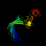
Top model
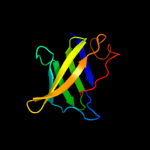
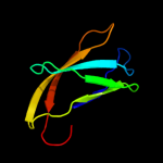
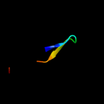
Image coloured by rainbow N → C terminus
Model (left) based on template c3rfzB_
Top template information
PDB header:cell adhesion/transport/chaperone
Chain: B: PDB Molecule:outer membrane usher protein, type 1 fimbrial synthesis;
PDBTitle: crystal structure of the fimd usher bound to its cognate fimc:fimh2 substrate
Confidence and coverage
Confidence:100.0% Coverage: 99%
379 residues ( 99% of your sequence) have been modelled with 100.0% confidence by the single highest scoring template.
3D viewing
Interactive 3D view in Jmol

Sequence analysis 

Secondary structure and disorder prediction 

   1........10.........20.........30.........40.........50.........60
Sequence  MSGYTVKPPTGDTNEQTQFIDYFNLFYSKRGQEQISISQQLGNYGTTFFSASRQSYWNTS
Secondary structure 


SS confidence 


Disorder  ??????


??????????????????????


?
?
???
Disorder confidence 


   .........70.........80.........90.........100.........110.........120
Sequence  RSDQQISFGLNVPFGDITTSLNYSYSNNIWQNDRDHLLAFTLNVPFSHWMRTDSQSAFRN
Secondary structure 


SS confidence 


Disorder  ???


?????????


?????????????
Disorder confidence 


   .........130.........140.........150.........160.........170.........180
Sequence  SNASYSMSNDLKGGMTNLSGVYGTLLPDNNLNYSVQVGNTHGGNTSSGTSGYSSLNYRGA
Secondary structure 


SS confidence 


Disorder  ?


??????


???????


?????????????


Disorder confidence 


   .........190.........200.........210.........220.........230.........240
Sequence  YGNTNVGYSRSGDSSQIYYGMSGGIIAHADGITFGQPLGDTMVLVKAPGADNVKIENQTG
Secondary structure 


SS confidence 


Disorder 


????


??
?

???
?
Disorder confidence 


   .........250.........260.........270.........280.........290.........300
Sequence  IHTDWRGYAILPFATEYRENRVALNANSLADNVELDETVVTVIPTHGAIARATFNAQIGG
Secondary structure 


SS confidence 


Disorder  ?


?


???


Disorder confidence 


   .........310.........320.........330.........340.........350.........360
Sequence  KVLMTLKYGNKSVPFGAIVTHGENKNGSIVAENGQVYLTGLPQSGQLQVSWGKDKNSNCI
Secondary structure 


SS confidence 


Disorder 


?

?


???


???


Disorder confidence 


   .........370.........380..
Sequence  VEYKLPEVSPGTLLNQQTAICR
Secondary structure 


SS confidence 


Disorder 


?????


??
Disorder confidence 


Confidence Key
High(9)                    Low (0)
?Disordered
Alpha helix
Beta strand

Domain analysis 

Hover over an aligned region to see model and summary info

Please note, only up to the top 20 hits are modelled to reduce computer load

RankAligned region

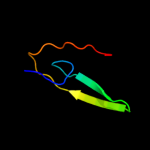
PDB 3rfz chain B

3D model

Region: 1 - 382
Aligned: 379
Modelled: 382
Confidence: 100.0%
Identity: 56%
PDB header:cell adhesion/transport/chaperone
Chain: B: PDB Molecule:outer membrane usher protein, type 1 fimbrial synthesis;
PDBTitle: crystal structure of the fimd usher bound to its cognate fimc:fimh2 substrate

Phyre2

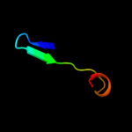
PDB 3ohn chain A

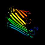
3D model

Region: 2 - 205
Aligned: 188
Modelled: 190
Confidence: 99.9%
Identity: 55%
PDB header:membrane protein
Chain: A: PDB Molecule:outer membrane usher protein fimd;
PDBTitle: crystal structure of the fimd translocation domain

Phyre2

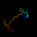
PDB 2xet chain B

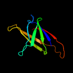
3D model

Region: 301 - 381
Aligned: 81
Modelled: 81
Confidence: 99.9%
Identity: 40%
PDB header:transport protein
Chain: B: PDB Molecule:f1 capsule-anchoring protein;
PDBTitle: conserved hydrophobic clusters on the surface of the caf1a usher2 c-terminal domain are important for f1 antigen assembly

Phyre2

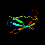
PDB 2vqi chain A

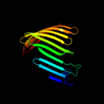
3D model

Region: 1 - 209
Aligned: 192
Modelled: 195
Confidence: 99.8%
Identity: 16%
PDB header:transport
Chain: A: PDB Molecule:outer membrane usher protein papc;
PDBTitle: structure of the p pilus usher (papc) translocation pore

Phyre2

PDB 3l48 chain B

3D model

Region: 299 - 382
Aligned: 78
Modelled: 82
Confidence: 99.8%
Identity: 29%
PDB header:transport protein
Chain: B: PDB Molecule:outer membrane usher protein papc;
PDBTitle: crystal structure of the c-terminal domain of the papc usher

Phyre2

PDB 2x5p chain A

3D model

Region: 287 - 350
Aligned: 60
Modelled: 64
Confidence: 37.0%
Identity: 18%
PDB header:protein binding
Chain: A: PDB Molecule:fibronectin binding protein;
PDBTitle: crystal structure of the streptococcus pyogenes fibronectin binding2 protein fbab-b

Phyre2

PDB 1aoz chain A domain 2

3D model

Region: 294 - 382
Aligned: 80
Modelled: 89
Confidence: 32.0%
Identity: 9%
Fold: Cupredoxin-like
Superfamily: Cupredoxins
Family: Multidomain cupredoxins

Phyre2

PDB 1uwy chain A domain 1

3D model

Region: 302 - 380
Aligned: 78
Modelled: 79
Confidence: 30.9%
Identity: 12%
Fold: Prealbumin-like
Superfamily: Carboxypeptidase regulatory domain-like
Family: Carboxypeptidase regulatory domain

Phyre2

PDB 2nsm chain A

3D model

Region: 302 - 370
Aligned: 67
Modelled: 69
Confidence: 27.3%
Identity: 13%
PDB header:hydrolase
Chain: A: PDB Molecule:carboxypeptidase n catalytic chain;
PDBTitle: crystal structure of the human carboxypeptidase n (kininase i)2 catalytic domain

Phyre2

PDB 3nad chain B

3D model

Region: 332 - 368
Aligned: 36
Modelled: 37
Confidence: 24.4%
Identity: 19%
PDB header:lyase
Chain: B: PDB Molecule:ferulate decarboxylase;
PDBTitle: crystal structure of phenolic acid decarboxylase from bacillus pumilus2 ui-670

Phyre2

PDB 2kyw chain A

3D model

Region: 261 - 309
Aligned: 48
Modelled: 49
Confidence: 21.9%
Identity: 25%
PDB header:cell adhesion
Chain: A: PDB Molecule:adhesion exoprotein;
PDBTitle: solution nmr structure of a domain of adhesion exoprotein from2 pediococcus pentosaceus, northeast structural genomics consortium3 target ptr41o

Phyre2

PDB 1nkg chain A domain 1

3D model

Region: 301 - 369
Aligned: 68
Modelled: 69
Confidence: 21.5%
Identity: 10%
Fold: Prealbumin-like
Superfamily: Starch-binding domain-like
Family: Rhamnogalacturonase B, RhgB, middle domain

Phyre2

PDB 2je6 chain I domain 3

3D model

Region: 329 - 340
Aligned: 12
Modelled: 12
Confidence: 18.8%
Identity: 33%
Fold: Eukaryotic type KH-domain (KH-domain type I)
Superfamily: Eukaryotic type KH-domain (KH-domain type I)
Family: Eukaryotic type KH-domain (KH-domain type I)

Phyre2

PDB 2ww8 chain A

3D model

Region: 243 - 350
Aligned: 107
Modelled: 108
Confidence: 18.8%
Identity: 12%
PDB header:cell adhesion
Chain: A: PDB Molecule:cell wall surface anchor family protein;
PDBTitle: structure of the pilus adhesin (rrga) from streptococcus2 pneumoniae

Phyre2

PDB 2ey4 chain C domain 1

3D model

Region: 304 - 332
Aligned: 29
Modelled: 29
Confidence: 18.2%
Identity: 14%
Fold: Reductase/isomerase/elongation factor common domain
Superfamily: Translation proteins
Family: Gar1-like SnoRNP

Phyre2

PDB 2hvy chain B

3D model

Region: 304 - 332
Aligned: 29
Modelled: 29
Confidence: 17.6%
Identity: 14%
PDB header:isomerase/biosynthetic protein/rna
Chain: B: PDB Molecule:small nucleolar rnp similar to gar1;
PDBTitle: crystal structure of an h/aca box rnp from pyrococcus furiosus

Phyre2

PDB 1ci3 chain M domain 2

3D model

Region: 317 - 353
Aligned: 37
Modelled: 37
Confidence: 17.5%
Identity: 27%
Fold: Barrel-sandwich hybrid
Superfamily: Rudiment single hybrid motif
Family: Cytochrome f, small domain

Phyre2

PDB 2ja9 chain A domain 2

3D model

Region: 329 - 347
Aligned: 19
Modelled: 19
Confidence: 15.5%
Identity: 21%
Fold: Eukaryotic type KH-domain (KH-domain type I)
Superfamily: Eukaryotic type KH-domain (KH-domain type I)
Family: Eukaryotic type KH-domain (KH-domain type I)

Phyre2

PDB 3ila chain G

3D model

Region: 337 - 378
Aligned: 42
Modelled: 42
Confidence: 15.4%
Identity: 21%
PDB header:signaling protein
Chain: G: PDB Molecule:ryanodine receptor 1;
PDBTitle: crystal structure of rabbit ryanodine receptor 1 n-terminal domain (9-2 205)

Phyre2

PDB 1h8l chain A

3D model

Region: 300 - 369
Aligned: 68
Modelled: 70
Confidence: 13.3%
Identity: 9%
PDB header:carboxypeptidase
Chain: A: PDB Molecule:carboxypeptidase gp180 residues 503-882;
PDBTitle: duck carboxypeptidase d domain ii in complex with gemsa

Phyre2
1

c3rfzB_
2

c3ohnA_
3

c2xetB_
4

c2vqiA_
5

c3l48B_
6

c2x5pA_
7

d1aoza2
8

d1uwya1
9

c2nsmA_
10

c3nadB_
11

c2kywA_
12

d1nkga1
13

d2je6i3
14

c2ww8A_
15

d2ey4c1
16

c2hvyB_
17

d1ci3m2
18

d2ja9a2
19

c3ilaG_
20

c1h8lA_
21


22


23


24


25


26


27


28


29


30


31


32


33


34


35


36


37


38


39


40


41


42


43


44


45


46


47


48


49


50


51


Detailed template information 

#
Template Alignment Coverage3D Model Confidence
% i.d. Template Information
1c3rfzB_


100.0 56 PDB header:cell adhesion/transport/chaperone
Chain: B: PDB Molecule:outer membrane usher protein, type 1 fimbrial synthesis;
PDBTitle: crystal structure of the fimd usher bound to its cognate fimc:fimh2 substrate
2c3ohnA_


99.9 55 PDB header:membrane protein
Chain: A: PDB Molecule:outer membrane usher protein fimd;
PDBTitle: crystal structure of the fimd translocation domain
3c2xetB_


99.9 40 PDB header:transport protein
Chain: B: PDB Molecule:f1 capsule-anchoring protein;
PDBTitle: conserved hydrophobic clusters on the surface of the caf1a usher2 c-terminal domain are important for f1 antigen assembly
4c2vqiA_


99.8 16 PDB header:transport
Chain: A: PDB Molecule:outer membrane usher protein papc;
PDBTitle: structure of the p pilus usher (papc) translocation pore
5c3l48B_


99.8 29 PDB header:transport protein
Chain: B: PDB Molecule:outer membrane usher protein papc;
PDBTitle: crystal structure of the c-terminal domain of the papc usher
6c2x5pA_


37.0 18 PDB header:protein binding
Chain: A: PDB Molecule:fibronectin binding protein;
PDBTitle: crystal structure of the streptococcus pyogenes fibronectin binding2 protein fbab-b
7d1aoza2


32.0 9 Fold:Cupredoxin-like
Superfamily:Cupredoxins
Family:Multidomain cupredoxins
8d1uwya1


30.9 12 Fold:Prealbumin-like
Superfamily:Carboxypeptidase regulatory domain-like
Family:Carboxypeptidase regulatory domain
9c2nsmA_


27.3 13 PDB header:hydrolase
Chain: A: PDB Molecule:carboxypeptidase n catalytic chain;
PDBTitle: crystal structure of the human carboxypeptidase n (kininase i)2 catalytic domain
10c3nadB_


24.4 19 PDB header:lyase
Chain: B: PDB Molecule:ferulate decarboxylase;
PDBTitle: crystal structure of phenolic acid decarboxylase from bacillus pumilus2 ui-670
11c2kywA_


21.9 25 PDB header:cell adhesion
Chain: A: PDB Molecule:adhesion exoprotein;
PDBTitle: solution nmr structure of a domain of adhesion exoprotein from2 pediococcus pentosaceus, northeast structural genomics consortium3 target ptr41o
12d1nkga1


21.5 10 Fold:Prealbumin-like
Superfamily:Starch-binding domain-like
Family:Rhamnogalacturonase B, RhgB, middle domain
13d2je6i3


18.8 33 Fold:Eukaryotic type KH-domain (KH-domain type I)
Superfamily:Eukaryotic type KH-domain (KH-domain type I)
Family:Eukaryotic type KH-domain (KH-domain type I)
14c2ww8A_


18.8 12 PDB header:cell adhesion
Chain: A: PDB Molecule:cell wall surface anchor family protein;
PDBTitle: structure of the pilus adhesin (rrga) from streptococcus2 pneumoniae
15d2ey4c1


18.2 14 Fold:Reductase/isomerase/elongation factor common domain
Superfamily:Translation proteins
Family:Gar1-like SnoRNP
16c2hvyB_


17.6 14 PDB header:isomerase/biosynthetic protein/rna
Chain: B: PDB Molecule:small nucleolar rnp similar to gar1;
PDBTitle: crystal structure of an h/aca box rnp from pyrococcus furiosus
17d1ci3m2


17.5 27 Fold:Barrel-sandwich hybrid
Superfamily:Rudiment single hybrid motif
Family:Cytochrome f, small domain
18d2ja9a2


15.5 21 Fold:Eukaryotic type KH-domain (KH-domain type I)
Superfamily:Eukaryotic type KH-domain (KH-domain type I)
Family:Eukaryotic type KH-domain (KH-domain type I)
19c3ilaG_


15.4 21 PDB header:signaling protein
Chain: G: PDB Molecule:ryanodine receptor 1;
PDBTitle: crystal structure of rabbit ryanodine receptor 1 n-terminal domain (9-2 205)
20c1h8lA_


13.3 9 PDB header:carboxypeptidase
Chain: A: PDB Molecule:carboxypeptidase gp180 residues 503-882;
PDBTitle: duck carboxypeptidase d domain ii in complex with gemsa
21d1hfua2


not modelled 13.1 14 Fold:Cupredoxin-like
Superfamily:Cupredoxins
Family:Multidomain cupredoxins
22d1e2wa2


not modelled 12.5 28 Fold:Barrel-sandwich hybrid
Superfamily:Rudiment single hybrid motif
Family:Cytochrome f, small domain
23d1h8la1


not modelled 12.0 8 Fold:Prealbumin-like
Superfamily:Carboxypeptidase regulatory domain-like
Family:Carboxypeptidase regulatory domain
24c1yukB_


not modelled 11.4 29 PDB header:cell adhesion
Chain: B: PDB Molecule:integrin beta-2 b chain;
PDBTitle: the crystal structure of the psi/hybrid domain/ i-egf12 segment from the human integrin beta2 at 1.8 resolution
25d2gc9a1


not modelled 11.0 14 Fold:Lipocalins
Superfamily:Lipocalins
Family:Phenolic acid decarboxylase (PAD)
26d1gyca2


not modelled 10.4 9 Fold:Cupredoxin-like
Superfamily:Cupredoxins
Family:Multidomain cupredoxins
27d1lnza1


not modelled 9.9 30 Fold:Obg-fold
Superfamily:Obg GTP-binding protein N-terminal domain
Family:Obg GTP-binding protein N-terminal domain
28d1zvca1


not modelled 8.5 18 Fold:AOC barrel-like
Superfamily:Allene oxide cyclase-like
Family:Allene oxide cyclase-like
29c1wsrA_


not modelled 8.4 11 PDB header:transferase
Chain: A: PDB Molecule:aminomethyltransferase;
PDBTitle: crystal structure of human t-protein of glycine cleavage2 system
30d1v8ha1


not modelled 8.2 13 Fold:Immunoglobulin-like beta-sandwich
Superfamily:E set domains
Family:SoxZ-like
31d1udxa1


not modelled 7.9 30 Fold:Obg-fold
Superfamily:Obg GTP-binding protein N-terminal domain
Family:Obg GTP-binding protein N-terminal domain
32d1wkqa_


not modelled 7.6 20 Fold:Cytidine deaminase-like
Superfamily:Cytidine deaminase-like
Family:Deoxycytidylate deaminase-like
33d1m1sa_


not modelled 7.5 11 Fold:Immunoglobulin-like beta-sandwich
Superfamily:PapD-like
Family:MSP-like
34d1v76a_


not modelled 7.5 15 Fold:Rof/RNase P subunit-like
Superfamily:Rof/RNase P subunit-like
Family:RNase P subunit p29-like
35c2zaeC_


not modelled 7.3 15 PDB header:hydrolase
Chain: C: PDB Molecule:ribonuclease p protein component 1;
PDBTitle: crystal structure of protein ph1601p in complex with protein ph1771p2 of archaeal ribonuclease p from pyrococcus horikoshii ot3
36d2nn6g3


not modelled 7.3 16 Fold:Eukaryotic type KH-domain (KH-domain type I)
Superfamily:Eukaryotic type KH-domain (KH-domain type I)
Family:Eukaryotic type KH-domain (KH-domain type I)
37d1fr5a_


not modelled 7.0 28 Fold:RNA bacteriophage capsid protein
Superfamily:RNA bacteriophage capsid protein
Family:RNA bacteriophage capsid protein
38d1kyaa2


not modelled 6.9 9 Fold:Cupredoxin-like
Superfamily:Cupredoxins
Family:Multidomain cupredoxins
39d2hqya1


not modelled 6.5 31 Fold:Acyl-CoA N-acyltransferases (Nat)
Superfamily:Acyl-CoA N-acyltransferases (Nat)
Family:FemXAB nonribosomal peptidyltransferases
40c1fw3A_


not modelled 6.0 31 PDB header:hydrolase, membrane protein
Chain: A: PDB Molecule:outer membrane phospholipase a;
PDBTitle: outer membrane phospholipase a from escherichia coli
41c2eqnA_


not modelled 5.6 10 PDB header:transcription
Chain: A: PDB Molecule:hypothetical protein loc92345;
PDBTitle: solution structure of the naf1 domain of hypothetical2 protein bc008207 [homo sapiens]
42c2jxmB_


not modelled 5.5 24 PDB header:electron transport
Chain: B: PDB Molecule:cytochrome f;
PDBTitle: ensemble of twenty structures of the prochlorothrix2 hollandica plastocyanin- cytochrome f complex
43c2gw4C_


not modelled 5.4 17 PDB header:luminescent protein
Chain: C: PDB Molecule:kaede;
PDBTitle: crystal structure of stony coral fluorescent protein kaede, red form
44d2q9oa2


not modelled 5.3 9 Fold:Cupredoxin-like
Superfamily:Cupredoxins
Family:Multidomain cupredoxins
45c2ja9A_


not modelled 5.3 13 PDB header:rna-binding protein
Chain: A: PDB Molecule:exosome complex exonuclease rrp40;
PDBTitle: structure of the n-terminal deletion of yeast exosome2 component rrp40
46d1rowa_


not modelled 5.3 9 Fold:Immunoglobulin-like beta-sandwich
Superfamily:PapD-like
Family:MSP-like
47c3mn8A_


not modelled 5.2 13 PDB header:hydrolase
Chain: A: PDB Molecule:lp15968p;
PDBTitle: structure of drosophila melanogaster carboxypeptidase d isoform 1b2 short
48c3nx2B_


not modelled 5.2 12 PDB header:lyase
Chain: B: PDB Molecule:ferulic acid decarboxylase;
PDBTitle: enterobacter sp. px6-4 ferulic acid decarboxylase in complex with2 substrate analogues
49d1gyva_


not modelled 5.2 18 Fold:Immunoglobulin-like beta-sandwich
Superfamily:Clathrin adaptor appendage domain
Family:gamma-adaptin C-terminal appendage domain-like
50c2brqA_


not modelled 5.1 7 PDB header:structural protein
Chain: A: PDB Molecule:filamin a;
PDBTitle: crystal structure of the filamin a repeat 21 complexed with2 the integrin beta7 cytoplasmic tail peptide
51c3iswB_


not modelled 5.1 7 PDB header:structural protein
Chain: B: PDB Molecule:filamin-a;
PDBTitle: crystal structure of filamin-a immunoglobulin-like repeat 21 bound to2 an n-terminal peptide of cftr

Binding site prediction 

Due to computational demand, binding site predictions are not run for batch jobs

If you want to predict binding sites, please manually submit your model of choice to 3DLigandSite


Phyre is for academic use only

Please cite: Protein structure prediction on the web: a case study using the Phyre server
Kelley LA and Sternberg MJE. Nature Protocols 4, 363 - 371 (2009) [pdf] [Import into BibTeX]
 
If you use the binding site predictions from 3DLigandSite, please also cite:
3DLigandSite: predicting ligand-binding sites using similar structures.
Wass MN, Kelley LA and Sternberg MJ Nucleic Acids Research 38, W469-73 (2010) [PubMed]
 
© Structural Bioinformatics Group
Imperial College London
Lawrence Kelley, Benjamin Jefferys
Disclaimer
Terms and Conditions
Component software
Template detection: HHpred 1.51
Secondary structure prediction: Psi-pred 2.5
Disorder prediction: Disopred 2.4
Transmembrane prediction: Memsat_SVM
Multi-template modelling and ab initio: Poing 1.0