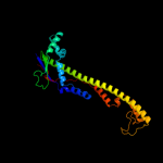
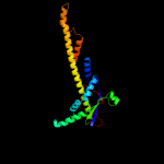
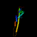
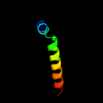
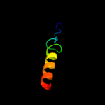
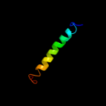
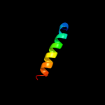
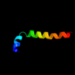
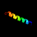
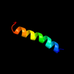
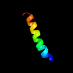
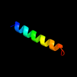
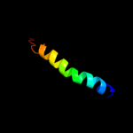
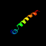
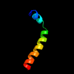
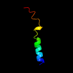
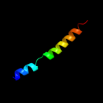
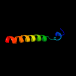
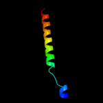
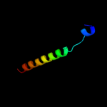
1 d3b8ma1
99.8
97
Fold: Ferredoxin-likeSuperfamily: Bacterial polysaccharide co-polymerase-likeFamily: FepE-like2 d3b8oa1
99.2
19
Fold: Ferredoxin-likeSuperfamily: Bacterial polysaccharide co-polymerase-likeFamily: FepE-like3 d3b8pa1
99.2
26
Fold: Ferredoxin-likeSuperfamily: Bacterial polysaccharide co-polymerase-likeFamily: FepE-like4 c2yl4A_
63.4
9
PDB header: membrane proteinChain: A: PDB Molecule: atp-binding cassette sub-family b member 10,PDBTitle: structure of the human mitochondrial abc transporter, abcb10
5 c2rmzA_
61.7
29
PDB header: cell adhesionChain: A: PDB Molecule: integrin beta-3;PDBTitle: bicelle-embedded integrin beta3 transmembrane segment
6 c2kluA_
56.6
13
PDB header: immune system, membrane proteinChain: A: PDB Molecule: t-cell surface glycoprotein cd4;PDBTitle: nmr structure of the transmembrane and cytoplasmic domains2 of human cd4
7 c2qtsA_
54.2
4
PDB header: membrane proteinChain: A: PDB Molecule: acid-sensing ion channel;PDBTitle: structure of an acid-sensing ion channel 1 at 1.9 a resolution and low2 ph
8 d2hyda2
50.5
9
Fold: ABC transporter transmembrane regionSuperfamily: ABC transporter transmembrane regionFamily: ABC transporter transmembrane region9 c2k1kA_
48.9
20
PDB header: signaling proteinChain: A: PDB Molecule: ephrin type-a receptor 1;PDBTitle: nmr structures of dimeric transmembrane domain of the2 receptor tyrosine kinase epha1 in lipid bicelles at ph 4.3
10 c2k1lA_
48.9
20
PDB header: signaling proteinChain: A: PDB Molecule: ephrin type-a receptor 1;PDBTitle: nmr structures of dimeric transmembrane domain of the2 receptor tyrosine kinase epha1 in lipid bicelles at ph 6.3
11 c2k1lB_
48.9
20
PDB header: signaling proteinChain: B: PDB Molecule: ephrin type-a receptor 1;PDBTitle: nmr structures of dimeric transmembrane domain of the2 receptor tyrosine kinase epha1 in lipid bicelles at ph 6.3
12 c2k1kB_
48.9
20
PDB header: signaling proteinChain: B: PDB Molecule: ephrin type-a receptor 1;PDBTitle: nmr structures of dimeric transmembrane domain of the2 receptor tyrosine kinase epha1 in lipid bicelles at ph 4.3
13 c2k21A_
43.3
10
PDB header: membrane proteinChain: A: PDB Molecule: potassium voltage-gated channel subfamily ePDBTitle: nmr structure of human kcne1 in lmpg micelles at ph 6.0 and2 40 degree c
14 c2hydB_
36.5
9
PDB header: transport proteinChain: B: PDB Molecule: abc transporter homolog;PDBTitle: multidrug abc transporter sav1866
15 d1pf4a2
35.8
9
Fold: ABC transporter transmembrane regionSuperfamily: ABC transporter transmembrane regionFamily: ABC transporter transmembrane region16 c2kncA_
33.2
18
PDB header: cell adhesionChain: A: PDB Molecule: integrin alpha-iib;PDBTitle: platelet integrin alfaiib-beta3 transmembrane-cytoplasmic2 heterocomplex
17 c3hd7A_
29.6
15
PDB header: exocytosisChain: A: PDB Molecule: vesicle-associated membrane protein 2;PDBTitle: helical extension of the neuronal snare complex into the membrane,2 spacegroup c 1 2 1
18 c3g5uB_
28.5
11
PDB header: membrane proteinChain: B: PDB Molecule: multidrug resistance protein 1a;PDBTitle: structure of p-glycoprotein reveals a molecular basis for2 poly-specific drug binding
19 d1v54d_
27.7
8
Fold: Single transmembrane helixSuperfamily: Mitochondrial cytochrome c oxidase subunit IVFamily: Mitochondrial cytochrome c oxidase subunit IV20 c2y69Q_
27.3
8
PDB header: electron transportChain: Q: PDB Molecule: cytochrome c oxidase subunit 4 isoform 1;PDBTitle: bovine heart cytochrome c oxidase re-refined with molecular2 oxygen
21 d3b60a2
not modelled
26.5
13
Fold: ABC transporter transmembrane regionSuperfamily: ABC transporter transmembrane regionFamily: ABC transporter transmembrane region22 d1rhzb_
not modelled
22.9
13
Fold: Single transmembrane helixSuperfamily: Preprotein translocase SecE subunitFamily: Preprotein translocase SecE subunit23 c3ij4A_
not modelled
22.7
13
PDB header: transport proteinChain: A: PDB Molecule: amiloride-sensitive cation channel 2, neuronal;PDBTitle: cesium sites in the crystal structure of a functional acid2 sensing ion channel in the desensitized state
24 c2l34B_
not modelled
21.6
21
PDB header: protein bindingChain: B: PDB Molecule: tyro protein tyrosine kinase-binding protein;PDBTitle: structure of the dap12 transmembrane homodimer
25 c2l34A_
not modelled
21.6
21
PDB header: protein bindingChain: A: PDB Molecule: tyro protein tyrosine kinase-binding protein;PDBTitle: structure of the dap12 transmembrane homodimer
26 d1y5ic1
not modelled
19.9
11
Fold: Heme-binding four-helical bundleSuperfamily: Respiratory nitrate reductase 1 gamma chainFamily: Respiratory nitrate reductase 1 gamma chain27 c3b5wE_
not modelled
17.5
13
PDB header: membrane proteinChain: E: PDB Molecule: lipid a export atp-binding/permease protein msba;PDBTitle: crystal structure of eschericia coli msba
28 d1v54m_
not modelled
17.1
17
Fold: Single transmembrane helixSuperfamily: Mitochondrial cytochrome c oxidase subunit VIIIb (aka IX)Family: Mitochondrial cytochrome c oxidase subunit VIIIb (aka IX)29 c2y69Z_
not modelled
16.5
17
PDB header: electron transportChain: Z: PDB Molecule: cytochrome c oxidase polypeptide 8h;PDBTitle: bovine heart cytochrome c oxidase re-refined with molecular2 oxygen
30 c3sc0A_
not modelled
16.1
32
PDB header: oxidoreductaseChain: A: PDB Molecule: methylmalonic aciduria and homocystinuria type c protein;PDBTitle: crystal structure of mmachc (1-238), a human b12 processing enzyme,2 complexed with methylcobalamin
31 c1vf5R_
not modelled
15.4
22
PDB header: photosynthesisChain: R: PDB Molecule: protein pet l;PDBTitle: crystal structure of cytochrome b6f complex from m.laminosus
32 d2e74e1
not modelled
15.4
22
Fold: Single transmembrane helixSuperfamily: PetL subunit of the cytochrome b6f complexFamily: PetL subunit of the cytochrome b6f complex33 c1vf5E_
not modelled
15.4
22
PDB header: photosynthesisChain: E: PDB Molecule: protein pet l;PDBTitle: crystal structure of cytochrome b6f complex from m.laminosus
34 c2e74E_
not modelled
15.4
22
PDB header: photosynthesisChain: E: PDB Molecule: cytochrome b6-f complex subunit 6;PDBTitle: crystal structure of the cytochrome b6f complex from m.laminosus
35 c2e75E_
not modelled
15.4
22
PDB header: photosynthesisChain: E: PDB Molecule: cytochrome b6-f complex subunit 6;PDBTitle: crystal structure of the cytochrome b6f complex with 2-nonyl-4-2 hydroxyquinoline n-oxide (nqno) from m.laminosus
36 c2e76E_
not modelled
15.4
22
PDB header: photosynthesisChain: E: PDB Molecule: cytochrome b6-f complex subunit 6;PDBTitle: crystal structure of the cytochrome b6f complex with tridecyl-2 stigmatellin (tds) from m.laminosus
37 d1v54g_
not modelled
15.0
8
Fold: Single transmembrane helixSuperfamily: Mitochondrial cytochrome c oxidase subunit VIaFamily: Mitochondrial cytochrome c oxidase subunit VIa38 c3k07A_
not modelled
13.1
13
PDB header: transport proteinChain: A: PDB Molecule: cation efflux system protein cusa;PDBTitle: crystal structure of cusa
39 d2r6gf1
not modelled
12.0
10
Fold: MalF N-terminal region-likeSuperfamily: MalF N-terminal region-likeFamily: MalF N-terminal region-like40 c3b5xB_
not modelled
11.2
9
PDB header: membrane proteinChain: B: PDB Molecule: lipid a export atp-binding/permease protein msba;PDBTitle: crystal structure of msba from vibrio cholerae
41 c2l35B_
not modelled
10.5
13
PDB header: protein bindingChain: B: PDB Molecule: tyro protein tyrosine kinase-binding protein;PDBTitle: structure of the dap12-nkg2c transmembrane heterotrimer
42 c2l2tA_
not modelled
10.4
15
PDB header: membrane proteinChain: A: PDB Molecule: receptor tyrosine-protein kinase erbb-4;PDBTitle: solution nmr structure of the erbb4 dimeric membrane domain
43 c2k1aA_
not modelled
10.4
9
PDB header: cell adhesionChain: A: PDB Molecule: integrin alpha-iib;PDBTitle: bicelle-embedded integrin alpha(iib) transmembrane segment
44 d1v54l_
not modelled
10.3
11
Fold: Single transmembrane helixSuperfamily: Mitochondrial cytochrome c oxidase subunit VIIc (aka VIIIa)Family: Mitochondrial cytochrome c oxidase subunit VIIc (aka VIIIa)45 c2l35A_
not modelled
10.2
21
PDB header: protein bindingChain: A: PDB Molecule: dap12-nkg2c_tm;PDBTitle: structure of the dap12-nkg2c transmembrane heterotrimer
46 c2yvxD_
not modelled
9.5
0
PDB header: transport proteinChain: D: PDB Molecule: mg2+ transporter mgte;PDBTitle: crystal structure of magnesium transporter mgte
47 d1xrda1
not modelled
9.4
19
Fold: Light-harvesting complex subunitsSuperfamily: Light-harvesting complex subunitsFamily: Light-harvesting complex subunits48 d1pw4a_
not modelled
9.2
16
Fold: MFS general substrate transporterSuperfamily: MFS general substrate transporterFamily: Glycerol-3-phosphate transporter49 c1kqfB_
not modelled
8.8
14
PDB header: oxidoreductaseChain: B: PDB Molecule: formate dehydrogenase, nitrate-inducible, iron-sulfurPDBTitle: formate dehydrogenase n from e. coli
50 d1rh5b_
not modelled
8.5
14
Fold: Single transmembrane helixSuperfamily: Preprotein translocase SecE subunitFamily: Preprotein translocase SecE subunit51 c2ht2B_
not modelled
8.3
9
PDB header: membrane proteinChain: B: PDB Molecule: h(+)/cl(-) exchange transporter clca;PDBTitle: structure of the escherichia coli clc chloride channel2 y445h mutant and fab complex
52 c2y69Y_
not modelled
8.3
10
PDB header: electron transportChain: Y: PDB Molecule: cytochrome c oxidase subunit 7c;PDBTitle: bovine heart cytochrome c oxidase re-refined with molecular2 oxygen
53 c2kb1A_
not modelled
8.0
13
PDB header: membrane proteinChain: A: PDB Molecule: wsk3;PDBTitle: nmr studies of a channel protein without membrane:2 structure and dynamics of water-solubilized kcsa
54 d1otsa_
not modelled
7.8
9
Fold: Clc chloride channelSuperfamily: Clc chloride channelFamily: Clc chloride channel55 c2wwbC_
not modelled
7.5
22
PDB header: ribosomeChain: C: PDB Molecule: protein transport protein sec61 subunit beta;PDBTitle: cryo-em structure of the mammalian sec61 complex bound to the2 actively translating wheat germ 80s ribosome
56 c2wwbB_
not modelled
7.5
16
PDB header: ribosomeChain: B: PDB Molecule: protein transport protein sec61 subunit gamma;PDBTitle: cryo-em structure of the mammalian sec61 complex bound to the2 actively translating wheat germ 80s ribosome
57 c1m2zE_
not modelled
7.3
25
PDB header: hormone/hormone activatorChain: E: PDB Molecule: nuclear receptor coactivator 2;PDBTitle: crystal structure of a dimer complex of the human2 glucocorticoid receptor ligand-binding domain bound to3 dexamethasone and a tif2 coactivator motif
58 c1ps9A_
not modelled
7.1
17
PDB header: oxidoreductaseChain: A: PDB Molecule: 2,4-dienoyl-coa reductase;PDBTitle: the crystal structure and reaction mechanism of e. coli 2,4-2 dienoyl coa reductase
59 c2jwaA_
not modelled
6.9
20
PDB header: transferaseChain: A: PDB Molecule: receptor tyrosine-protein kinase erbb-2;PDBTitle: erbb2 transmembrane segment dimer spatial structure
60 d2e74d2
not modelled
6.9
13
Fold: Single transmembrane helixSuperfamily: ISP transmembrane anchorFamily: ISP transmembrane anchor61 d1o73a_
not modelled
6.7
19
Fold: Thioredoxin foldSuperfamily: Thioredoxin-likeFamily: Glutathione peroxidase-like62 c3ipdB_
not modelled
6.7
12
PDB header: exocytosisChain: B: PDB Molecule: syntaxin-1a;PDBTitle: helical extension of the neuronal snare complex into the2 membrane, spacegroup i 21 21 21
63 c2kvlA_
not modelled
6.6
19
PDB header: viral proteinChain: A: PDB Molecule: major outer capsid protein vp7;PDBTitle: nmr structure of the c-terminal domain of vp7
64 c1m2zB_
not modelled
6.6
25
PDB header: hormone/hormone activatorChain: B: PDB Molecule: nuclear receptor coactivator 2;PDBTitle: crystal structure of a dimer complex of the human2 glucocorticoid receptor ligand-binding domain bound to3 dexamethasone and a tif2 coactivator motif
65 c2k9yA_
not modelled
6.5
14
PDB header: transferaseChain: A: PDB Molecule: ephrin type-a receptor 2;PDBTitle: epha2 dimeric structure in the lipidic bicelle at ph 5.0
66 c2k9yB_
not modelled
6.5
14
PDB header: transferaseChain: B: PDB Molecule: ephrin type-a receptor 2;PDBTitle: epha2 dimeric structure in the lipidic bicelle at ph 5.0
67 c1xl6B_
not modelled
6.5
13
PDB header: metal transportChain: B: PDB Molecule: inward rectifier potassium channel;PDBTitle: intermediate gating structure 2 of the inwardly rectifying k+ channel2 kirbac3.1
68 c2j7aC_
not modelled
6.5
5
PDB header: oxidoreductaseChain: C: PDB Molecule: cytochrome c quinol dehydrogenase nrfh;PDBTitle: crystal structure of cytochrome c nitrite reductase nrfha2 complex from desulfovibrio vulgaris
69 c1iijA_
not modelled
6.4
13
PDB header: signaling proteinChain: A: PDB Molecule: erbb-2 receptor protein-tyrosine kinase;PDBTitle: solution structure of the neu/erbb-2 membrane spanning2 segment
70 d1nwaa_
not modelled
6.3
13
Fold: Ferredoxin-likeSuperfamily: Peptide methionine sulfoxide reductaseFamily: Peptide methionine sulfoxide reductase71 c1nwaA_
not modelled
6.3
13
PDB header: oxidoreductaseChain: A: PDB Molecule: peptide methionine sulfoxide reductase msra;PDBTitle: structure of mycobacterium tuberculosis methionine2 sulfoxide reductase a in complex with protein-bound3 methionine
72 c2o01L_
not modelled
6.3
13
PDB header: photosynthesisChain: L: PDB Molecule: photosystem i reaction center subunit xi,PDBTitle: the structure of a plant photosystem i supercomplex at 3.42 angstrom resolution
73 c3p5nA_
not modelled
6.1
8
PDB header: transport proteinChain: A: PDB Molecule: riboflavin uptake protein;PDBTitle: structure and mechanism of the s component of a bacterial ecf2 transporter
74 d2yvxa3
not modelled
6.1
8
Fold: MgtE membrane domain-likeSuperfamily: MgtE membrane domain-likeFamily: MgtE membrane domain-like75 d1fftb2
not modelled
6.0
10
Fold: Transmembrane helix hairpinSuperfamily: Cytochrome c oxidase subunit II-like, transmembrane regionFamily: Cytochrome c oxidase subunit II-like, transmembrane region76 c2ks1B_
not modelled
6.0
29
PDB header: transferaseChain: B: PDB Molecule: epidermal growth factor receptor;PDBTitle: heterodimeric association of transmembrane domains of erbb1 and erbb22 receptors enabling kinase activation
77 c1fvaA_
not modelled
5.8
25
PDB header: oxidoreductaseChain: A: PDB Molecule: peptide methionine sulfoxide reductase;PDBTitle: crystal structure of bovine methionine sulfoxide reductase
78 c2gqcA_
not modelled
5.7
31
PDB header: hydrolaseChain: A: PDB Molecule: rhomboid intramembrane protease;PDBTitle: solution structure of the n-terminal domain of rhomboid intramembrane2 protease from p. aeruginosa
79 c3a0hx_
not modelled
5.7
11
PDB header: electron transportChain: X: PDB Molecule: photosystem ii reaction center protein x;PDBTitle: crystal structure of i-substituted photosystem ii complex
80 c3a0bX_
not modelled
5.7
11
PDB header: electron transportChain: X: PDB Molecule: photosystem ii reaction center protein x;PDBTitle: crystal structure of br-substituted photosystem ii complex
81 c3a0hX_
not modelled
5.7
11
PDB header: electron transportChain: X: PDB Molecule: photosystem ii reaction center protein x;PDBTitle: crystal structure of i-substituted photosystem ii complex
82 c3a0bx_
not modelled
5.7
11
PDB header: electron transportChain: X: PDB Molecule: photosystem ii reaction center protein x;PDBTitle: crystal structure of br-substituted photosystem ii complex
83 c1q2iA_
not modelled
5.7
26
PDB header: antitumor proteinChain: A: PDB Molecule: pnc27;PDBTitle: nmr solution structure of a peptide from the mdm-2 binding2 domain of the p53 protein that is selectively cytotoxic to3 cancer cells
84 d1kqfb2
not modelled
5.7
6
Fold: Single transmembrane helixSuperfamily: Iron-sulfur subunit of formate dehydrogenase N, transmembrane anchorFamily: Iron-sulfur subunit of formate dehydrogenase N, transmembrane anchor85 c3jycA_
not modelled
5.5
7
PDB header: metal transportChain: A: PDB Molecule: inward-rectifier k+ channel kir2.2;PDBTitle: crystal structure of the eukaryotic strong inward-rectifier2 k+ channel kir2.2 at 3.1 angstrom resolution
86 c3ghfA_
not modelled
5.5
9
PDB header: cell cycleChain: A: PDB Molecule: septum site-determining protein minc;PDBTitle: crystal structure of the septum site-determining protein2 minc from salmonella typhimurium
87 c2h4bC_
not modelled
5.4
29
PDB header: de novo proteinChain: C: PDB Molecule: pancreatic hormone;PDBTitle: cis-4-aminomethylphenylazobenzoic acid-avian pancreatic2 polypeptide
88 c2h3sB_
not modelled
5.4
29
PDB header: de novo proteinChain: B: PDB Molecule: pancreatic hormone;PDBTitle: cis-azobenzene-avian pancreatic polypeptide bound to dpc2 micelles
89 c2h4bD_
not modelled
5.4
29
PDB header: de novo proteinChain: D: PDB Molecule: pancreatic hormone;PDBTitle: cis-4-aminomethylphenylazobenzoic acid-avian pancreatic2 polypeptide
90 d1q90d_
not modelled
5.4
14
Fold: a domain/subunit of cytochrome bc1 complex (Ubiquinol-cytochrome c reductase)Superfamily: a domain/subunit of cytochrome bc1 complex (Ubiquinol-cytochrome c reductase)Family: a domain/subunit of cytochrome bc1 complex (Ubiquinol-cytochrome c reductase)91 c2j89A_
not modelled
5.4
38
PDB header: oxidoreductaseChain: A: PDB Molecule: methionine sulfoxide reductase a;PDBTitle: functional and structural aspects of poplar cytosolic and2 plastidial type a methionine sulfoxide reductases
92 d1reoa1
not modelled
5.4
14
Fold: FAD/NAD(P)-binding domainSuperfamily: FAD/NAD(P)-binding domainFamily: FAD-linked reductases, N-terminal domain93 c2kncB_
not modelled
5.4
18
PDB header: cell adhesionChain: B: PDB Molecule: integrin beta-3;PDBTitle: platelet integrin alfaiib-beta3 transmembrane-cytoplasmic2 heterocomplex
94 c1xopA_
not modelled
5.3
30
PDB header: viral proteinChain: A: PDB Molecule: hemagglutinin;PDBTitle: nmr structure of g1v mutant of influenza hemagglutinin2 fusion peptide in dpc micelles at ph 5
95 c1s5lx_
not modelled
5.3
11
PDB header: photosynthesisChain: X: PDB Molecule: photosystem ii psbx protein;PDBTitle: architecture of the photosynthetic oxygen evolving center
96 c3bqhA_
not modelled
5.3
13
PDB header: oxidoreductaseChain: A: PDB Molecule: peptide methionine sulfoxide reductase msra/msrb;PDBTitle: structure of the central domain (msra) of neisseria meningitidis pilb2 (oxidized form)
97 d3cmna1
not modelled
5.2
26
Fold: Zincin-likeSuperfamily: Metalloproteases ("zincins"), catalytic domainFamily: Caur0242-like98 d1lfma_
not modelled
5.2
8
Fold: Cytochrome cSuperfamily: Cytochrome cFamily: monodomain cytochrome c99 d2pila_
not modelled
5.2
6
Fold: Pili subunitsSuperfamily: Pili subunitsFamily: Pilin


