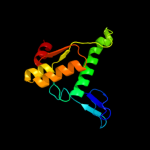
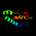
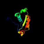
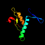
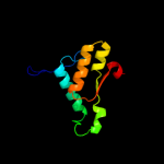
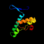
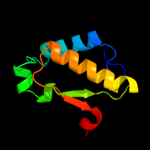
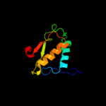
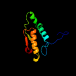
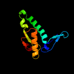
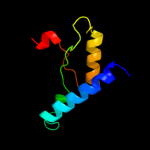
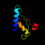
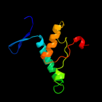
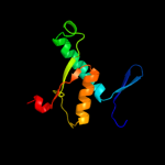
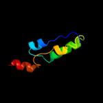
1 d1jcea1
98.8
19
Fold: Ribonuclease H-like motifSuperfamily: Actin-like ATPase domainFamily: Actin/HSP702 c3h1qB_
97.5
15
PDB header: structural proteinChain: B: PDB Molecule: ethanolamine utilization protein eutj;PDBTitle: crystal structure of ethanolamine utilization protein eutj from2 carboxydothermus hydrogenoformans
3 c1o1f4_
97.0
6
PDB header: contractile proteinChain: 4: PDB Molecule: skeletal muscle actin;PDBTitle: molecular models of averaged rigor crossbridges from2 tomograms of insect flight muscle
4 d1bupa1
96.8
11
Fold: Ribonuclease H-like motifSuperfamily: Actin-like ATPase domainFamily: Actin/HSP705 c1jcgA_
96.8
17
PDB header: structural proteinChain: A: PDB Molecule: rod shape-determining protein mreb;PDBTitle: mreb from thermotoga maritima, amppnp
6 d2e8aa1
96.8
10
Fold: Ribonuclease H-like motifSuperfamily: Actin-like ATPase domainFamily: Actin/HSP707 c3dwlB_
96.1
7
PDB header: structural proteinChain: B: PDB Molecule: actin-related protein 3;PDBTitle: crystal structure of fission yeast arp2/3 complex lacking the arp22 subunit
8 c2p9lA_
95.9
4
PDB header: structural proteinChain: A: PDB Molecule: actin-like protein 3;PDBTitle: crystal structure of bovine arp2/3 complex
9 c3qb0C_
95.6
12
PDB header: structural proteinChain: C: PDB Molecule: actin-related protein 4;PDBTitle: crystal structure of actin-related protein arp4 from s. cerevisiae2 complexed with atp
10 c2v7yA_
94.8
11
PDB header: chaperoneChain: A: PDB Molecule: chaperone protein dnak;PDBTitle: crystal structure of the molecular chaperone dnak from2 geobacillus kaustophilus hta426 in post-atp hydrolysis3 state
11 d1dkgd1
93.8
13
Fold: Ribonuclease H-like motifSuperfamily: Actin-like ATPase domainFamily: Actin/HSP7012 c2khoA_
91.8
13
PDB header: chaperoneChain: A: PDB Molecule: heat shock protein 70;PDBTitle: nmr-rdc / xray structure of e. coli hsp70 (dnak) chaperone2 (1-605) complexed with adp and substrate
13 c3d2fC_
91.1
8
PDB header: chaperoneChain: C: PDB Molecule: heat shock protein homolog sse1;PDBTitle: crystal structure of a complex of sse1p and hsp70
14 d2hf3a1
87.4
6
Fold: Ribonuclease H-like motifSuperfamily: Actin-like ATPase domainFamily: Actin/HSP7015 d2fxua1
86.7
8
Fold: Ribonuclease H-like motifSuperfamily: Actin-like ATPase domainFamily: Actin/HSP7016 d1yaga1
86.0
6
Fold: Ribonuclease H-like motifSuperfamily: Actin-like ATPase domainFamily: Actin/HSP7017 d1c0fa1
73.9
9
Fold: Ribonuclease H-like motifSuperfamily: Actin-like ATPase domainFamily: Actin/HSP7018 c2v7zA_
73.0
11
PDB header: chaperoneChain: A: PDB Molecule: heat shock cognate 71 kda protein;PDBTitle: crystal structure of the 70-kda heat shock cognate protein2 from rattus norvegicus in post-atp hydrolysis state
19 c3iucC_
64.3
13
PDB header: chaperoneChain: C: PDB Molecule: heat shock 70kda protein 5 (glucose-regulatedPDBTitle: crystal structure of the human 70kda heat shock protein 52 (bip/grp78) atpase domain in complex with adp
20 d1dn1a_
58.7
19
Fold: Sec1/munc18-like (SM) proteinsSuperfamily: Sec1/munc18-like (SM) proteinsFamily: Sec1/munc18-like (SM) proteins21 c1dkgD_
not modelled
57.9
13
PDB header: complex (hsp24/hsp70)Chain: D: PDB Molecule: molecular chaperone dnak;PDBTitle: crystal structure of the nucleotide exchange factor grpe2 bound to the atpase domain of the molecular chaperone dnak
22 c1hpmA_
not modelled
54.2
11
PDB header: hydrolase (acting on acid anhydrides)Chain: A: PDB Molecule: 44k atpase fragment (n-terminal) of 7o kd heat-PDBTitle: how potassium affects the activity of the molecular2 chaperone hsc70. ii. potassium binds specifically in the3 atpase active site
23 d1k8ka1
not modelled
49.2
5
Fold: Ribonuclease H-like motifSuperfamily: Actin-like ATPase domainFamily: Actin/HSP7024 d1epua_
not modelled
33.3
21
Fold: Sec1/munc18-like (SM) proteinsSuperfamily: Sec1/munc18-like (SM) proteinsFamily: Sec1/munc18-like (SM) proteins25 c2xheA_
not modelled
27.6
14
PDB header: exocytosisChain: A: PDB Molecule: unc18;PDBTitle: crystal structure of the unc18-syntaxin 1 complex from monosiga2 brevicollis
26 c1xofA_
not modelled
19.5
50
PDB header: de novo proteinChain: A: PDB Molecule: bbahett1;PDBTitle: heterooligomeric beta beta alpha miniprotein
27 c1bmtB_
not modelled
17.8
15
PDB header: methyltransferaseChain: B: PDB Molecule: methionine synthase;PDBTitle: how a protein binds b12: a 3.o angstrom x-ray structure of2 the b12-binding domains of methionine synthase
28 d1sqsa_
not modelled
15.9
13
Fold: Flavodoxin-likeSuperfamily: FlavoproteinsFamily: Hypothetical protein SP195129 d1mqsa_
not modelled
14.6
13
Fold: Sec1/munc18-like (SM) proteinsSuperfamily: Sec1/munc18-like (SM) proteinsFamily: Sec1/munc18-like (SM) proteins30 c2kk4A_
not modelled
14.5
18
PDB header: structural genomics, unknown functionChain: A: PDB Molecule: uncharacterized protein af_2094;PDBTitle: solution nmr structure of protein af2094 from archaeoglobus2 fulgidus. northeast structural genomics consotium (nesg)3 target gt2
31 c2pjxB_
not modelled
11.9
15
PDB header: endocytosis/exocytosisChain: B: PDB Molecule: syntaxin-binding protein 3;PDBTitle: crystal structure of the munc18c/syntaxin4 n-peptide complex
32 d1wdea_
not modelled
11.3
27
Fold: Tetrapyrrole methylaseSuperfamily: Tetrapyrrole methylaseFamily: Tetrapyrrole methylase33 d1xsza2
not modelled
10.2
7
Fold: TBP-likeSuperfamily: RalF, C-terminal domainFamily: RalF, C-terminal domain34 c2phjA_
not modelled
10.1
11
PDB header: hydrolaseChain: A: PDB Molecule: 5'-nucleotidase sure;PDBTitle: crystal structure of sure protein from aquifex aeolicus
35 d1mixa2
not modelled
9.7
19
Fold: PH domain-like barrelSuperfamily: PH domain-likeFamily: Third domain of FERM36 d1vhka2
not modelled
9.3
33
Fold: alpha/beta knotSuperfamily: alpha/beta knotFamily: YggJ C-terminal domain-like37 d3d37a2
not modelled
9.0
11
Fold: Phage tail proteinsSuperfamily: Phage tail proteinsFamily: Baseplate protein-like38 c3itqB_
not modelled
8.8
23
PDB header: oxidoreductaseChain: B: PDB Molecule: prolyl 4-hydroxylase, alpha subunit domain protein;PDBTitle: crystal structure of a prolyl 4-hydroxylase from bacillus anthracis
39 c3cq9C_
not modelled
8.7
7
PDB header: transferaseChain: C: PDB Molecule: uncharacterized protein lp_1622;PDBTitle: crystal structure of the lp_1622 protein from lactobacillus2 plantarum. northeast structural genomics consortium target3 lpr114
40 d1omwa2
not modelled
8.5
18
Fold: PH domain-like barrelSuperfamily: PH domain-likeFamily: Pleckstrin-homology domain (PH domain)41 d1gyxa_
not modelled
7.9
13
Fold: Tautomerase/MIFSuperfamily: Tautomerase/MIFFamily: 4-oxalocrotonate tautomerase-like42 c2wj8N_
not modelled
7.5
29
PDB header: rna binding protein/rnaChain: N: PDB Molecule: nucleoprotein;PDBTitle: respiratory syncitial virus ribonucleoprotein
43 c3lmmA_
not modelled
7.1
16
PDB header: structural genomics, unknown functionChain: A: PDB Molecule: uncharacterized protein;PDBTitle: crystal structure of the dip2311 protein from2 corynebacterium diphtheriae, northeast structural genomics3 consortium target cdr35
44 c3kw2A_
not modelled
6.5
26
PDB header: transferaseChain: A: PDB Molecule: probable r-rna methyltransferase;PDBTitle: crystal structure of probable rrna-methyltransferase from2 porphyromonas gingivalis
45 c1mszA_
not modelled
6.4
15
PDB header: dna binding proteinChain: A: PDB Molecule: dna-binding protein smubp-2;PDBTitle: solution structure of the r3h domain from human smubp-2
46 d1msza_
not modelled
6.4
15
Fold: IF3-likeSuperfamily: R3H domainFamily: R3H domain47 d1cqxa3
not modelled
6.1
18
Fold: Ferredoxin reductase-like, C-terminal NADP-linked domainSuperfamily: Ferredoxin reductase-like, C-terminal NADP-linked domainFamily: Flavohemoglobin, C-terminal domain48 d2dw4a1
not modelled
6.1
10
Fold: DNA/RNA-binding 3-helical bundleSuperfamily: Homeodomain-likeFamily: SWIRM domain49 c2pihA_
not modelled
6.1
17
PDB header: structural genomics, unknown functionChain: A: PDB Molecule: protein ymca;PDBTitle: crystal structure of protein ymca from bacillus subtilis,2 northeast structural genomics target sr375
50 d2piha1
not modelled
6.1
17
Fold: YheA-likeSuperfamily: YheA/YmcA-likeFamily: YmcA-like51 d2bz2a1
not modelled
5.9
12
Fold: Ferredoxin-likeSuperfamily: RNA-binding domain, RBDFamily: Canonical RBD52 d1gph11
not modelled
5.9
9
Fold: PRTase-likeSuperfamily: PRTase-likeFamily: Phosphoribosyltransferases (PRTases)53 c2rm4A_
not modelled
5.7
26
PDB header: protein bindingChain: A: PDB Molecule: cg6311-pb;PDBTitle: solution structure of the lsm domain of dm edc3 (enhancer2 of decapping 3)
54 d2nu7a1
not modelled
5.5
13
Fold: NAD(P)-binding Rossmann-fold domainsSuperfamily: NAD(P)-binding Rossmann-fold domainsFamily: CoA-binding domain55 c3bt6B_
not modelled
5.4
18
PDB header: viral proteinChain: B: PDB Molecule: influenza b hemagglutinin (ha);PDBTitle: crystal structure of influenza b virus hemagglutinin
56 c3melC_
not modelled
5.3
12
PDB header: structural genomics, unknown functionChain: C: PDB Molecule: thiamin pyrophosphokinase family protein;PDBTitle: crystal structure of thiamin pyrophosphokinase family protein from2 enterococcus faecalis, northeast structural genomics consortium3 target efr150


