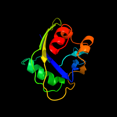
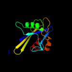
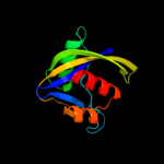
| 1 | d1puna_
|
|
 |
100.0 |
31 |
Fold:Nudix
Superfamily:Nudix
Family:MutT-like |
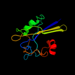
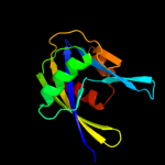
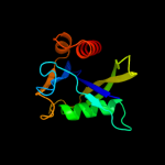
| 2 | c3gwyA_
|
|
 |
100.0 |
36 |
PDB header:hydrolase
Chain: A: PDB Molecule:putative ctp pyrophosphohydrolase;
PDBTitle: crystal structure of putative ctp pyrophosphohydrolase from2 bacteroides fragilis
|
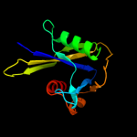
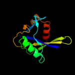
| 3 | c3ef5A_
|
|
 |
100.0 |
37 |
PDB header:hydrolase
Chain: A: PDB Molecule:probable pyrophosphohydrolase;
PDBTitle: structure of the rna pyrophosphohydrolase bdrpph in complex with dgtp
|
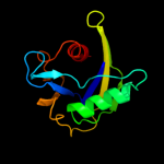
| 4 | c3hhjA_
|
|
 |
99.9 |
33 |
PDB header:hydrolase
Chain: A: PDB Molecule:mutator mutt protein;
PDBTitle: crystal structure of mutator mutt from bartonella henselae
|
| 5 | c3grnB_
|
|
 |
99.9 |
30 |
PDB header:hydrolase
Chain: B: PDB Molecule:mutt related protein;
PDBTitle: crystal structure of mutt protein from methanosarcina mazei go1
|
| 6 | c3exqA_
|
|
 |
99.9 |
18 |
PDB header:hydrolase
Chain: A: PDB Molecule:nudix family hydrolase;
PDBTitle: crystal structure of a nudix family hydrolase from2 lactobacillus brevis
|
| 7 | d1vk6a2
|
|
 |
99.9 |
24 |
Fold:Nudix
Superfamily:Nudix
Family:NADH pyrophosphatase |
| 8 | c3n77B_
|
|
 |
99.9 |
27 |
PDB header:hydrolase
Chain: B: PDB Molecule:nucleoside triphosphatase nudi;
PDBTitle: crystal structure of idp01880, putative ntp pyrophosphohydrolase of2 salmonella typhimurium lt2
|
| 9 | c2gb5B_
|
|
 |
99.9 |
24 |
PDB header:hydrolase
Chain: B: PDB Molecule:nadh pyrophosphatase;
PDBTitle: crystal structure of nadh pyrophosphatase (ec 3.6.1.22) (1790429) from2 escherichia coli k12 at 2.30 a resolution
|
| 10 | d1irya_
|
|
 |
99.9 |
19 |
Fold:Nudix
Superfamily:Nudix
Family:MutT-like |
| 11 | c3r03B_
|
|
 |
99.9 |
39 |
PDB header:hydrolase
Chain: B: PDB Molecule:nudix hydrolase;
PDBTitle: the crystal structure of nudix hydrolase from rhodospirillum rubrum
|
| 12 | c2pq1B_
|
|
 |
99.9 |
23 |
PDB header:hydrolase
Chain: B: PDB Molecule:ap4a hydrolase;
PDBTitle: crystal structure of ap4a hydrolase complexed with amp and2 atp (aq_158) from aquifex aeolicus vf5
|
| 13 | d2b06a1
|
|
 |
99.9 |
25 |
Fold:Nudix
Superfamily:Nudix
Family:MutT-like |
| 14 | c3cngC_
|
|
 |
99.9 |
22 |
PDB header:hydrolase
Chain: C: PDB Molecule:nudix hydrolase;
PDBTitle: crystal structure of nudix hydrolase from nitrosomonas europaea
|
| 15 | c3fk9B_
|
|
 |
99.9 |
25 |
PDB header:hydrolase
Chain: B: PDB Molecule:mutator mutt protein;
PDBTitle: crystal structure of mmutator mutt protein from bacillus2 halodurans
|
| 16 | d2b0va1
|
|
 |
99.9 |
24 |
Fold:Nudix
Superfamily:Nudix
Family:MutT-like |
| 17 | c1rrqA_
|
|
 |
99.9 |
17 |
PDB header:hydrolase/dna
Chain: A: PDB Molecule:muty;
PDBTitle: muty adenine glycosylase in complex with dna containing an2 a:oxog pair
|
| 18 | c3dkuB_
|
|
 |
99.9 |
25 |
PDB header:hydrolase
Chain: B: PDB Molecule:putative phosphohydrolase;
PDBTitle: crystal structure of nudix hydrolase orf153, ymfb, from2 escherichia coli k-1
|
| 19 | d1vcda1
|
|
 |
99.9 |
27 |
Fold:Nudix
Superfamily:Nudix
Family:MutT-like |
| 20 | c3h95A_
|
|
 |
99.9 |
18 |
PDB header:gene regulation
Chain: A: PDB Molecule:nucleoside diphosphate-linked moiety x motif 6;
PDBTitle: crystal structure of the nudix domain of nudt6
|
| 21 | d1rrqa2 |
|
not modelled |
99.9 |
16 |
Fold:Nudix
Superfamily:Nudix
Family:MutY C-terminal domain-like |
| 22 | c3o8sA_ |
|
not modelled |
99.9 |
25 |
PDB header:hydrolase
Chain: A: PDB Molecule:adp-ribose pyrophosphatase;
PDBTitle: crystal structure of an adp-ribose pyrophosphatase (ssu98_1448) from2 streptococcus suis 89-1591 at 2.27 a resolution
|
| 23 | d2fkba1 |
|
not modelled |
99.9 |
23 |
Fold:Nudix
Superfamily:Nudix
Family:IPP isomerase-like |
| 24 | c3gz8C_ |
|
not modelled |
99.9 |
22 |
PDB header:dna binding protein
Chain: C: PDB Molecule:mutt/nudix family protein;
PDBTitle: cocrystal structure of nudix domain of shewanella oneidensis2 nrtr complexed with adp ribose
|
| 25 | c3q4iA_ |
|
not modelled |
99.9 |
18 |
PDB header:hydrolase
Chain: A: PDB Molecule:phosphohydrolase (mutt/nudix family protein);
PDBTitle: crystal structure of cdp-chase in complex with gd3+
|
| 26 | c3gg6A_ |
|
not modelled |
99.9 |
21 |
PDB header:hydrolase
Chain: A: PDB Molecule:nucleoside diphosphate-linked moiety x motif 18;
PDBTitle: crystal structure of the nudix domain of human nudt18
|
| 27 | d1x51a1 |
|
not modelled |
99.9 |
17 |
Fold:Nudix
Superfamily:Nudix
Family:MutY C-terminal domain-like |
| 28 | d2azwa1 |
|
not modelled |
99.9 |
19 |
Fold:Nudix
Superfamily:Nudix
Family:MutT-like |
| 29 | c3fjyB_ |
|
not modelled |
99.9 |
26 |
PDB header:hydrolase
Chain: B: PDB Molecule:probable mutt1 protein;
PDBTitle: crystal structure of a probable mutt1 protein from bifidobacterium2 adolescentis
|
| 30 | d1k2ea_ |
|
not modelled |
99.9 |
19 |
Fold:Nudix
Superfamily:Nudix
Family:MutT-like |
| 31 | c2yyhC_ |
|
not modelled |
99.9 |
19 |
PDB header:hydrolase
Chain: C: PDB Molecule:8-oxo-dgtpase domain;
PDBTitle: crystal structure of nudix family protein from aquifex aeolicus
|
| 32 | c2kdvA_ |
|
not modelled |
99.9 |
23 |
PDB header:hydrolase
Chain: A: PDB Molecule:rna pyrophosphohydrolase;
PDBTitle: solution structure of rna pyrophosphohydrolase rpph from2 escherichia coli
|
| 33 | d1ryaa_ |
|
not modelled |
99.9 |
18 |
Fold:Nudix
Superfamily:Nudix
Family:GDP-mannose mannosyl hydrolase NudD |
| 34 | c2qjoB_ |
|
not modelled |
99.9 |
18 |
PDB header:transferase, hydrolase
Chain: B: PDB Molecule:bifunctional nmn adenylyltransferase/nudix hydrolase;
PDBTitle: crystal structure of a bifunctional nmn adenylyltransferase/adp ribose2 pyrophosphatase (nadm) complexed with adprp and nad from3 synechocystis sp.
|
| 35 | c2o1cB_ |
|
not modelled |
99.9 |
23 |
PDB header:hydrolase
Chain: B: PDB Molecule:datp pyrophosphohydrolase;
PDBTitle: structure of the e. coli dihydroneopterin triphosphate2 pyrophosphohydrolase
|
| 36 | d2fb1a2 |
|
not modelled |
99.9 |
18 |
Fold:Nudix
Superfamily:Nudix
Family:BT0354 N-terminal domain-like |
| 37 | d1ktga_ |
|
not modelled |
99.9 |
27 |
Fold:Nudix
Superfamily:Nudix
Family:MutT-like |
| 38 | d1sjya_ |
|
not modelled |
99.8 |
24 |
Fold:Nudix
Superfamily:Nudix
Family:MutT-like |
| 39 | d2fvva1 |
|
not modelled |
99.8 |
20 |
Fold:Nudix
Superfamily:Nudix
Family:MutT-like |
| 40 | c2fvvA_ |
|
not modelled |
99.8 |
20 |
PDB header:hydrolase
Chain: A: PDB Molecule:diphosphoinositol polyphosphate phosphohydrolase
PDBTitle: human diphosphoinositol polyphosphate phosphohydrolase 1
|
| 41 | c3rh7A_ |
|
not modelled |
99.8 |
18 |
PDB header:oxidoreductase
Chain: A: PDB Molecule:hypothetical oxidoreductase;
PDBTitle: crystal structure of a hypothetical oxidoreductase (sma0793) from2 sinorhizobium meliloti 1021 at 3.00 a resolution
|
| 42 | c3i9xA_ |
|
not modelled |
99.8 |
22 |
PDB header:hydrolase
Chain: A: PDB Molecule:mutt/nudix family protein;
PDBTitle: crystal structure of a mutt/nudix family protein from listeria innocua
|
| 43 | d1xsba_ |
|
not modelled |
99.8 |
25 |
Fold:Nudix
Superfamily:Nudix
Family:MutT-like |
| 44 | c2r5wA_ |
|
not modelled |
99.8 |
23 |
PDB header:hydrolase, transferase
Chain: A: PDB Molecule:nicotinamide-nucleotide adenylyltransferase;
PDBTitle: crystal structure of a bifunctional nmn2 adenylyltransferase/adp ribose pyrophosphatase from3 francisella tularensis
|
| 45 | c2pqvA_ |
|
not modelled |
99.8 |
24 |
PDB header:structural genomics, unknown function
Chain: A: PDB Molecule:mutt/nudix family protein;
PDBTitle: crystal structure of mutt/nudix family protein from streptococcus2 pneumoniae
|
| 46 | c3gz6A_ |
|
not modelled |
99.8 |
22 |
PDB header:dna binding protein/dna
Chain: A: PDB Molecule:mutt/nudix family protein;
PDBTitle: crystal structure of shewanella oneidensis nrtr complexed2 with a 27mer dna
|
| 47 | d1vhza_ |
|
not modelled |
99.8 |
19 |
Fold:Nudix
Superfamily:Nudix
Family:MutT-like |
| 48 | c2jvbA_ |
|
not modelled |
99.8 |
22 |
PDB header:hydrolase
Chain: A: PDB Molecule:mrna-decapping enzyme subunit 2;
PDBTitle: solution structure of catalytic domain of ydcp2
|
| 49 | c2fb1A_ |
|
not modelled |
99.8 |
18 |
PDB header:structural genomics, unknown function
Chain: A: PDB Molecule:conserved hypothetical protein;
PDBTitle: crystal structure of protein bt0354 from bacteroides thetaiotaomicron
|
| 50 | c3f6aA_ |
|
not modelled |
99.8 |
21 |
PDB header:hydrolase
Chain: A: PDB Molecule:hydrolase, nudix family;
PDBTitle: crystal structure of a hydrolase, nudix family from2 clostridium perfringens
|
| 51 | c3fcmA_ |
|
not modelled |
99.8 |
18 |
PDB header:hydrolase
Chain: A: PDB Molecule:hydrolase, nudix family;
PDBTitle: crystal structure of a nudix hydrolase from clostridium2 perfringens
|
| 52 | d1jkna_ |
|
not modelled |
99.8 |
20 |
Fold:Nudix
Superfamily:Nudix
Family:MutT-like |
| 53 | c3sonB_ |
|
not modelled |
99.8 |
20 |
PDB header:hydrolase
Chain: B: PDB Molecule:hypothetical nudix hydrolase;
PDBTitle: crystal structure of a hypothetical nudix hydrolase (lmof2365_2679)2 from listeria monocytogenes (atcc 19115) at 1.70 a resolution
|
| 54 | c2w4eA_ |
|
not modelled |
99.8 |
21 |
PDB header:hydrolase
Chain: A: PDB Molecule:mutt/nudix family protein;
PDBTitle: structure of an n-terminally truncated nudix hydrolase2 dr2204 from deinococcus radiodurans
|
| 55 | c3id9B_ |
|
not modelled |
99.8 |
21 |
PDB header:hydrolase
Chain: B: PDB Molecule:mutt/nudix family protein;
PDBTitle: crystal structure of a mutt/nudix family protein from2 bacillus thuringiensis
|
| 56 | c3f13A_ |
|
not modelled |
99.8 |
23 |
PDB header:hydrolase
Chain: A: PDB Molecule:putative nudix hydrolase family member;
PDBTitle: crystal structure of putative nudix hydrolase family member2 from chromobacterium violaceum
|
| 57 | d1ppva_ |
|
not modelled |
99.8 |
20 |
Fold:Nudix
Superfamily:Nudix
Family:IPP isomerase-like |
| 58 | d1hzta_ |
|
not modelled |
99.8 |
20 |
Fold:Nudix
Superfamily:Nudix
Family:IPP isomerase-like |
| 59 | c2yvoA_ |
|
not modelled |
99.8 |
24 |
PDB header:hydrolase
Chain: A: PDB Molecule:mutt/nudix family protein;
PDBTitle: crystal structure of ndx2 in complex with mg2+ and amp from thermus2 thermophilus hb8
|
| 60 | d2a6ta2 |
|
not modelled |
99.8 |
17 |
Fold:Nudix
Superfamily:Nudix
Family:mRNA decapping enzyme-like |
| 61 | c3edsA_ |
|
not modelled |
99.8 |
24 |
PDB header:hydrolase
Chain: A: PDB Molecule:mutt/nudix family protein;
PDBTitle: crystal structure of a mut/nudix family protein from bacillus2 thuringiensis
|
| 62 | d1v8ya_ |
|
not modelled |
99.8 |
28 |
Fold:Nudix
Superfamily:Nudix
Family:MutT-like |
| 63 | d1nqza_ |
|
not modelled |
99.8 |
23 |
Fold:Nudix
Superfamily:Nudix
Family:MutT-like |
| 64 | c2qkmF_ |
|
not modelled |
99.8 |
20 |
PDB header:hydrolase
Chain: F: PDB Molecule:spac19a8.12 protein;
PDBTitle: the crystal structure of fission yeast mrna decapping enzyme dcp1-dcp22 complex
|
| 65 | d1g0sa_ |
|
not modelled |
99.8 |
22 |
Fold:Nudix
Superfamily:Nudix
Family:MutT-like |
| 66 | d2fmla2 |
|
not modelled |
99.8 |
19 |
Fold:Nudix
Superfamily:Nudix
Family:BT0354 N-terminal domain-like |
| 67 | c3bm4B_ |
|
not modelled |
99.8 |
19 |
PDB header:hydrolase
Chain: B: PDB Molecule:adp-sugar pyrophosphatase;
PDBTitle: crystal structure of human adp-ribose pyrophosphatase nudt52 in complex with magnesium and ampcpr
|
| 68 | d1mqea_ |
|
not modelled |
99.8 |
20 |
Fold:Nudix
Superfamily:Nudix
Family:MutT-like |
| 69 | d2o5fa1 |
|
not modelled |
99.7 |
23 |
Fold:Nudix
Superfamily:Nudix
Family:IPP isomerase-like |
| 70 | c2fmlB_ |
|
not modelled |
99.7 |
19 |
PDB header:structural genomics, unknown function
Chain: B: PDB Molecule:mutt/nudix family protein;
PDBTitle: crystal structure of mutt/nudix family protein from enterococcus2 faecalis
|
| 71 | c3e57A_ |
|
not modelled |
99.7 |
14 |
PDB header:structural genomics, unknown function
Chain: A: PDB Molecule:uncharacterized protein tm1382;
PDBTitle: crystal structure of tm1382, a putative nudix hydrolase
|
| 72 | c2i6kA_ |
|
not modelled |
99.7 |
19 |
PDB header:isomerase
Chain: A: PDB Molecule:isopentenyl-diphosphate delta-isomerase 1;
PDBTitle: crystal structure of human type i ipp isomerase complexed2 with a substrate analog
|
| 73 | c2pnyA_ |
|
not modelled |
99.7 |
18 |
PDB header:isomerase
Chain: A: PDB Molecule:isopentenyl-diphosphate delta-isomerase 2;
PDBTitle: structure of human isopentenyl-diphosphate delta-isomerase 2
|
| 74 | d1viua_ |
|
not modelled |
99.7 |
16 |
Fold:Nudix
Superfamily:Nudix
Family:MutT-like |
| 75 | c3q91D_ |
|
not modelled |
99.6 |
16 |
PDB header:hydrolase
Chain: D: PDB Molecule:uridine diphosphate glucose pyrophosphatase;
PDBTitle: crystal structure of human uridine diphosphate glucose pyrophosphatase2 (nudt14)
|
| 76 | d1q33a_ |
|
not modelled |
99.6 |
21 |
Fold:Nudix
Superfamily:Nudix
Family:MutT-like |
| 77 | c3dupB_ |
|
not modelled |
99.5 |
15 |
PDB header:hydrolase
Chain: B: PDB Molecule:mutt/nudix family protein;
PDBTitle: crystal structure of mutt/nudix family hydrolase from rhodospirillum2 rubrum atcc 11170
|
| 78 | c3qsjA_ |
|
not modelled |
99.4 |
27 |
PDB header:hydrolase
Chain: A: PDB Molecule:nudix hydrolase;
PDBTitle: crystal structure of nudix hydrolase from alicyclobacillus2 acidocaldarius
|
| 79 | c2j8qB_ |
|
not modelled |
99.4 |
21 |
PDB header:nuclear protein
Chain: B: PDB Molecule:cleavage and polyadenylation specificity factor 5;
PDBTitle: crystal structure of human cleavage and polyadenylation2 specificity factor 5 (cpsf5) in complex with a sulphate3 ion.
|
| 80 | d1u20a1 |
|
not modelled |
99.4 |
21 |
Fold:Nudix
Superfamily:Nudix
Family:MutT-like |
| 81 | c3couA_ |
|
not modelled |
98.7 |
32 |
PDB header:hydrolase
Chain: A: PDB Molecule:nucleoside diphosphate-linked moiety x motif 16;
PDBTitle: crystal structure of human nudix motif 16 (nudt16)
|
| 82 | c3kvhA_ |
|
not modelled |
97.9 |
27 |
PDB header:rna binding protein
Chain: A: PDB Molecule:protein syndesmos;
PDBTitle: crystal structure of human protein syndesmos (nudt16-like protein)
|
| 83 | c3p5tE_ |
|
not modelled |
97.0 |
22 |
PDB header:rna binding protein
Chain: E: PDB Molecule:cleavage and polyadenylation specificity factor subunit 5;
PDBTitle: cfim25-cfim68 complex
|
| 84 | c2vldA_ |
|
not modelled |
16.0 |
31 |
PDB header:hydrolase
Chain: A: PDB Molecule:upf0286 protein pyrab01260;
PDBTitle: crystal structure of a repair endonuclease from pyrococcus2 abyssi
|
| 85 | c3enoB_ |
|
not modelled |
15.2 |
19 |
PDB header:hydrolase/unknown function
Chain: B: PDB Molecule:putative o-sialoglycoprotein endopeptidase;
PDBTitle: crystal structure of pyrococcus furiosus pcc1 in complex2 with thermoplasma acidophilum kae1
|
| 86 | c1zwvA_ |
|
not modelled |
12.5 |
21 |
PDB header:transferase
Chain: A: PDB Molecule:lipoamide acyltransferase component of branched-
PDBTitle: solution structure of the subunit binding domain (hbsbd) of2 the human mitochondrial branched-chain alpha-ketoacid3 dehydrogenase
|
| 87 | c3fpvC_ |
|
not modelled |
12.3 |
27 |
PDB header:heme binding protein
Chain: C: PDB Molecule:extracellular haem-binding protein;
PDBTitle: crystal structure of hbps
|
| 88 | d2a2la1 |
|
not modelled |
9.5 |
36 |
Fold:Profilin-like
Superfamily:GlcG-like
Family:GlcG-like |
| 89 | c2ivoC_ |
|
not modelled |
8.2 |
16 |
PDB header:hydrolase
Chain: C: PDB Molecule:up1;
PDBTitle: structure of up1 protein
|
| 90 | d1nc7a_ |
|
not modelled |
6.8 |
27 |
Fold:Hypothetical protein TM1070
Superfamily:Hypothetical protein TM1070
Family:Hypothetical protein TM1070 |
| 91 | d2dexx3 |
|
not modelled |
5.7 |
26 |
Fold:Pentein, beta/alpha-propeller
Superfamily:Pentein
Family:Peptidylarginine deiminase Pad4, catalytic C-terminal domain |
| 92 | c3kngA_ |
|
not modelled |
5.5 |
22 |
PDB header:oxidoreductase
Chain: A: PDB Molecule:snoab;
PDBTitle: crystal structure of snoab, a cofactor-independent oxygenase2 from streptomyces nogalater, determined to 1.9 resolution
|
| 93 | c2eq8C_ |
|
not modelled |
5.4 |
35 |
PDB header:oxidoreductase
Chain: C: PDB Molecule:pyruvate dehydrogenase complex, dihydrolipoamide
PDBTitle: crystal structure of lipoamide dehydrogenase from thermus thermophilus2 hb8 with psbdp
|