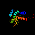
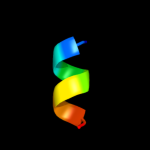
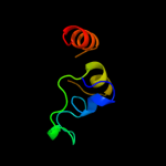
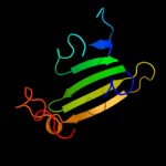
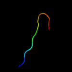
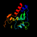
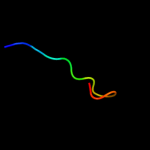
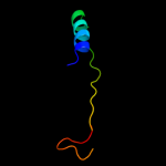
1 d2prva1
83.6
10
Fold: SMI1/KNR4-likeSuperfamily: SMI1/KNR4-likeFamily: SMI1/KNR4-like2 c3dsmA_
64.3
24
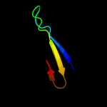
PDB header: structural genomics, unknown functionChain: A: PDB Molecule: uncharacterized protein bacuni_02894;PDBTitle: crystal structure of the surface layer protein bacuni_02894 from2 bacteroides uniformis, northeast structural genomics consortium3 target btr193d.
3 d2paga1
49.3
23
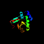
Fold: SMI1/KNR4-likeSuperfamily: SMI1/KNR4-likeFamily: SMI1/KNR4-like4 c3d5pB_
23.1
10
PDB header: gene regulationChain: B: PDB Molecule: putative glucan synthesis regulator of smi1/knr4 family;PDBTitle: crystal structure of a putative glucan synthesis regulator of2 smi1/knr4 family (bf1740) from bacteroides fragilis nctc 9343 at 1.453 a resolution
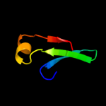
5 c1jrjA_
23.1
33
PDB header: hormone/growth factorChain: A: PDB Molecule: exendin-4;PDBTitle: solution structure of exendin-4 in 30-vol% trifluoroethanol
6 c1d0rA_
21.7
40
PDB header: hormone/growth factorChain: A: PDB Molecule: glucagon-like peptide-1-(7-36)-amide;PDBTitle: solution structure of glucagon-like peptide-1-(7-36)-amide2 in trifluoroethanol/water
7 d2hy5c1
17.7
27
Fold: DsrEFH-likeSuperfamily: DsrEFH-likeFamily: DsrH-like8 c2vt8B_
15.4
20
PDB header: hydrolase inhibitorChain: B: PDB Molecule: proteasome inhibitor pi31 subunit;PDBTitle: structure of a conserved dimerisation domain within fbox72 and pi31
9 c2dk7A_
14.8
25
PDB header: transcriptionChain: A: PDB Molecule: transcription elongation regulator 1;PDBTitle: solution structure of ww domain in transcription elongation2 regulator 1
10 c3mbqC_
14.3
22
PDB header: hydrolaseChain: C: PDB Molecule: deoxyuridine 5'-triphosphate nucleotidohydrolase;PDBTitle: crystal structure of deoxyuridine 5-triphosphate nucleotidohydrolase2 from brucella melitensis, orthorhombic crystal form
11 c1r0lD_
13.9
28
PDB header: oxidoreductaseChain: D: PDB Molecule: 1-deoxy-d-xylulose 5-phosphate reductoisomerase;PDBTitle: 1-deoxy-d-xylulose 5-phosphate reductoisomerase from2 zymomonas mobilis in complex with nadph
12 c1nauA_
13.6
38
PDB header: hormone/growth factorChain: A: PDB Molecule: glucagon;PDBTitle: nmr solution structure of the glucagon antagonist [deshis1,2 desphe6, glu9]glucagon amide in the presence of3 perdeuterated dodecylphosphocholine micelles
13 d2rm0w1
13.6
42
Fold: WW domain-likeSuperfamily: WW domainFamily: WW domain14 c1zr7A_
13.1
33
PDB header: signaling proteinChain: A: PDB Molecule: huntingtin-interacting protein hypa/fbp11;PDBTitle: solution structure of the first ww domain of fbp11
15 d2icga1
13.0
13
Fold: SMI1/KNR4-likeSuperfamily: SMI1/KNR4-likeFamily: SMI1/KNR4-like16 c2uuvC_
12.9
33
PDB header: transferaseChain: C: PDB Molecule: alkyldihydroxyacetonephosphate synthase;PDBTitle: alkyldihydroxyacetonephosphate synthase in p1
17 d1ywia1
12.5
33
Fold: WW domain-likeSuperfamily: WW domainFamily: WW domain18 c1ywjA_
12.4
33
PDB header: structural proteinChain: A: PDB Molecule: formin-binding protein 3;PDBTitle: structure of the fbp11ww1 domain
19 c3iuzA_
12.2
28
PDB header: lyaseChain: A: PDB Molecule: putative glyoxalase superfamily protein;PDBTitle: crystal structure of putative glyoxalase superfamily protein2 (yp_299723.1) from ralstonia eutropha jmp134 at 1.90 a resolution
20 d1zxoa1
12.1
11
Fold: Ribonuclease H-like motifSuperfamily: Actin-like ATPase domainFamily: BadF/BadG/BcrA/BcrD-like21 d1zr7a1
not modelled
11.5
33
Fold: WW domain-likeSuperfamily: WW domainFamily: WW domain22 c1bh0A_
not modelled
11.3
50
PDB header: synthetic hormoneChain: A: PDB Molecule: glucagon;PDBTitle: structure of a glucagon analog
23 d1o6wa2
not modelled
11.2
50
Fold: WW domain-likeSuperfamily: WW domainFamily: WW domain24 d1qw9a2
not modelled
11.1
47
Fold: TIM beta/alpha-barrelSuperfamily: (Trans)glycosidasesFamily: beta-glycanases25 c3ks8D_
not modelled
11.1
16
PDB header: viral protein/rnaChain: D: PDB Molecule: polymerase cofactor vp35;PDBTitle: crystal structure of reston ebolavirus vp35 rna binding2 domain in complex with 18bp dsrna
26 d1zbsa2
not modelled
10.9
18
Fold: Ribonuclease H-like motifSuperfamily: Actin-like ATPase domainFamily: BadF/BadG/BcrA/BcrD-like27 c3fkeB_
not modelled
10.7
16
PDB header: rna binding proteinChain: B: PDB Molecule: polymerase cofactor vp35;PDBTitle: structure of the ebola vp35 interferon inhibitory domain
28 c2eghA_
not modelled
10.7
24
PDB header: oxidoreductaseChain: A: PDB Molecule: 1-deoxy-d-xylulose 5-phosphate reductoisomerase;PDBTitle: crystal structure of 1-deoxy-d-xylulose 5-phosphate reductoisomerase2 complexed with a magnesium ion, nadph and fosmidomycin
29 c3mcaA_
not modelled
10.7
22
PDB header: translation regulation/hydrolaseChain: A: PDB Molecule: elongation factor 1 alpha-like protein;PDBTitle: structure of the dom34-hbs1 complex and implications for its role in2 no-go decay
30 d1o6wa1
not modelled
10.5
27
Fold: WW domain-likeSuperfamily: WW domainFamily: WW domain31 c3a14B_
not modelled
10.4
31
PDB header: oxidoreductaseChain: B: PDB Molecule: 1-deoxy-d-xylulose 5-phosphate reductoisomerase;PDBTitle: crystal structure of dxr from thermotoga maritima, in complex with2 nadph
32 c3e53A_
not modelled
10.2
24
PDB header: ligaseChain: A: PDB Molecule: fatty-acid-coa ligase fadd28;PDBTitle: crystal structure of n-terminal domain of a fatty acyl amp2 ligase faal28 from mycobacterium tuberculosis
33 d1xdpa3
not modelled
9.8
18
Fold: Phospholipase D/nucleaseSuperfamily: Phospholipase D/nucleaseFamily: Polyphosphate kinase C-terminal domain34 d1iloa_
not modelled
9.8
23
Fold: Thioredoxin foldSuperfamily: Thioredoxin-likeFamily: Thioltransferase35 c2zxqA_
not modelled
9.7
21
PDB header: hydrolaseChain: A: PDB Molecule: endo-alpha-n-acetylgalactosaminidase;PDBTitle: crystal structure of endo-alpha-n-acetylgalactosaminidase2 from bifidobacterium longum (engbf)
36 c3gafF_
not modelled
9.6
22
PDB header: oxidoreductaseChain: F: PDB Molecule: 7-alpha-hydroxysteroid dehydrogenase;PDBTitle: 2.2a crystal structure of 7-alpha-hydroxysteroid2 dehydrogenase from brucella melitensis
37 d2c7fa2
not modelled
9.3
40
Fold: TIM beta/alpha-barrelSuperfamily: (Trans)glycosidasesFamily: beta-glycanases38 d1r0ka3
not modelled
9.0
30
Fold: FwdE/GAPDH domain-likeSuperfamily: Glyceraldehyde-3-phosphate dehydrogenase-like, C-terminal domainFamily: Dihydrodipicolinate reductase-like39 d2dk1a1
not modelled
8.8
25
Fold: WW domain-likeSuperfamily: WW domainFamily: WW domain40 c2qkmG_
not modelled
8.4
22
PDB header: hydrolaseChain: G: PDB Molecule: spbc3b9.21 protein;PDBTitle: the crystal structure of fission yeast mrna decapping enzyme dcp1-dcp22 complex
41 d1q67a_
not modelled
8.3
22
Fold: PH domain-like barrelSuperfamily: PH domain-likeFamily: Dcp142 c2dk1A_
not modelled
8.0
25
PDB header: gene regulationChain: A: PDB Molecule: ww domain-binding protein 4;PDBTitle: solution structure of ww domain in ww domain binding2 protein 4 (wbp-4)
43 c2w4lC_
not modelled
8.0
21
PDB header: hydrolaseChain: C: PDB Molecule: deoxycytidylate deaminase;PDBTitle: human dcmp deaminase
44 c1q67B_
not modelled
7.9
22
PDB header: transcriptionChain: B: PDB Molecule: decapping protein involved in mrna degradation-PDBTitle: crystal structure of dcp1p
45 c2y3rC_
not modelled
7.9
11
PDB header: oxidoreductaseChain: C: PDB Molecule: taml;PDBTitle: structure of the tirandamycin-bound fad-dependent2 tirandamycin oxidase taml in p21 space group
46 c3muwE_
not modelled
7.9
67
PDB header: virusChain: E: PDB Molecule: structural polyprotein;PDBTitle: pseudo-atomic structure of the e2-e1 protein shell in sindbis virus
47 c1bmxA_
not modelled
7.9
44
PDB header: viral proteinChain: A: PDB Molecule: human immunodeficiency virus type 1 capsid;PDBTitle: hiv-1 capsid protein major homology region peptide analog,2 nmr, 8 structures
48 c1z8yE_
not modelled
7.8
67
PDB header: virusChain: E: PDB Molecule: spike glycoprotein e1;PDBTitle: mapping the e2 glycoprotein of alphaviruses
49 c1ld4O_
not modelled
7.8
67
PDB header: virusChain: O: PDB Molecule: spike glycoprotein e1;PDBTitle: placement of the structural proteins in sindbis virus
50 c2alaA_
not modelled
7.4
67
PDB header: viral proteinChain: A: PDB Molecule: structural polyprotein (p130);PDBTitle: crystal structure of the semliki forest virus envelope protein e1 in2 its monomeric conformation.
51 d2alaa2
not modelled
7.4
67
Fold: Viral glycoprotein, central and dimerisation domainsSuperfamily: Viral glycoprotein, central and dimerisation domainsFamily: Viral glycoprotein, central and dimerisation domains52 c1t5qA_
not modelled
7.3
38
PDB header: hormone/growth factorChain: A: PDB Molecule: gastric inhibitory polypeptide;PDBTitle: solution structure of gip(1-30)amide in tfe/water
53 c3n42F_
not modelled
7.3
67
PDB header: viral proteinChain: F: PDB Molecule: e1 envelope glycoprotein;PDBTitle: crystal structures of the mature envelope glycoprotein complex (furin2 cleavage) of chikungunya virus.
54 c2xfcD_
not modelled
7.3
67
PDB header: virusChain: D: PDB Molecule: e1 envelope glycoprotein;PDBTitle: the chikungunya e1 e2 envelope glycoprotein complex fit into2 the semliki forest virus cryo-em map
55 c2xfbF_
not modelled
7.2
67
PDB header: virusChain: F: PDB Molecule: e1 envelope glycoprotein;PDBTitle: the chikungunya e1 e2 envelope glycoprotein complex fit into2 the sindbis virus cryo-em map
56 d1q0qa3
not modelled
7.2
24
Fold: FwdE/GAPDH domain-likeSuperfamily: Glyceraldehyde-3-phosphate dehydrogenase-like, C-terminal domainFamily: Dihydrodipicolinate reductase-like57 c3tu8A_
not modelled
7.2
35
PDB header: unknown functionChain: A: PDB Molecule: burkholderia lethal factor 1 (blf1);PDBTitle: crystal structure of the burkholderia lethal factor 1 (blf1)
58 c3j0cG_
not modelled
7.1
67
PDB header: virusChain: G: PDB Molecule: e1 envelope glycoprotein;PDBTitle: models of e1, e2 and cp of venezuelan equine encephalitis virus tc-832 strain restrained by a near atomic resolution cryo-em map
59 d1ccwb_
not modelled
6.7
37
Fold: TIM beta/alpha-barrelSuperfamily: Cobalamin (vitamin B12)-dependent enzymesFamily: Glutamate mutase, large subunit60 c3mzlH_
not modelled
6.6
22
PDB header: protein transportChain: H: PDB Molecule: protein transport protein sec31;PDBTitle: sec13/sec31 edge element, loop deletion mutant
61 c2xhfA_
not modelled
6.6
15
PDB header: oxidoreductaseChain: A: PDB Molecule: peroxiredoxin 5;PDBTitle: crystal structure of peroxiredoxin 5 from alvinella pompejana
62 c3muuA_
not modelled
6.5
67
PDB header: viral proteinChain: A: PDB Molecule: structural polyprotein;PDBTitle: crystal structure of the sindbis virus e2-e1 heterodimer at low ph
63 c6rlxB_
not modelled
6.5
58
PDB header: hormone(muscle relaxant)Chain: B: PDB Molecule: relaxin, b-chain;PDBTitle: x-ray structure of human relaxin at 1.5 angstroms. comparison to2 insulin and implications for receptor binding determinants
64 d3er7a1
not modelled
6.5
24
Fold: Cystatin-likeSuperfamily: NTF2-likeFamily: Exig0174-like65 c2hvwC_
not modelled
6.5
29
PDB header: hydrolaseChain: C: PDB Molecule: deoxycytidylate deaminase;PDBTitle: crystal structure of dcmp deaminase from streptococcus2 mutans
66 d1y9ia_
not modelled
6.3
19
Fold: YutG-likeSuperfamily: YutG-likeFamily: YutG-like67 d1yt3a2
not modelled
6.2
13
Fold: SAM domain-likeSuperfamily: HRDC-likeFamily: RNase D C-terminal domains68 d1cqqa_
not modelled
6.2
36
Fold: Trypsin-like serine proteasesSuperfamily: Trypsin-like serine proteasesFamily: Viral cysteine protease of trypsin fold69 d1twfb_
not modelled
6.1
50
Fold: beta and beta-prime subunits of DNA dependent RNA-polymeraseSuperfamily: beta and beta-prime subunits of DNA dependent RNA-polymeraseFamily: RNA-polymerase beta70 c3eukL_
not modelled
5.9
14
PDB header: cell cycleChain: L: PDB Molecule: chromosome partition protein muke;PDBTitle: crystal structure of muke-mukf(residues 292-443)-mukb(head2 domain)-atpgammas complex, asymmetric dimer
71 d1vq2a_
not modelled
5.6
33
Fold: Cytidine deaminase-likeSuperfamily: Cytidine deaminase-likeFamily: Deoxycytidylate deaminase-like72 c2l5aA_
not modelled
5.6
20
PDB header: nuclear proteinChain: A: PDB Molecule: histone h3-like centromeric protein cse4, protein scm3,PDBTitle: structural basis for recognition of centromere specific histone h32 variant by nonhistone scm3
73 d2bsya1
not modelled
5.5
25
Fold: beta-clipSuperfamily: dUTPase-likeFamily: dUTPase-like74 c2kruA_
not modelled
5.3
21
PDB header: oxidoreductaseChain: A: PDB Molecule: light-independent protochlorophyllide reductasePDBTitle: solution nmr structure of the pcp_red domain of light-2 independent protochlorophyllide reductase subunit b from3 chlorobium tepidum. northeast structural genomics4 consortium target ctr69a (casp target)
75 c2obuA_
not modelled
5.2
38
PDB header: hormone/growth factorChain: A: PDB Molecule: gastric inhibitory polypeptide;PDBTitle: solution structure of gip in tfe/water
76 d2arca_
not modelled
5.1
15
Fold: Double-stranded beta-helixSuperfamily: Regulatory protein AraCFamily: Regulatory protein AraC77 c3fqgA_
not modelled
5.1
22
PDB header: protein bindingChain: A: PDB Molecule: protein din1;PDBTitle: crystal structure of the s. pombe rai1
78 d2a8na1
not modelled
5.1
24
Fold: Cytidine deaminase-likeSuperfamily: Cytidine deaminase-likeFamily: Deoxycytidylate deaminase-like79 d1rfza_
not modelled
5.1
42
Fold: YutG-likeSuperfamily: YutG-likeFamily: YutG-like80 c1zxoB_
not modelled
5.1
16
PDB header: unknown functionChain: B: PDB Molecule: conserved hypothetical protein q8a1p1;PDBTitle: x-ray crystal structure of protein q8a1p1 from bacteroides2 thetaiotaomicron. northeast structural genomics consortium3 target btr25.


