| 1 |
|
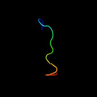
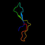
PDB 1zup chain A domain 1
Region: 65 - 80
Aligned: 16
Modelled: 16
Confidence: 51.0%
Identity: 38%
Fold: Ribonuclease H-like motif
Superfamily: Ribonuclease H-like
Family: TM1739-like
Phyre2
| 2 |
|
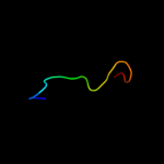
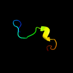
PDB 2jln chain A
Region: 51 - 68
Aligned: 18
Modelled: 18
Confidence: 38.1%
Identity: 28%
PDB header:membrane protein
Chain: A: PDB Molecule:mhp1;
PDBTitle: structure of mhp1, a nucleobase-cation-symport-1 family2 transporter
Phyre2
| 3 |
|
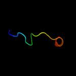
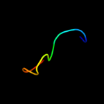
PDB 3mcu chain F
Region: 28 - 66
Aligned: 39
Modelled: 39
Confidence: 14.6%
Identity: 26%
PDB header:oxidoreductase
Chain: F: PDB Molecule:dipicolinate synthase, b chain;
PDBTitle: crystal structure of the dipicolinate synthase chain b from2 bacillus cereus. northeast structural genomics consortium3 target bcr215.
Phyre2
| 4 |
|
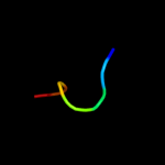
PDB 1zc1 chain A
Region: 21 - 70
Aligned: 44
Modelled: 50
Confidence: 11.9%
Identity: 30%
PDB header:protein turnover
Chain: A: PDB Molecule:ubiquitin fusion degradation protein 1;
PDBTitle: ufd1 exhibits the aaa-atpase fold with two distinct2 ubiquitin interaction sites
Phyre2
| 5 |
|
PDB 2yuj chain A
Region: 21 - 70
Aligned: 44
Modelled: 50
Confidence: 10.9%
Identity: 36%
PDB header:protein binding
Chain: A: PDB Molecule:ubiquitin fusion degradation 1-like;
PDBTitle: solution structure of human ubiquitin fusion degradation2 protein 1 homolog ufd1
Phyre2
| 6 |
|
PDB 3pvc chain A
Region: 53 - 87
Aligned: 35
Modelled: 35
Confidence: 8.9%
Identity: 26%
PDB header:oxidoreductase, transferase
Chain: A: PDB Molecule:trna 5-methylaminomethyl-2-thiouridine biosynthesis
PDBTitle: crystal structure of apo mnmc from yersinia pestis
Phyre2
| 7 |
|
PDB 3lqk chain A
Region: 28 - 62
Aligned: 35
Modelled: 35
Confidence: 8.9%
Identity: 29%
PDB header:oxidoreductase
Chain: A: PDB Molecule:dipicolinate synthase subunit b;
PDBTitle: crystal structure of dipicolinate synthase subunit b from bacillus2 halodurans c
Phyre2
| 8 |
|
PDB 2l14 chain B
Region: 40 - 61
Aligned: 22
Modelled: 22
Confidence: 6.7%
Identity: 41%
PDB header:protein binding
Chain: B: PDB Molecule:cellular tumor antigen p53;
PDBTitle: structure of cbp nuclear coactivator binding domain in complex with2 p53 tad
Phyre2
| 9 |
|
PDB 1q3j chain A
Region: 79 - 92
Aligned: 14
Modelled: 14
Confidence: 6.5%
Identity: 50%
Fold: Knottins (small inhibitors, toxins, lectins)
Superfamily: Gurmarin-like
Family: Antifungal peptide
Phyre2
| 10 |
|
PDB 1c99 chain A
Region: 55 - 61
Aligned: 7
Modelled: 7
Confidence: 5.3%
Identity: 86%
Fold: Transmembrane helix hairpin
Superfamily: F1F0 ATP synthase subunit C
Family: F1F0 ATP synthase subunit C
Phyre2