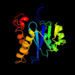
1 c3f9kV_
99.8
18
PDB header: viral protein, recombinationChain: V: PDB Molecule: integrase;PDBTitle: two domain fragment of hiv-2 integrase in complex with ledgf ibd
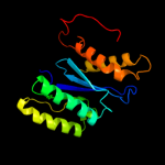
2 d1c0ma2
99.8
15
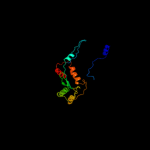
Fold: Ribonuclease H-like motifSuperfamily: Ribonuclease H-likeFamily: Retroviral integrase, catalytic domain3 c1c0mA_
99.8
15
PDB header: transferaseChain: A: PDB Molecule: protein (integrase);PDBTitle: crystal structure of rsv two-domain integrase
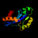
4 d1bcoa2
99.8
17
Fold: Ribonuclease H-like motifSuperfamily: Ribonuclease H-likeFamily: mu transposase, core domain5 d1asua_
99.8
15
Fold: Ribonuclease H-like motifSuperfamily: Ribonuclease H-likeFamily: Retroviral integrase, catalytic domain6 d1exqa_
99.8
14
Fold: Ribonuclease H-like motifSuperfamily: Ribonuclease H-likeFamily: Retroviral integrase, catalytic domain7 c3nf9A_
99.7
17
PDB header: hydrolase/hydrolase inhibitorChain: A: PDB Molecule: integrase;PDBTitle: structural basis for a new mechanism of inhibition of hiv integrase2 identified by fragment screening and structure based design
8 c1ex4A_
99.7
18
PDB header: viral proteinChain: A: PDB Molecule: integrase;PDBTitle: hiv-1 integrase catalytic core and c-terminal domain
9 c1bcoA_
99.7
16
PDB header: transposaseChain: A: PDB Molecule: bacteriophage mu transposase;PDBTitle: bacteriophage mu transposase core domain
10 c3kksB_
99.7
15
PDB header: dna binding proteinChain: B: PDB Molecule: integrase;PDBTitle: crystal structure of catalytic core domain of biv integrase in crystal2 form ii
11 d1c6va_
99.7
18
Fold: Ribonuclease H-like motifSuperfamily: Ribonuclease H-likeFamily: Retroviral integrase, catalytic domain12 d1cxqa_
99.7
18
Fold: Ribonuclease H-like motifSuperfamily: Ribonuclease H-likeFamily: Retroviral integrase, catalytic domain13 c1k6yB_
99.7
19
PDB header: transferaseChain: B: PDB Molecule: integrase;PDBTitle: crystal structure of a two-domain fragment of hiv-1 integrase
14 d1hyva_
99.6
18
Fold: Ribonuclease H-like motifSuperfamily: Ribonuclease H-likeFamily: Retroviral integrase, catalytic domain15 c3hpgC_
99.6
18
PDB header: transferaseChain: C: PDB Molecule: integrase;PDBTitle: visna virus integrase (residues 1-219) in complex with ledgf2 ibd: examples of open integrase dimer-dimer interfaces
16 c3l2tB_
99.6
12
PDB header: recombination/dnaChain: B: PDB Molecule: integrase;PDBTitle: crystal structure of the prototype foamy virus (pfv) intasome in2 complex with magnesium and mk0518 (raltegravir)
17 c3dlrA_
99.4
12
PDB header: transferaseChain: A: PDB Molecule: integrase;PDBTitle: crystal structure of the catalytic core domain from pfv2 integrase
18 c3hosA_
98.7
17
PDB header: transferase, dna binding protein/dnaChain: A: PDB Molecule: transposable element mariner, complete cds;PDBTitle: crystal structure of the mariner mos1 paired end complex with mg
19 c2f7tA_
97.4
15
PDB header: dna binding proteinChain: A: PDB Molecule: mos1 transposase;PDBTitle: crystal structure of the catalytic domain of mos1 mariner2 transposase
20 c3f2kB_
95.6
20
PDB header: transferaseChain: B: PDB Molecule: histone-lysine n-methyltransferase setmar;PDBTitle: structure of the transposase domain of human histone-lysine2 n-methyltransferase setmar
21 c3mwmA_
not modelled
44.9
25
PDB header: transcriptionChain: A: PDB Molecule: putative metal uptake regulation protein;PDBTitle: graded expression of zinc-responsive genes through two regulatory2 zinc-binding sites in zur
22 d1mzba_
not modelled
43.2
25
Fold: DNA/RNA-binding 3-helical bundleSuperfamily: "Winged helix" DNA-binding domainFamily: FUR-like23 c2fe3B_
not modelled
41.5
30
PDB header: dna binding proteinChain: B: PDB Molecule: peroxide operon regulator;PDBTitle: the crystal structure of bacillus subtilis perr-zn reveals a novel2 zn(cys)4 structural redox switch
24 c2o03A_
not modelled
38.0
27
PDB header: gene regulationChain: A: PDB Molecule: probable zinc uptake regulation protein furb;PDBTitle: crystal structure of furb from m. tuberculosis- a zinc uptake2 regulator
25 c2fu4B_
not modelled
36.5
29
PDB header: dna binding proteinChain: B: PDB Molecule: ferric uptake regulation protein;PDBTitle: crystal structure of the dna binding domain of e.coli fur (ferric2 uptake regulator)
26 c2w57A_
not modelled
36.3
33
PDB header: metal transportChain: A: PDB Molecule: ferric uptake regulation protein;PDBTitle: crystal structure of the vibrio cholerae ferric uptake2 regulator (fur) reveals structural rearrangement of the3 dna-binding domains
27 c2xigA_
not modelled
34.2
27
PDB header: transcriptionChain: A: PDB Molecule: ferric uptake regulation protein;PDBTitle: the structure of the helicobacter pylori ferric uptake2 regulator fur reveals three functional metal binding sites
28 c3e7lD_
not modelled
34.0
17
PDB header: transcription regulatorChain: D: PDB Molecule: transcriptional regulator (ntrc family);PDBTitle: crystal structure of sigma54 activator ntrc4's dna binding2 domain
29 c3eyyA_
not modelled
33.3
31
PDB header: transportChain: A: PDB Molecule: putative iron uptake regulatory protein;PDBTitle: structural basis for the specialization of nur, a nickel-2 specific fur homologue, in metal sensing and dna3 recognition
30 c2kkmA_
not modelled
28.5
15
PDB header: translationChain: A: PDB Molecule: translation machinery-associated protein 16;PDBTitle: solution nmr structure of yeast protein yor252w [residues2 38-178]: northeast structural genomics consortium target3 yt654
31 d2aq0a1
not modelled
21.4
35
Fold: SAM domain-likeSuperfamily: RuvA domain 2-likeFamily: Hef domain-like32 d1f1ea_
not modelled
19.4
20
Fold: Histone-foldSuperfamily: Histone-foldFamily: Archaeal histone33 c1htrP_
not modelled
12.4
26
PDB header: aspartyl proteaseChain: P: PDB Molecule: progastricsin (pro segment);PDBTitle: crystal and molecular structures of human progastricsin at 1.622 angstroms resolution
34 c1u78A_
not modelled
10.8
7
PDB header: dna binding protein/dnaChain: A: PDB Molecule: transposable element tc3 transposase;PDBTitle: structure of the bipartite dna-binding domain of tc32 transposase bound to transposon dna
35 c1yshE_
not modelled
10.7
19
PDB header: structural protein/rnaChain: E: PDB Molecule: 40s ribosomal protein s13;PDBTitle: localization and dynamic behavior of ribosomal protein l30e
36 d1gdta1
not modelled
8.6
20
Fold: DNA/RNA-binding 3-helical bundleSuperfamily: Homeodomain-likeFamily: Recombinase DNA-binding domain37 c1s1hO_
not modelled
8.4
31
PDB header: ribosomeChain: O: PDB Molecule: 40s ribosomal protein s13;PDBTitle: structure of the ribosomal 80s-eef2-sordarin complex from2 yeast obtained by docking atomic models for rna and protein3 components into a 11.7 a cryo-em map. this file, 1s1h,4 contains 40s subunit. the 60s ribosomal subunit is in file5 1s1i.
38 c3iwfA_
not modelled
7.6
7
PDB header: transcription regulatorChain: A: PDB Molecule: transcription regulator rpir family;PDBTitle: the crystal structure of the n-terminal domain of a rpir2 transcriptional regulator from staphylococcus epidermidis to 1.4a
39 c3u5gK_
not modelled
7.6
14
PDB header: ribosomeChain: K: PDB Molecule: 40s ribosomal protein s10-a;PDBTitle: the structure of the eukaryotic ribosome at 3.0 a resolution
40 c3oioA_
not modelled
7.4
47
PDB header: transcription regulatorChain: A: PDB Molecule: transcriptional regulator (arac-type dna-binding domain-PDBTitle: crystal structure of transcriptional regulator (arac-type dna-binding2 domain-containing proteins) from chromobacterium violaceum
41 d1pdnc_
not modelled
6.5
14
Fold: DNA/RNA-binding 3-helical bundleSuperfamily: Homeodomain-likeFamily: Paired domain42 d2gtad1
not modelled
6.1
4
Fold: all-alpha NTP pyrophosphatasesSuperfamily: all-alpha NTP pyrophosphatasesFamily: MazG-like